📅 Published on January 1, 2026 | 🕒 Last updated on June 10, 2026
Overview of Cervical Spine Anatomy
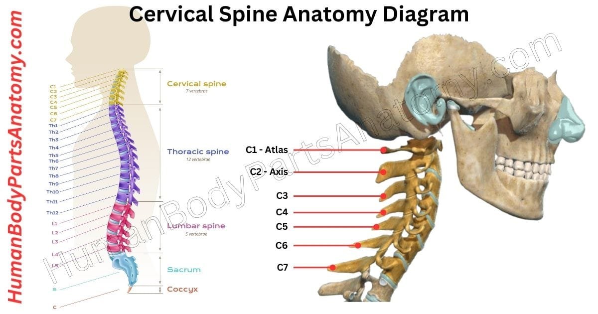
The cervical spine, also called the neck, is the upper part of your spine.[1] The cervical spine anatomy consists of seven small bones called cervical vertebrae, labeled C1-C7.[1][2] These bones support your head and allow your neck to move.[3][1] The first two cervical vertebrae are special. The first one, C1, is called the atlas.[2] It sits at the base of the skull and supports the weight of your head, helping you keep it upright.[2][9] The second vertebra, C2, is called the axis. It works with the atlas to let you turn your head from side to side, like when you say “no.”[2][9]
All seven cervical vertebrae are connected at the back by small joints called facet joints.[1][3] These joints help your neck bend forward, bend backward, and twist smoothly.[3][9]
The cervical spine is supported by muscles, ligaments, tendons, and nerves.[1][3] Together, they provide strength, flexibility, and stability.[1][3] Between each vertebra is a soft cushion called an intervertebral disc.[1][8] These discs act like shock absorbers and reduce stress when you move.[1][8]
The spinal cord runs through the center of the cervical spine.[1][6] It carries messages between your brain and the rest of your body, helping control movement, feeling, and important body functions.[1][6]
Anatomy of the Cervical Spine

Anatomy of Cervical Spine
Vertebrae (C1–C7)
- C1 (Atlas)
- C2 (Axis)
- C3–C6 (Typical Cervical Vertebrae)
- C7 (Vertebra Prominens)
Joints of the Cervical Spine
- Atlanto-occipital joints (C0–C1)
- Atlanto-axial joints (C1–C2)
- Median atlanto-axial joint
- Lateral atlanto-axial joints
- Facet (zygapophyseal) joints (C2–C7)
- Uncovertebral joints (Joints of Luschka) (C3–C7)
- Intervertebral joints (via discs)
Intervertebral Discs
Neural Structures
- Cervical spinal cord
- Cervical spinal nerves (C1–C8)
- Dorsal and ventral nerve roots
- Dorsal root ganglia
- Cervical plexus (C1–C4)
- Brachial plexus roots (C5–T1)
Vascular Structures
- Vertebral arteries
- Vertebral veins
- Deep cervical artery
- Ascending cervical artery
- Internal carotid artery (anterior to spine)
Ligaments of the Cervical Spine
Longitudinal Ligaments
- Anterior longitudinal ligament (ALL)
- Posterior longitudinal ligament (PLL)
Posterior & Supporting Ligaments
- Ligamentum flavum
- Interspinous ligaments
- Supraspinous ligament
- Ligamentum nuchae
Upper Cervical Ligaments
- Transverse ligament of atlas
- Alar ligaments
- Apical ligament
- Tectorial membrane
Spinal Canal & Foramina
- Vertebral canal
- Intervertebral foramina
- Transverse foramina
Supporting & Protective Structures
- Spinal meninges
- Dura mater
- Arachnoid mater
- Pia mater
- Epidural space
- Subarachnoid space (CSF)
Muscles of the Cervical Region
Superficial Muscles
- Sternocleidomastoid
- Trapezius
Suprahyoid & Infrahyoid Muscles
- Digastric
- Mylohyoid
- Omohyoid
- Sternohyoid
- Sternothyroid
Deep Neck Muscles
- Longus colli
- Longus capitis
- Rectus capitis anterior
- Rectus capitis lateralis
Posterior Cervical Muscles
- Splenius capitis
- Splenius cervicis
- Semispinalis capitis
- Semispinalis cervicis
- Suboccipital muscles
- Rectus capitis posterior major/minor
- Obliquus capitis superior/inferior
Key Anatomical Landmarks
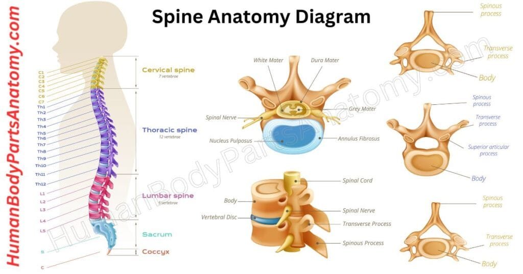
- Spinous processes
- Transverse processes
- Vertebral body
- Uncinate processes
- Dens (odontoid process)
Cervical Spine Anatomy
Vertebrae (C1–C7)
The cervical spine is the top part of the spinal column, located in the neck.[1] It is made up of seven vertebrae, labeled C1 through C7.[1][2] These bones support the head, protect the spinal cord, and allow the neck to move in different directions.[1][2][3]
The cervical spine is divided into two main sections based on structure and function:[2]
- Upper cervical spine: C1 and C2
- Lower cervical spine: C3 to C7
The upper cervical spine works closely with the base of the skull and plays a major role in head movement, balance, and stability.[2][9]
Upper Cervical Spine (C1 and C2)
The first two cervical vertebrae—C1 (atlas) and C2 (axis)—have a unique shape that allows the head to nod, rotate, and stay stable. Together, they provide most of the neck’s rotational movement.[2][9]
1. Atlas (C1)
The atlas is the first cervical vertebra and directly supports the weight of the head. Unlike most vertebrae, it lacksa vertebral body. Instead, its body becomes part of the axis below.[2]
Key features of the atlas include:
- Front (anterior) and back (posterior) arches are connected by strong side portions called lateral masses.[2]
- Upper joint surfaces that connect to the base of the skull, forming the atlanto-occipital joint, which allows the head to nod up and down (the “yes” motion)[2][9]
- Lower joint surfaces that connect with the axis.[2]
- A large central opening (vertebral foramen) that gives the spinal cord extra space.[1][2]
- Transverse foramina in the side projections, which allow the vertebral arteries to pass through and supply blood to the brain.[1][2]
- A small inner joint surface on the front arch that connects with the dens of the axis and is held in place by a strong ligament.[2]
2. Axis (C2)
The axis is the second cervical vertebra and serves as the main pivot point for head rotation, such as turning the head left and right.[2][9]
Important features of the axis include:
- The dens (odontoid process), a vertical bony projection that fits into the atlas and allows rotational movement.[2][9]
- A strong, split (bifid) spinous process that provides attachment for neck muscles.[2]
- Transverse foramina on both sides for the vertebral arteries.[1][2]
- Upper joint surfaces that connect with the atlas.[2]
- Lower joint surfaces that connect with the third cervical vertebra (C3).[2]
Lower Cervical Spine (C3–C7) – Simple Explanation
The lower cervical spine includes the vertebrae from C3 to C7.[2] These bones support the head while allowing the neck to move smoothly in different directions.[2][3]
- The vertebral bodies are small and slightly taller at the back than at the front.[2] This shape helps maintain the normal inward curve of the neck (cervical lordosis).[2][8]
- The top surface of each vertebral body is gently hollowed, while the bottom surface is slightly rounded, allowing the bones to stack and move efficiently.[2]
- Along the upper sides of the vertebral bodies are uncinate processes, which form uncovertebral joints (joints of Luschka). These joints guide neck motion and add stability, especially during side-to-side movement.[2]
- Each vertebra has transverse processes with two bumps (tubercles) on the front and back. Between them is a groove that allows the spinal nerve to pass safely.[2]
- Most cervical vertebrae contain a transverse foramen, an opening that carries the vertebral artery and vein supplying blood to the brain.[1][2]
C7 (Vertebra Prominens)
- The C7 vertebra is different because its transverse foramen usually carries only a small vein, not the vertebral artery.[2]
- The intervertebral foramina are openings formed between neighboring vertebrae.[2] These spaces allow spinal nerves to exit the spine and travel to the shoulders, arms, and hands.[2][4]
- The spinous processes of C3 to C5 are typically split into two tips (bifid).[2]
- C6 and C7 have longer spinous processes.[2]
- C7 has a large, easily felt spinous process at the base of the neck, which is why it is called the vertebra prominens.[2]
This structure of the lower cervical spine balances strength, flexibility, and nerve protection, making normal neck movement and posture possible.[2][3]
Read More – Spine Anatomy: Complete Guide with Parts, Names, Functions & Diagram
Joints of the Cervical Spine
The cervical spine (neck) is the most flexible part of the spine.[3][9] It allows the head and neck to move in many directions while still protecting the spinal cord and supporting the weight of the head.[1][3][9]
Compared to the thoracic (mid-back) and lumbar (lower-back) regions, the neck is specially designed for movement.[3][9] However, not all cervical vertebrae move in the same way or contribute equally to each motion.[3][9]
Occipitoatlantoaxial joints (Upper Cervical Spine)
The joints between the skull and the first two cervical vertebrae are mainly responsible for head movements.[3][9]
1. Skull to C1 (Occiput–Atlas joint)
This joint allows you to nod your head.[3][9]
It mainly provides:
- Forward and backward movement (flexion and extension, like saying “yes”)[3][9]
- A small amount of side bending.[3]
- No rotation.[3][9]
Rotation does not happen here because the joint shape is designed for stability rather than twisting.[3][9]
2. C1 to C2 (Atlas–Axis joint)
This joint allows most of the head’s rotation. In fact, about half of all neck rotation happens here.[3][9]
It allows:
- Large amounts of rotation (like saying “no”).[3][9]
- Small amounts of bending forward and backward.[3][9]
- No side bending.[3]
This rotation is possible because of a bony projection on C2 called the dens, which acts like a pivot for C1.[2][3][9]
Joints between C2 and C7 (Lower Cervical Spine)
Most daily neck movements happen in the lower cervical spine.[3][9]
- Bending forward and backward is greatest at C3–C4, C4–C5, and especially C5–C6.[3][9]
- Side bending and rotation mainly occur at C2–C3, C3–C4, and C4–C5.[3][9]
- Movement gradually decreases as you move down toward the base of the neck.[3][9]
A special feature of the lower cervical spine is coupled motion. This means movements happen together. For example, when you bend your neck to one side, it also rotates to the same side naturally.[3][9]
This effect is stronger in the upper neck and reduces lower down. This is important because stiffness or injury at one level can affect movement in other areas.[3][9]
Main Areas Where Cervical Movement Occurs
Neck movement happens in two main regions:[3]
- Front (anterior) structures: vertebral bodies, intervertebral discs, and uncovertebral joints.[2][3]
- Back (posterior) structures: facet joints, arches, and spinous and transverse processes.[2][3]
1. Anterior Structures of the Cervical Spine
Intervertebral joints
Intervertebral joints are formed between each pair of vertebrae by an intervertebral disc.[1][2][3] In the cervical spine, these discs allow-
- Allow smooth movement between bones.[1][3]
- Help share loads during bending and lifting.[1][3]
- Act as shock absorbers when the spine is compressed.[1][3]
Stability of these joints is mainly provided by the anterior and posterior longitudinal ligaments, along with the uncovertebral joints.[1][3]
Uncovertebral Joints
The uncovertebral joints develop during childhood as small clefts form at the outer edges of the cervical intervertebral discs.[2] Although they are not true synovial joints, they play an important role in-
- Improve side-to-side stability.[2][3]
- Guide neck movement.[2][3]
- Limit excessive side bending.[2][3]
With aging, these joints are commonly affected by degenerative changes, which can contribute to neck pain and, in some cases, nerve compression.[2][8]
2. Posterior Structures and Facet Joints
At the back of the neck, stability is provided by strong ligaments and the facet joints, which are true synovial joints.[1][3]
3. Facet joints:
- Are covered with cartilage.[1][3]
- Contain synovial fluid.[1][3]
- Are enclosed by a joint capsule.[1][3]
The facet joints in the cervical spine are angled in a way that allows more movement than in the mid- or lower back.[3][9]
Because of this joint orientation:
- Rotation always happens with side bending to the same side.[3][9]
- During rotation, one facet slides backward while the other slides forward.[3][9]
- During extension, the facets move backward and downward, closing the joint space.[3][9]
- During flexion, the facets move forward and upward, opening the joint space.[3][9]
This coordinated movement allows the neck to move smoothly while staying stable.[3][9]
Intervertebral Discs
The cervical spine (neck) has six intervertebral discs. There is no disc between the first two neck bones, called the atlas (C1) and axis (C2). The first disc starts between C2 and C3, and the discs continue down to C7–T1, where the neck meets the upper back.[1][2]
Each disc is named after the bone above it. For example, the C4 disc is located between the C4 and C5 vertebrae.[2] These discs act as cushions between the vertebrae, allowing the neck to move smoothly while performing everyday actions such as bending, turning, and supporting the head.[1][2][3]
Like the discs in the lower back, each cervical disc has three main parts:
- Annulus fibrosus – a tough outer layer.[1]
- Nucleus pulposus – a soft, gel-like center.[1]
- Endplates – cartilage layers that attach the disc to the bones.[1]
Although cervical discs work in a similar way to lumbar (lower back) discs, they also have some key differences.[1]
How Cervical Discs Are Different
- Better fit in the spine: The shape of the neck bones helps the discs sit more securely in place.[2]
- Thicker in front: Cervical discs are thicker at the front than the back, which helps form the natural inward curve of the neck and supports good posture.[1][2][8]
- Stronger back portion: The back part of the disc’s outer layer is thicker than in the lower back, adding extra stability to the neck.[1]
- Early changes with age: In the lower cervical spine, the soft center of the disc sits more toward the front and tends to shrink or disappear earlier in life compared to discs in the lower back.[1][8]
Ligaments of the Cervical Spine
The cervical spine is supported by six major ligaments that provide stability while still allowing a wide range of neck movement. Most of these ligaments are found throughout the entire vertebral column, while a few are specific to the cervical (neck) region.[1]
1. Ligaments Present Throughout the Vertebral Column
- Anterior longitudinal ligament (ALL):
This ligament runs along the front (anterior) surface of the vertebral bodies and intervertebral discs. Its main function is to prevent excessive backward bending of the spine (hyperextension).[1] - Posterior longitudinal ligament (PLL):
It is located along the back (posterior) surface of the vertebral bodies and discs. This ligament helps limit excessive forward bending of the spine (hyperflexion) and supports spinal stability.[1] - Ligamentum flavum:
This highly elastic ligament connects the laminae of neighboring vertebrae. It plays an important role in maintaining an upright posture and helps the spine return to an extended position after flexion.[1] - Interspinous ligament:
Found between adjacent spinous processes, this ligament helps control forward bending movements and resists excessive flexion of the spine.[1]
2. Ligaments Unique to the Cervical Spine
- Nuchal ligament:
The nuchal ligament is an extension of the supraspinous ligament in the neck. It attaches to the tips of the spinous processes from C1 to C7 and provides important attachment points for muscles such as the trapezius and deep neck muscles.[1] - Transverse ligament of the atlas:
This strong ligament stretches between the lateral masses of the atlas (C1). It holds the dens of the axis (C2) in place, ensuring stability of the atlanto-axial joint and protecting the spinal cord.[1]
Together, these cervical spine ligaments help maintain neck stability, protect the spinal cord, and allow smooth, controlled movement of the head and neck.[1][3]
Neural Structures
The cervical spinal cord gives rise to many important nerves that control movement and sensation in the head, neck, shoulders, and arms.[4] These nerves are grouped into two main networks called plexuses: the cervical plexus and the brachial plexus.[4][5][7][10] Plexuses allow nerve fibers to mix and redistribute, which helps the body coordinate movements and sensations more effectively.[4][5][10]
1. Cervical Plexus
The cervical plexus is formed from spinal nerves C1 to C4 (and sometimes part of C5). It lies deep in the neck, under the sternocleidomastoid (SCM) muscle, close to other neck muscles.[4]
This plexus supplies nerves to the neck muscles, the diaphragm, and the skin of the neck and upper chest. Its branches are divided into motor (muscle-controlling) and sensory (skin-sensing) nerves.[4]
Motor (Muscle) Branches
These nerves control important functions such as breathing, swallowing, and neck movement:
- Phrenic nerve (C3–C5): Supplies the diaphragm and is essential for breathing.[4]
- Ansa cervicalis (C1–C3): Supplies the infrahyoid muscles, which help with swallowing and speech.[4]
- Suboccipital nerve (C1): Controls small muscles at the base of the skull that move the head.[4]
- C1–C3 branches: Supply deep neck muscles that stabilize and bend the neck.[4]
Contributions to Other Nerves
Some nerve fibers from the cervical region also help form nerves that affect shoulder movement:[4]
- Dorsal scapular nerve (C4–C5): Supplies muscles that stabilize the shoulder blade.[4][5][10]
- Long thoracic nerve (C5–C7): Supplies the serratus anterior muscle, which prevents winging of the shoulder blade.[4][5][10]
Sensory (Skin) Branches
These nerves carry sensation from the skin of the neck, shoulder, and upper chest:[4]
- Lesser occipital nerve (C2): Skin of the upper neck and scalp behind the ear.[4]
- Great auricular nerve (C2–C3): Skin around the ear and jaw.[4]
- Transverse cervical nerve (C2–C3): Skin over the front of the neck.[4]
- Supraclavicular nerves (C3–C4): Skin over the collarbone, shoulder, and upper chest.[4]
2. Brachial Plexus
The brachial plexus supplies the upper limb and is formed by spinal nerves C5 to T1.[4][5][7][10] These nerves pass between neck muscles and then reorganize into a structured network that controls the arm and hand.[5][7][10]
Structure of the Brachial Plexus
- Roots: C5–T1
- Trunks:
- Upper trunk (C5–C6)
- Middle trunk (C7)
- Lower trunk (C8–T1)
- Divisions: Each trunk splits into an anterior and posterior division.[5][10]
- Cords: Lateral, medial, and posterior.[5][10]
- Terminal branches: The main nerves of the arm and hand.[5][7][10]
Major Nerves of the Brachial Plexus
These five main nerves control most arm and hand movements and sensations:
- Musculocutaneous nerve (C5–C7): Bends the elbow and supplies sensation to the outer forearm.[5][7][10]
- Median nerve (C5–T1): Controls many forearm muscles and provides sensation to the thumb, index, middle, and part of the ring finger (palm side).[5][7][10]
- Ulnar nerve (C8–T1): Controls most small hand muscles and supplies sensation to the little finger and part of the ring finger.[5][7][10]
- Radial nerve (C5–T1): Controls arm and forearm extension and supplies sensation to the back of the hand.[5][7][10]
- Axillary nerve (C5–C6): Supplies the shoulder muscles and skin over the shoulder.[5][7][10]
Additional (Smaller) Branches
Several smaller nerves branch off to supply specific muscles and skin areas:
- Nerve to subclavius (C5–C6): Subclavius muscle.[5][10]
- Suprascapular nerve (C5–C6): Rotator cuff muscles.[5][10]
- Lateral pectoral nerve (C5–C7): Pectoralis major.[5][10]
- Medial pectoral nerve (C8–T1): Pectoralis major and minor.[5][10]
- Medial cutaneous nerves of arm and forearm (C8–T1): Sensation to the inner arm and forearm.[5][10]
- Thoracodorsal nerve (C6–C8): Latissimus dorsi.[5][10]
- Upper and lower subscapular nerves (C5–C6): Subscapularis; the lower branch also supplies teres major.[5][10]
Muscles That Attach to the Cervical Spine (Neck)
The cervical spine is made up of seven vertebrae (C1–C7). It supports the head and allows movements such as bending, turning, and tilting. Many muscles attach to these vertebrae to control head and neck motion and to keep the spine stable.[1][3]
- Muscles that attach to the skull mainly move the head.[3]
- Muscles that attach only to the cervical vertebrae mainly move and stabilize the neck.[3]
Below is a clear overview of the main muscle groups connected to the cervical spine.[3]
1. Anterior (Front) Neck Muscles
Main functions: Bend the head and neck forward, stabilize the cervical spine, and assist breathing by lifting the upper ribs.[3]
- Rectus capitis anterior – Attaches to C1 and helps bend the head forward.[3]
- Rectus capitis lateralis – Attaches to C1 and helps side-bend the head and stabilize the upper neck.[3]
- Longus capitis – Runs from C3–C6 to the skull and bends the head and neck forward.[3]
- Longus colli – A deep stabilizing muscle that runs along the front of the cervical spine and helps maintain neck posture.[3]
- Anterior scalene – Attaches from C3–C6 and helps flex the neck and lift the first rib during breathing.[3]
- Middle scalene – Attaches from C1–C7 and helps side-bend the neck and elevate the ribs.[3]
- Posterior scalene – Attaches from C5–C7 and helps move the neck and lift the second rib.[3]
2. Superficial Back Muscles
Main functions: Extend the head and neck, move the shoulder blades, and assist breathing.[3]
- Trapezius – Extends the neck and supports shoulder movement and posture.[3]
- Levator scapulae – Raises the shoulder blade and helps side-bend the neck.[3]
- Rhomboid minor – Stabilizes and retracts the shoulder blade.[3]
- Serratus posterior superior – Assists breathing by lifting the upper ribs.[3]
3. Suboccipital Muscles (Upper Neck Muscles)
Main functions: Provide fine control of head movement and maintain posture.[3]
- Rectus capitis posterior major
- Rectus capitis posterior minor
- Obliquus capitis superior
- Obliquus capitis inferior
These small muscles connect C1 and C2 to the skull and are essential for precise head extension and rotation.[3]
4. Deep Back Muscles – Splenius Group
Main functions: Extend the neck and rotate the head.[3]
- Splenius capitis – Extends and rotates the head.[3]
- Splenius cervicis – Extends and rotates the neck.[3]
5. Deep Back Muscles – Erector Spinae Group
Main functions: Maintain upright posture and extend the head and neck.[3]
- Iliocostalis cervicis – Extends and stabilizes the cervical spine.[3]
- Longissimus capitis – Extends and rotates the head.[3]
- Longissimus cervicis – Extends and stabilizes the neck.[3]
- Spinalis cervicis – Supports neck extension and posture.[3]
6. Deep Back Muscles – Transversospinalis Group
Main functions: Stabilize the cervical spine, extend the neck, and control rotation.[3]
- Semispinalis capitis – Extends and rotates the head.[3]
- Semispinalis cervicis – Extends and stabilizes the neck.[3]
- Multifidus – Provides deep spinal stability and supports posture.[3]
- Rotatores cervicis – Assist with spinal rotation and fine movement control.[3]
The cervical spine is the upper part of the spinal column located in the neck.[1] It consists of 7 vertebrae (C1–C7) that support the head, protect the spinal cord, and allow movements such as bending, rotating, and tilting the neck.[1][2][3]
The cervical spine has seven vertebrae, labeled C1 to C7.[1][2]
C1 (Atlas) supports the skull.[2]
C2 (Axis) allows head rotation.[2]
C3–C7 provide stability, flexibility, and protect nerves.[2]
The cervical spine gives rise to eight cervical nerves (C1–C8) that control sensation and movement in the neck, shoulders, arms, and hands. Compression of these nerves can cause pain, tingling, or weakness in the upper limbs.[4][6]
Common cervical spine conditions include:
Cervical spondylosis (age-related wear and tear).[8]
Herniated or bulging discs.[8]
Cervical radiculopathy (nerve compression).[4][8]
Neck strain or poor posture.[3][8]
Cervical spinal stenosis.[8]
These conditions often cause neck pain, stiffness, or arm numbness.[8]
Problems in the cervical spine can cause cervicogenic headaches, which start in the neck and radiate to the head.[3] Muscle tension, joint dysfunction, or nerve irritation in the upper cervical region are common causes.[3][4]
You should consult a doctor if you experience:
Persistent neck pain lasting more than a few days.[8]
Numbness or weakness in arms or hands.[4][8]
Severe pain after an injury.[8]
Loss of balance or coordination.[4][8]
Early diagnosis helps prevent long-term nerve damage.[4][8]
The human neck contains 7 cervical vertebrae, labeled C1 to C7.[1][2] These vertebrae form the uppermost part of the spine, supporting the head and allowing a wide range of motion, including rotation, flexion, and extension.[1][2][3] The first cervical vertebra (C1) is called the atlas, and the second (C2) is the axis, which play a key role in head movement.[2][9] Understanding the cervical vertebrae is essential for studying human anatomy, spinal health, and posture.[1][2]
The cervical spine is located in the neck region, extending from the base of the skull to the upper chest. It consists of the first seven vertebrae (C1–C7) and supports the head while allowing neck movement such as rotation and bending.[1][2]
Straightening of the cervical spine is not usually dangerous, but it may indicate muscle spasm, poor posture, or neck strain. In some cases, it can be associated with neck pain, stiffness, or nerve compression, especially if symptoms persist.[8][9]
Spondylosis is a degenerative condition of the spine caused by age-related wear and tear of spinal discs, joints, and bones. It commonly affects the cervical (neck) and lumbar (lower back) spine and may cause pain, stiffness, or nerve symptoms.[1][8]
There are seven cervical bones, also called cervical vertebrae, labeled C1 through C7. These bones protect the spinal cord, support the head, and enable neck movement.[1][2]
References-
- Rahman S, Das J. Anatomy, Head and Neck: Cervical Spine. StatPearls Publishing; 2023. https://www.ncbi.nlm.nih.gov/books/NBK557516 PMID: 32491448
- Kaiser JT, Reddy V, Lugo-Pico JG. Anatomy, Head and Neck: Cervical Vertebrae. StatPearls Publishing; 2023. https://www.ncbi.nlm.nih.gov/books/NBK539734 PMID: 31194344
- Bordoni B, Varacallo M. Anatomy, Head and Neck, Neck Movements. StatPearls Publishing; 2023. https://www.ncbi.nlm.nih.gov/books/NBK557555 PMID: 32491677
- Walker C, Uribe JS. Anatomy, Head and Neck: Cervical Nerves. StatPearls Publishing; 2025. https://www.ncbi.nlm.nih.gov/books/NBK538136 PMID: 30725827
- Saker E, O’Rourke DJ, Harrington S, et al. Anatomy, Head and Neck: Brachial Plexus. StatPearls Publishing; 2023. https://www.ncbi.nlm.nih.gov/books/NBK531473 PMID: 30285393
- Cervical Myelopathy. Johns Hopkins Medicine. https://www.hopkinsmedicine.org/health/conditions-and-diseases/cervical-myelopathy
- Brachial Plexus Injury. Johns Hopkins Medicine. https://www.hopkinsmedicine.org/health/conditions-and-diseases/brachial-plexus-injuries
- Cervical spondylosis – Symptoms & causes. Mayo Clinic; 2024. https://www.mayoclinic.org/diseases-conditions/cervical-spondylosis/symptoms-causes/syc-20370787
- Swartz EE, Floyd RT, Cendoma M. Cervical spine functional anatomy and the biomechanics of injury due to compressive loading. J Athl Train. 2005;40(3):155-61. PMID: 16284634 PMCID: PMC1250253 https://pmc.ncbi.nlm.nih.gov/articles/PMC1250253/
- Mitchell BS, Humphreys BK, O’Sullivan E. Anatomy, Shoulder and Upper Limb, Brachial Plexus. StatPearls Publishing; 2023. https://www.ncbi.nlm.nih.gov/books/NBK500016 PMID: 29763192
Medical Disclaimer
All content on HumanBodyPartsAnatomy.com is educational and based on verified, peer-reviewed medical sources. Articles are authored or reviewed by qualified medical or biomedical professionals to ensure accuracy.
This website does not provide medical advice, diagnosis, or treatment. Always consult a licensed healthcare professional for personal medical guidance.
No commercial or promotional interests influence the medical content published on this site.