📅 Published on July 13, 2025 | 🕒 Last Reviewed & Updated on August 1, 2026
Overview of Brain Anatomy
The brain is a part of the control center of your body. It manages everything from your thoughts and emotions to your movements and memories. It also controls functions like breathing, heartbeat, and body temperature, running without you even noticing. Every part of who you are—your personality, reactions, and decisions—starts in your brain.[1][2]
Different parts of the brain have specific roles, but they work together as one powerful network. It is a complex organ composed of approximately 86 billion neurons,[3] interconnected by trillions of synapses.[4] These neurons send and receive signals to share information quickly and precisely. The brain translates these signals into instructions that your body can understand and respond to.[1]
The brain is directly linked to your spinal cord, forming the central nervous system (CNS).2] Together, they act as the body’s communication highway, helping you think, feel, and move the way you do.
How does the brain grow?
At birth, the human brain weighs around 350 to 400 grams, which is only about 25% of its full adult size.[5] By adulthood, the brain typically weighs between 1.4 and 1.45 kilograms.[6]
Even though it starts small, the brain grows very quickly in early childhood. It experiences its fastest growth during the first three years, and by the age of five, it reaches nearly 90% of its adult volume.[7]
In adults, the brain’s dimensions average about 140 mm in width, 167 mm in length, and 93 mm in height.[1]
Brain development doesn’t stop after childhood—it continues throughout life, although the most significant changes happen in the early years.[8]
During the first four years, the brain’s structure and functions undergo dramatic transformations that shape how we learn, process emotions, and respond to the world around us.[7]
Although the brain accounts for roughly 2% of total body weight, it consumes about 20% of the body’s oxygen and energy.[9] It is a vital organ that controls everything from thinking and memory to movement and senses.[1]
Its growth and development in the early years lay the foundation for lifelong learning, health, and behavior.[8]
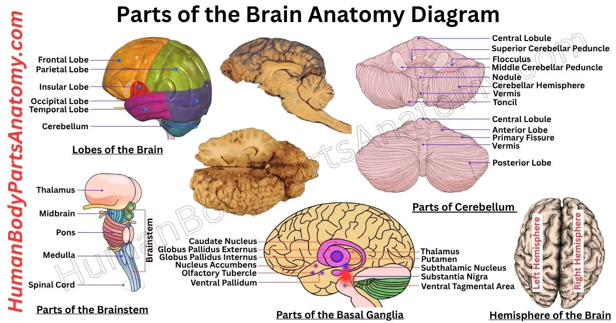
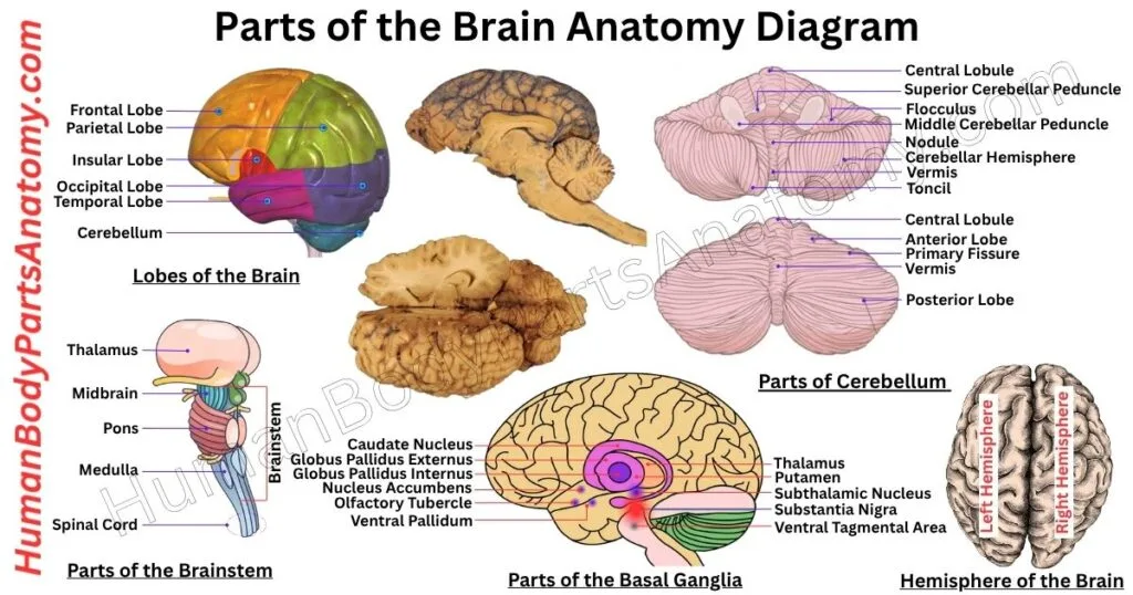
Parts of the Brain Diagram
Parts of the Brain and Their Functions
1. Cerebrum
a. Cerebral Hemispheres
- Left Hemisphere
- Right Hemisphere
b. Cerebral Cortex
- Frontal Lobe
- Prefrontal Cortex
- Premotor Cortex
- Primary Motor Cortex
- Broca’s Area
- Parietal Lobe
- Primary Somatosensory Cortex
- Posterior Parietal Cortex
- Temporal Lobe
- Auditory processing
- Wernicke’s area
- Medial Temporal Lobe
- Occipital Lobe
- Primary Visual Cortex (V1)
- Visual Association Areas (V2–V5)
d. White Matter
e. Corpus Callosum
2. Diencephalon
- Thalamus
- Hypothalamus
- Epithalamus
- Subthalamus
3. Limbic System
- Hippocampus
- Amygdala
- Fornix
- Cingulate Gyrus
- Parahippocampal Gyrus
- Mammillary Bodies
- Septal Nuclei
4. Basal Ganglia
- Striatum
- Caudate nucleus
- Putamen
- Nucleus accumbens & olfactory tubercle
- Globus pallidus
- Subthalamic nucleus
- Substantia nigra
5. Cerebellum
- Cerebellar Hemisphere
- Relations
- Superior
- Anterior
- Posterior and Lateral
- Surfaces
- Lobes
- Anterior Lobe
- Posterior Lobe
- Flocculonodular Lobe
- Fissures
- Primary Fissure
- Posterolateral Fissure
- Horizontal Fissure
- Zones
- Vermal Zone
- Paravermal (Intermediate) Zone
- Lateral Zones
- Cerebellar Cortex
- Deep Nuclei
- Fastigial Nucleus
- Globose Nucleus
- Emboliform Nucleus
- Dentate Nucleus
- Cerebellar Peduncles
- Superior
- Middle
- Inferior
- White Matter (Arbor Vitae)
6. Brainstem
a. Midbrain
- Tectum (posterior part)
- Superior colliculi
- Inferior colliculi
- Tegmentum (central part)
- Red nucleus
- Substantia nigra
- Reticular formation
- Cerebral aqueduct
- Cerebral peduncles (crura cerebri)
b. Pons
- Basilar (ventral) part
- Corticospinal and corticobulbar tracts
- Pontine nuclei
- Tegmentum (dorsal part)
- Reticular formation
- Cranial nerve nuclei
- Medial lemniscus
- Middle cerebellar peduncles
c. Medulla Oblongata
- Pyramids
- Olives (olivary bodies)
- Gracile and cuneate nuclei
- Reticular formation
- Vital centers
- Cranial nerve nuclei
7. Ventricular System
- Lateral ventricles
- Third ventricle
- Fourth ventricle
- Cerebral aqueduct (connects 3rd and 4th ventricles)
- Central Canal (spinal cord)
- Choroid plexus (produces CSF)
8. Meninges (Protective Layers)
a. Dura Mater
- Periosteal Layer
- Meningeal Layer
- Dural Sinuses (e.g., Superior Sagittal Sinus)
b. Arachnoid Mater
- Subarachnoid space (contains CSF)
c. Pia Mater
9. Blood Supply
a. Arterial Supply
- Internal Carotid Arteries
- Vertebral Arteries
- Basilar Artery
- Circle of Willis (anastomosis of arteries)
- Anterior Cerebral Arteries (ACA)
- Middle Cerebral Arteries (MCA)
- Posterior Cerebral Arteries (PCA)
- Communicating arteries (anterior & posterior)
b. Venous Drainage
- Cerebral veins
- Dural venous sinuses (superior sagittal, transverse, sigmoid)
- Internal Jugular Vein
Parts of the Brain Anatomy
The brain is divided into four main regions: the cerebrum, cerebellum, diencephalon, and brainstem. Each region has specialized functions and contains multiple substructures.[2][10]
1. Cerebrum
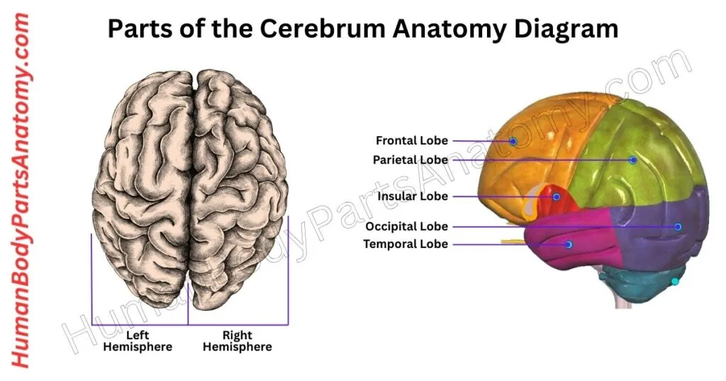
The cerebrum is the largest part of the brain and is located at the front and top of the head. Its name comes from the Latin word for “brain.”[10]
The cerebrum is responsible for many important functions that affect everyday life. It helps you think, feel emotions, control movement, and communicate through speech.[1]
This part of the brain also manages voluntary actions—those things you do consciously—such as making decisions, planning tasks, solving problems, and responding to your surroundings.[1][10]
Functions of the Cerebrum
- Senses: Your cerebrum processes all the information from your five senses — sight, hearing, smell, taste, and touch.[1][10]
- Language: It helps you read, write, and speak by managing the parts of the brain linked to communication.[1]
- Short-term memory: It helps you remember little things for a short time — like keeping a grocery item in mind until you buy it.[1][10]
- Personality and behavior: The front part of the cerebrum, called the frontal lobe, shapes your personality and controls your behavior. It helps you think before you act or speak.[1][10]
- Movement: Certain areas in the cerebrum send signals to your muscles, letting you move your body as needed.[1]
- Learning and thinking: The cerebrum works when you are learning something new, solving problems, or making decisions.[1]
Read More – Cerebrum Anatomy: Guide with Parts, Lobes, Hemispheres, Functions & Diagram
2. Diencephalon
The diencephalon is a centrally located part of the brain, positioned just above the brainstem. It consists of four key structures: the thalamus, hypothalamus, epithalamus, and subthalamus.[2][11]
Each of these components plays a crucial role in maintaining the body’s internal balance. The diencephalon helps relay sensory and motor signals to the cerebral cortex.[11]
It works with the endocrine system to control hormone release and helps regulate the body’s circadian rhythm-our natural sleep-wake cycle.[11]
In addition, the diencephalon plays a crucial role in regulating important functions, including controlling body temperature, managing hunger and thirst, and maintaining emotional balance.[11]
When any part of the diencephalon becomes damaged or dysfunctional, it can lead to serious health issues, affecting both physical and mental well-being.[11]
a. Thalamus
The thalamus is a pair of oval-shaped structures made mostly of gray matter, located on either side of the brain’s third ventricle.[11]
These two halves are joined by a small connection called the interthalamic adhesion. It sits just above the subthalamus and is linked to the epithalamus. The thalamus acts as a vital relay center in the brain.[1][11]
A network of nerve fibers connects the thalamus to the cerebral cortex, allowing it to process and transmit a wide range of information.[11]
Blood supply to the thalamus comes from four main arteries: the tuberothalamic, paramedian, thalamogeniculate, and the medial and lateral posterior choroidal arteries.[11]
Functionally, the thalamus plays a key role in receiving and sending out sensory and motor signals. Except for the smell, all sensory information—including vision, sound, touch, pain, and temperature—passes through the thalamus before reaching the brain’s outer layer for interpretation. It also helps regulate consciousness, alertness, and attention.[1][11]
Additionally, the thalamus is connected to the limbic system, which ties it to emotions and motivation. It contributes to motor control, language processing, and cognitive functions, making it one of the brain’s central hubs for communication and coordination.[11]
- Cognitive Control Functions of the Human Thalamus
- Thalamus Seizure Detection With a Deep Brain Stimulator System
b. Hypothalamus
The hypothalamus plays a key role in maintaining homeostasis, which means it helps keep the body’s internal environment stable and balanced.[2][12]
It serves as a control center for many automatic (autonomic) functions, such as regulating hunger, thirst, blood pressure, body temperature, and more.[1][12]
Besides these roles, the hypothalamus also influences sexual behavior, motivation, and how the body reacts to stress. It responds to a variety of signals like light, emotional arousal, stress levels, and smells to carry out its functions effectively.[12]
If the hypothalamus becomes damaged, it can lead to a range of problems, including-
- Increased aggression
- Chronic stress
- Constant fatigue
- Abnormal body temperatures (either too low or too high)
- Fluctuations in body weight
- Disturbances in sexual desire, either increasing or decreasing it[12]
c. Epithalamus
The epithalamus forms the rear segment of the diencephalon and is built from three main parts:
- Habenula: A small paired nucleus that links to other diencephalic regions through the habenular commissure, a bundle of nerve fibers.[11]
- Stria medullaris: Another fiber tract that carries signals from the septal area, lateral preoptic region of the hypothalamus, and anterior thalamic nuclei into the habenula.[11]
- Pineal gland: A single, pea-sized structure (about 5–7 mm across) that produces melatonin, the hormone that helps regulate our daily sleep-wake rhythm.[2][11]
Together, these elements of the epithalamus not only coordinate with each other but also send and receive connections to the limbic system and basal ganglia, integrating emotional, motivational, and motor information.[11]
d. Subthalamus
The subthalamus is located just below the thalamus in the diencephalon. It acts as a key control center that helps coordinate brain activity.[11] Its main structure is the subthalamic nucleus, which is divided into three specialized zones:
- Dorsolateral (motor) region – fine-tunes movement by adjusting motor signals.[11]
- Ventromedial (associative) region – supports thinking, learning, and decision-making.[11]
- Medial (limbic) region – connects movement with emotions and motivation.[11]
Around the subthalamic nucleus are several important structures:
- The zona incerta influences a wide range of functions, including attention, reflex control, smooth and cardiac muscle activity, and even glandular responses.[11]
- The reticular nucleus manages the flow of signals from the thalamus to the brain’s cortex, helping maintain alertness and focus.[11]
- The perigeniculate nucleus plays a role in shaping visual information before it reaches the visual cortex.[11]
- The subthalamus also serves as a busy communication hub, with many nerve fibers linking it to other brain regions. It forms critical connections between the nervous, endocrine, and limbic systems. It allows emotions and hormones to affect movement, sensation, and body regulation.[11]
3. Limbic System
a. Hippocampus
The hippocampus exists as a pair, with one located in each half of the brain. Its name comes from its resemblance to a seahorse, and it plays a central role in memory.[1][13]
It is where our personal experiences, known as episodic memories, are first formed and organized before being transferred to long-term storage in other areas of the brain.[13]
Beyond memory, the hippocampus helps link memories to our senses. For example, the smell of gingerbread might remind someone of Christmas because of the connections formed here.[13]
It also plays a crucial role in helping us understand where we are and how to move through our environment, supporting spatial awareness and navigation.[13]
Uniquely, the hippocampus is one of the few regions in the adult brain where new neurons continue to grow from stem cells—a process called neurogenesis.[13]
This ability to create new brain cells supports learning and adaptation, making the hippocampus a key player in brain flexibility or plasticity.[13]
b. Amygdala
The amygdala is shaped like an almond and sits right next to the hippocampus in the brain. While both structures help form new memories, the amygdala’s key role is to process emotions and link them to these memories.[2][13]
This close partnership helps explain why emotionally charged experiences are often remembered more vividly than neutral ones.[13]
The amygdala governs emotional reactions such as fear, anger, and happiness. Its strong tie to emotions allows it to convert emotionally intense moments into lasting memories, while less emotional events may fade over time.[13]
Beyond memory, the amygdala is also involved in emotional learning — helping the brain recognize patterns that are rewarding or threatening based on past experiences.[13]
Imbalances in amygdala function can contribute to mental health conditions, including social anxiety and addiction, where emotional processing becomes disrupted.[13]
c. Fornix
The fornix is a curved bundle of white matter located deep inside the brain, just below the corpus callosum. It is within the central part of the cerebral hemispheres.[13]
It is a crucial structure of the limbic system, serving as the primary communication route between the hippocampus and various subcortical regions.[13]
The fornix begins in the hippocampus, which is found in the inner part of the temporal lobe. From there, it travels forward and upward, curving over the thalamus and extending toward the diencephalon and the basal forebrain.[13]
Through these connections, the fornix helps coordinate several essential brain functions, including learning, memory storage, emotional processing, and sexual behavior.[13]
d. Cingulate Gyrus
The cingulate gyrus plays a key role in controlling emotions, managing certain behaviors, processing pain, and regulating some automatic body functions. It is especially important in how the brain reacts to fear and stressful or negative situations.[13]
If the cingulate gyrus is damaged, it can lead to problems such as inappropriate emotional responses, difficulties with learning, reduced fear reactions, and other behavioral issues.[13]
e. Parahippocampal Gyrus
The parahippocampal gyrus is found along the inner surface of the cerebral hemisphere and is visible from the underside of the brain. It plays an important role in processing, storing, and retrieving memories.[13]
At its front, this gyrus curves inward to form a structure known as the uncus. Moving backward, it continues and blends with the lingual gyrus as it approaches the medial part of the occipital lobe.[13]
On either side, the parahippocampal gyrus is bordered by the collateral sulcus and the rhinal sulcus, which separate it from the fusiform (or occipitotemporal) gyrus.[13]
The subiculum is positioned above the parahippocampal gyrus, which acts as a bridge connecting it to other structures within the medial temporal lobe, especially those involved in hippocampal formation. This formation includes the hippocampus, subiculum, and dentate gyrus.[13]
During early brain development, the medial temporal lobe undergoes two folds, creating an S-shaped curve. This folding arrangement places the subiculum directly above the parahippocampal gyrus.[13]
The hippocampus sits above and slightly lateral to the subiculum, while the dentate gyrus lies in between, nestled against both the hippocampus and subiculum.[13]
f. Mammillary Bodies
The mammillary bodies are two small, round structures found on the underside of the brain. It forms part of the diencephalon and is included in the limbic system.[13]
It is positioned at the ends of the anterior arches of the fornix and contains two groups of nerve cell clusters: the medial and lateral mammillary nuclei.[13]
Although structurally distinct, these bodies are often considered part of the hypothalamus. They serve as important relay centers, receiving signals from the hippocampus and amygdala and sending them to the thalamus through the mammillothalamic tract.[13]
This pathway contributes to the Papez circuit, a network involved in controlling emotions and memory. Specifically, the mammillary bodies help process recollective memory—our ability to remember specific experiences.[13]
Damage to the medial mammillary nucleus has been shown to impair spatial memory, particularly in animal studies.[13]
g. Septal Nuclei
The septal area, also called the medial olfactory area, is a small but important region of the brain located deep inside the frontal lobe, near a thin membrane called the septum pellucidum. It contains two main parts: the lateral septum and the medial septum.[13]
Within this region are clusters of nerve cells known as the septal nuclei. These nuclei are divided into groups—dorsal, ventral, medial, and caudal—and are well-connected to various brain structures.[13]
They receive inputs from areas such as the olfactory bulb, hippocampus, amygdala, hypothalamus, midbrain, habenula, thalamus, and the cingulate gyrus.[13]
One of their most important roles is helping to produce theta waves, a type of brain rhythm closely linked to learning and memory in the hippocampus.[13]
Despite its name, the septal area is not directly involved in detecting smells. Instead, it plays a key role in emotion, motivation, and reward.[13]
Along with the nucleus accumbens, it forms part of the brain’s reward system, influencing how we experience pleasure and drive.[13]
4. Basal Ganglia
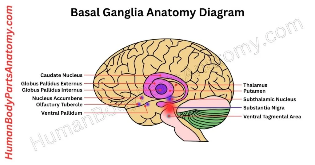
The basal ganglia are a group of deep brain structures made up of several subcortical nuclei. These nuclei are grouped based on shared functions rather than physical location. They are spread across different regions of the brain, mainly within the forebrain.[1]
The primary role of the basal ganglia is to fine-tune movements. They act as a regulatory system for motor control by sending feedback to the cerebral cortex.[1]
This feedback helps suppress unwanted movements and ensures that body motions are smooth and controlled. One of the key ways it achieves this is by reducing excessive excitatory signals going to the motor cortex.[1]
Besides movement, the basal ganglia also help process emotions and cognitive functions. Different parts of the basal ganglia receive and send information from specific brain regions:
- The putamen gets input from motor and sensory areas and connects back to motor regions. It makes it crucial for the motor loop.[1]
- The caudate nucleus gets signals from association areas and connects with the prefrontal cortex. It plays a role in planning and decision-making.[1]
- The ventral striatum includes the nucleus accumbens, receives input from emotional centers (limbic system), and is involved in motivation and emotional behavior.[1]
Although the term “basal ganglia” is commonly used, it is technically incorrect. In neuroanatomy, “ganglia” refer to clusters of neuron cell bodies located outside the central nervous system. Since the basal ganglia lie within the brain, the more accurate term is “basal nuclei.”[1]
The basal ganglia are organized into three functional groups:
- Input nuclei – receive information from other brain areas.[1]
- Intrinsic nuclei – process this information internally.[1]
- Output nuclei – send the final signals to other brain regions.[1]
Key Structures of the Basal Ganglia:
- Striatum: It is made up of the caudate nucleus and putamen. This is the main entry point for signals coming into the basal ganglia.[1]
- Nucleus accumbens: It is found where the caudate and putamen meet at the front. It is part of the ventral striatum and is involved in emotional and reward pathways.[1]
- Globus pallidus: It is split into external (GPe) and internal (GPi) segments. These help filter and relay signals during movement processing.[1]
- Subthalamic nucleus (STN): It is located in the diencephalon and works closely with the globus pallidus to fine-tune motor commands.[1]
- Substantia nigra: It is found in the midbrain and has two parts—
- Pars compacta (SNc): Produces dopamine, a key chemical for movement regulation.[1]
- Pars reticulata (SNr): Acts as an output station, sending signals to other motor centers.[1]
Together, these components form a highly organized and balanced system that controls how we move, feel, and think.[1]
Read More – Basal Ganglia Anatomy: Guide with Parts, Structure & Role in Movement
5. Cerebellum
The cerebellum is a key part of the hindbrain that plays a crucial role in controlling movement and maintaining balance. Though small in size, it holds more than half of all the nerve cells of the brain.[1][14]
The cerebellum gets signals from other brain regions and uses this information to fine-tune our movements and behaviors.[14]
Together with the cerebrum and brainstem, the cerebellum forms one of the three major parts of the brain. While it doesn’t directly make movements happen, it helps make them smooth, accurate, and well-timed.[14]
Here are some important roles of the cerebellum:
- Keeps balance and posture so you can stand and move steadily.[14]
- Coordinates muscle actions, helping your arms and legs move together.[14]
- Guides eye movements, allowing smooth tracking of objects.[14]
- Supports motor learning, like learning to ride a bike or play an instrument.[14]
- Contributes to speech, helping you speak clearly and fluently.[14]
- Influences thinking and emotions, playing a role in focus, planning, and mood regulation.[14]

Read More – Cerebellum Anatomy: Guide with Parts, Structure, Functions & Diagram
6. Brainstem
The brainstem is a small but crucial structure at the base of the brain that connects it to the spinal cord. Though it accounts for just about 2.6% of the total brain weight, it plays a vital role in keeping us alive by controlling many automatic body functions.[1][15]
It consists of three major parts:
- Midbrain: It is located at the top of the brainstem and helps coordinate body movements, especially eye movements. It also processes visual and auditory signals, helping us respond to what we see and hear.[15]
- Pons: It is positioned in the middle and acts as a communication hub between different parts of the brain. It supports balance, hearing, facial movements, and eye coordination.[15]
- Medulla Oblongata: It is found at the bottom, just above the spinal cord, and it regulates essential functions like breathing, heartbeat, blood pressure, and swallowing—functions we don’t have to think about.[15]
The brainstem also serves as a vital pathway for messages between the brain and the rest of the body.[15] It contains several important nerve tracts:
- Corticospinal tract: Sends movement signals from the brain to the muscles.[15]
- Dorsal column–medial lemniscus pathway: Carries messages related to touch, position, and vibration.[15]
- Spinothalamic tract: Transmits signals of pain, temperature, and crude touch.[15]
Out of the twelve cranial nerves, ten originate from the brainstem. These nerves control key abilities like facial expressions, eye movement, swallowing, and taste.[15]
The brainstem also plays a key role in maintaining wakefulness and regulating the sleep-wake cycle.[15]

Read More – Brainstem Anatomy: Complete Guide with Midbrain, Pons & Medulla Explained
7. Ventricular System
The cerebral ventricles are a group of connected, fluid-filled spaces located deep inside the brain. They are leftover hollow areas from early brain development and are found in the forebrain and brainstem.[16]
These ventricles don’t do any work themselves, but they help protect the brain and are useful for doctors to study brain structure.[16]
- The largest are the lateral ventricles, one in each half of the brain. In cross-sections, their bottom side is next to the basal ganglia, the top is near the corpus callosum, and the inner wall is lined by a thin sheet called the septum pellucidum.[16]
- The third ventricle lies between the two thalamus areas and connects to the lateral ventricles through a small hole called the interventricular foramen. This ventricle continues down into the cerebral aqueduct, a narrow tube running through the midbrain.[16]
- The aqueduct leads into the fourth ventricle, which sits behind the pons and medulla. From there, the space narrows to form the central canal of the spinal cord.[16]
These ventricles are filled with cerebrospinal fluid (CSF), a clear liquid that protects and supports the brain and spinal cord. The fluid is made in a tissue called the choroid plexus, found inside the lateral, third, and fourth ventricles.[16]
CSF flows through the ventricles and then into the space around the brain and spine. It finally gets absorbed into the blood through small structures called arachnoid granulations.[16]
8. Meninges (Protective Layers)
Dura Mater
The dura mater is the outermost protective layer of the brain and spinal cord.[17] It is a strong, fibrous membrane made of two closely joined sheets:
- Periosteal layer – the outer part, firmly attached to the inside of the skull.[17]
- Meningeal layer – the inner part, also known as the dural border cell layer.[17]
These layers usually stay fused, forming a single tough cover. But in certain spots, they split apart to create dural venous sinuses—channels that drain blood from the brain back toward the heart.[17]
Beneath the dura are two more protective layers: the arachnoid mater and the pia mater, which lies directly on the brain and spinal cord.[17]
In specific areas, the dura mater folds inward to help divide the brain and support its weight:
- The falx cerebri separates the two sides of the brain (right and left hemispheres).[17]
- The tentorium cerebelli lies between the cerebrum and the cerebellum, forming a supportive roof over the lower brain.[17]
During early development, the dura mostly forms from neural crest cells, with additional support from mesodermal tissue as growth continues.[17]
Overall, this tough, folded structure anchors the brain in place and helps control pressure inside the skull by directing blood flow through its drainage channels.[17]
Cranial Arachnoid Mater
The cranial arachnoid mater is a delicate, web-like membrane that lies between two other protective brain layers—the dura mater on the outside and the pia mater on the inside.[17]
Between the arachnoid and the dura is the subdural space, which may contain a thin film of fluid, though this is debated.[17]
Below the arachnoid lies the subarachnoid space, a crucial area filled with cerebrospinal fluid (CSF) that cushions the brain and also houses all major cerebral blood vessels.[17]
The outer side of the arachnoid tightly adheres to the dura, forming a barrier that stops CSF from leaking into the subdural space.[17]
Where the dura mater creates venous sinuses, the arachnoid mater extends into them as small knob-like structures called arachnoid granulations, which help absorb CSF into the bloodstream.[17]
On its inner side, the arachnoid sends out fine threads called arachnoid trabeculae. These fibers stretch across the subarachnoid space and anchor to the pia mater, which closely hugs the brain’s surface.[17]
Because of their shared origin and structure, the arachnoid and pia mater are together known as the leptomeninges.[17]
Pia Mater
The cranial pia mater is a very thin, soft layer that covers the brain and follows all its curves and folds. Although it seems to touch the brain, there’s actually a tiny space between them called the subpial space, formed by special cells called astrocytes.[17]
This layer has many small blood vessels. Some of these vessels run inside the pia mater, while others are held in place by thin strands called arachnoid trabeculae from the layer above.[17]
The pia mater helps protect the brain by keeping its tissue separate from blood vessels. It also helps control chemicals like neurotransmitters so they don’t affect the brain for too long.[17]
9. Blood Supply
The brain relies on a constant and rich supply of oxygen and nutrients, which are delivered through a dense network of blood vessels.[18]
This blood flow primarily originates from four key arteries: the right and left internal carotid arteries, and the right and left vertebral arteries.[18]
These arteries work together to form two main circulatory routes in the brain: the anterior circulation and the posterior circulation.[18]
Internal Carotid Arteries
The internal carotid arteries originate in the neck and enter the skull through the carotid canal. From there, they travel through the cavernous sinus and pass beside the optic chiasm in the middle cranial fossa.[18]
Once inside the brain, each internal carotid artery splits into three major branches:[18]
- Anterior cerebral artery
- Middle cerebral artery
- Posterior communicating artery
These branches supply blood to the front and middle portions of the brain, including the cerebral hemispheres.[18]
Vertebral Arteries
The vertebral arteries branch off from the subclavian arteries in the chest. They ascend through small openings in the cervical vertebrae and enter the skull via the foramen magnum.[18]
Inside the skull, both vertebral arteries join at the brainstem to form the basilar artery. This artery then continues upward and splits into the posterior cerebral arteries, which supply the back portions of the brain.[18]
On their way to forming the basilar artery, the vertebral arteries give off several important branches:[18]
- Posterior spinal artery
- Anterior spinal artery
- Posterior inferior cerebellar artery (PICA)
These arteries supply the spinal cord, medulla oblongata, and parts of the cerebellum. Damage to these vessels can lead to problems with movement or sensation, depending on the area affected.[18]
Together, the anterior and posterior circulations form a complete loop known as the Circle of Willis. It helps maintain blood flow even if one part gets blocked.[18]
Basilar Artery
The basilar artery is created where the left and right vertebral arteries meet at the base of the skull. As it travels upward along the brainstem, it gives off several key branches:[18]
- Anterior inferior cerebellar arteries (AICA): supply the lower parts of the cerebellum and portions of the pons.[18]
- Labyrinthine (internal auditory) arteries: nourish the inner ear structures.[18]
- Pontine branches: feed the pons itself.[18]
- Superior cerebellar arteries (SCA): serve the upper regions of the cerebellum and the midbrain.[18]
- Together, these vessels ensure the brainstem and cerebellum receive the oxygen-rich blood they need to coordinate movement, posture, and vital reflexes.[18]
Anterior Cerebral Arteries (ACA)
Each anterior cerebral artery begins as a continuation of the internal carotid artery, curving forward and inward above the optic nerve toward the top of the brain. The ACAs run along the midline in the longitudinal fissure and wrap around the corpus callosum.[18] They primarily supply blood to:
- The medial (inner) surfaces of the frontal lobes govern decision-making, personality, and voluntary movement of the lower limbs.[18]
- The medial parts of the superior parietal lobes are involved in sensation and spatial orientation.[18]
By delivering oxygenated blood to these central brain regions, the ACAs support critical functions like leg movement, bladder control, and aspects of cognition.[18]
Middle Cerebral Arteries (MCAs)
The middle cerebral arteries are large blood vessels that branch from the internal carotid arteries. They move out through a groove in the brain called the Sylvian fissure and spread across the outer parts of the brain.[18]
These arteries supply blood to the sides of the frontal, parietal, and temporal lobes. These are the areas that help control movement, touch, and speech.[18]
They also have small branches called lenticulostriate arteries that go deeper to supply the basal ganglia (which help with movement) and the internal capsule (a major path for nerve signals).[18]
Posterior Cerebral Arteries (PCAs)
The posterior cerebral arteries start from the top of the basilar artery and curve around the midbrain toward the back of the brain.[18]
They supply the occipital lobe (which handles vision), the underside of the temporal lobe (used for recognizing faces and objects), and the back of the parietal lobe (important for understanding space and direction).[18]
These arteries also send branches to the thalamus and midbrain, which are key for passing on sensory information and helping with eye movement. They also carry visual signals to the brain through pathways called optic radiations.[18]
Venous Drainage
The venous circulation of the brain is unique compared to the rest of the body. Instead of following the usual pattern where veins run alongside arteries, the brain uses a specialized drainage system.[19]
In this system, large veins are located within the layers of the dura mater, the tough outer covering of the brain. These veins form wide channels called dural venous sinuses.[19]
The superior and inferior sagittal sinuses, running along the top and inner surfaces of the brain, collect blood mainly from the cerebrum. The cavernous sinuses, found near the base of the skull behind the eyes, drain blood from the front parts of the brain and face.[19]
All these sinuses eventually direct blood into the sigmoid sinuses, which then flow into the internal jugular veins—the main exit routes for blood leaving the brain.[19]
Because the brain relies almost entirely on the internal jugular veins for venous drainage, any blockage can severely disrupt this flow. When blood cannot drain properly, pressure builds up inside the skull.[19]
This increase in pressure is known as raised intracranial pressure and can lead to serious health problems if not treated.[19]
FAQ’s
The human brain contains approximately 86 billion neurons (nerve cells). In addition to neurons, it has a similar number of glial cells, which support, protect, and nourish neurons.[3]
No, the brain is not a muscle. It is a complex organ made of neurons, glial cells, blood vessels, and connective tissue, and it does not contract or relax like muscle tissue.[2]
The adult human brain has about 86 billion neurons, which communicate through electrical and chemical signals to control thought, movement, sensation, and bodily functions.[3]
Humans use virtually all parts of the brain, even during simple tasks or rest. The idea that people use only 10% of their brain is a myth and is not supported by neuroscience.[1]
Yes, the brain is a vital organ and is the central control system of the nervous system. It regulates cognition, movement, emotions, sensory processing, and automatic functions like breathing and heart rate.[1]
Memory is primarily controlled by the hippocampus, located in the temporal lobe. Other areas, including the amygdala and prefrontal cortex, also play key roles in forming and retrieving memories.[1][13]
The brain continues developing into a person’s mid-to-late 20s, with the prefrontal cortex (responsible for decision-making and impulse control) being one of the last areas to mature.[8]
The living human brain is typically pinkish-gray due to rich blood supply. It appears lighter or darker depending on blood flow and whether white or gray matter is visible.[2]
Brain ventricles are fluid-filled cavities that produce and circulate cerebrospinal fluid (CSF). CSF cushions the brain, removes waste, and helps maintain stable pressure inside the skull.[16]
White matter consists of myelinated nerve fibers that connect different brain regions. It allows fast communication between neurons and is essential for learning, movement, and coordination.[2]
The right hemisphere is mainly involved in spatial awareness, visual processing, creativity, and emotional interpretation, and it controls the left side of the body.[1]
The left hemisphere primarily controls language, speech, logic, and analytical thinking, and it governs movement and sensation on the right side of the body.[1]
Balance and coordination are controlled mainly by the cerebellum, located at the back of the brain. It works with the inner ear and sensory systems to maintain posture and stability.[14]
References-
- National Institute of Neurological Disorders and Stroke (NINDS). (2023). Brain Basics: Know Your Brain. National Institutes of Health (NIH). URL: https://www.ninds.nih.gov/health-information/public-education/brain-basics/know-your-brain
- Maldonado KA, Alsayouri K. (2023). Physiology, Brain. StatPearls Publishing.
URL: https://www.ncbi.nlm.nih.gov/books/NBK551718/ - Azevedo FA, et al. (2009). Equal numbers of neuronal and nonneuronal cells make the human brain an isometrically scaled-up primate brain. Journal of Comparative Neurology.
PMCID: PMC2776484, URL: https://www.ncbi.nlm.nih.gov/pmc/articles/PMC2776484/ - Obi-Nagata K, et al. (2023). Bringing synapses into focus: Recent advances in synaptic imaging and mass-spectrometry for studying synaptopathy. Frontiers in Synaptic Neuroscience.
PMCID: PMC10050382, PMID: 37008517, DOI: 10.3389/fnsyn.2023.1135592
URL: https://pmc.ncbi.nlm.nih.gov/articles/PMC10050382/ - Holland D, et al. (2014). Structural Growth Trajectories and Rates of Change in the First 3 Months of Infant Brain Development. JAMA Neurology, 71(10), 1266–1274. PMCID: PMC4940157
URL: https://pmc.ncbi.nlm.nih.gov/articles/PMC4940157/ - Presti MF. (2024). The uniqueness of the human brain: A review. Frontiers in Neuroscience.
PMCID: PMC11019715, PMID: 38660193, DOI: 10.3389/fnins.2024.1360576
URL: https://pmc.ncbi.nlm.nih.gov/articles/PMC11019715/ - Garner B, et al. (2024). Early Brain Development and Public Health.
PMCID: PMC11526699, PMID: 39493243
URL: https://pmc.ncbi.nlm.nih.gov/articles/PMC11526699/ - Arain M, et al. (2013). Maturation of the adolescent brain. Neuropsychiatric Disease and Treatment.
PMCID: PMC3621648, PMID: 23579318, DOI: 10.2147/NDT.S39776
URL: https://pmc.ncbi.nlm.nih.gov/articles/PMC3621648/ - Mergenthaler P, et al. (2013). Sugar for the brain: The role of glucose in physiological and pathological brain function. Trends in Neurosciences. PMCID: PMC3900881
URL: https://pmc.ncbi.nlm.nih.gov/articles/PMC3900881/ - Johns Hopkins Medicine. (2024). Brain Anatomy and How the Brain Works. Johns Hopkins University School of Medicine.
URL: https://www.hopkinsmedicine.org/health/conditions-and-diseases/anatomy-of-the-brain - Torrico J, Munakomi S. (2024). Neuroanatomy, Thalamus. StatPearls Publishing.
URL: https://www.ncbi.nlm.nih.gov/books/NBK542184/ - Shahid Z, Asuka E, Singh G. (2023). Physiology, Hypothalamus. StatPearls Publishing.
URL: https://www.ncbi.nlm.nih.gov/books/NBK535380/ - Torrico J, Abdijadid S. (2023). Neuroanatomy, Limbic System. StatPearls Publishing.
URL: https://www.ncbi.nlm.nih.gov/books/NBK538491/ - Jimsheleishvili S, Dididze M. (2023). Neuroanatomy, Cerebellum. StatPearls Publishing.
URL: https://www.ncbi.nlm.nih.gov/books/NBK538167/ - Basinger H, Hogg JP. (2023). Neuroanatomy, Brainstem. StatPearls Publishing.
URL: https://www.ncbi.nlm.nih.gov/books/NBK544297/ - Shenoy SS, Lui F. (2023). Neuroanatomy, Ventricular System. StatPearls Publishing.
URL: https://www.ncbi.nlm.nih.gov/books/NBK532932/ - Ghannam JY, Al Kharazi KA. (2023). Neuroanatomy, Cranial Meninges. StatPearls Publishing.
URL: https://www.ncbi.nlm.nih.gov/books/NBK539882/ - Konan L, Reddy V, Mesfin FB. (2023). Neuroanatomy, Cerebral Blood Supply. StatPearls Publishing.
URL: https://www.ncbi.nlm.nih.gov/books/NBK532297/ - Behnia F, et al. (2023). Anatomy, Head and Neck: Cerebral Venous System. StatPearls Publishing.
URL: https://www.ncbi.nlm.nih.gov/books/NBK560496/
Read More-
Human Body-
- Human Anatomy: Guide to Bones, Muscles, Organs, Systems, Functions & Diagram
- Human Skeleton Anatomy: All 206 Bones Explained with Functions & Diagrams
- Human Muscle Anatomy: Validated Guide to Every Major Muscles & Functions
Head, Face & Senses-
- Nose Anatomy: Parts of the Nose, Structure, Nasal Cavity & Sinuses Explained
- Skull Anatomy: Parts of the Skull, Structure, Cranial, Facial Bones & Functions
- Mouth Anatomy: Guide on Parts of Mouth, Lips, Palate, Gums & Oral Cavity
- Eye Anatomy: Parts of the Eye, Cornea, Lens, Retina, Optic Nerve & Diagram
- Ear Anatomy: Parts of the Ear, Outer, Middle & Inner Ear & Structures
Brain & Nervous System-
Spine & Back-
- Cervical Spine Anatomy: C1–C7 Vertebrae, Muscles & Nerves Explained
- Spine Anatomy: Parts of the Spine, Vertebrae, Curves, Spinal Cord & Diagram
- Neck Muscle Anatomy: Guide with Key Muscles, Groups, Functions & Diagrams
- Rib Cage Anatomy: Ribs, Sternum, Thoracic, Vertebrae & Functions Explained
Organs-
- Pancreas Anatomy: Parts of Pancreas, Structure, Location, Functions & Role
- Stomach Anatomy: Parts of Stomach, Regions, Layers & Digestive Function
- Heart Anatomy: Guide on Parts of Heart, Chambers, Valves & Blood Flow
- Liver Anatomy: Key Parts of Liver, Functions, Lobes, Segments & Diagram
- Kidney Anatomy: Guide on Parts of Kidney, Structure, Functions & Diagram
Upper Limb-
- Shoulder Anatomy: Parts of the Shoulder, Bones, Joint Structure & Diagram
- Hand Anatomy: Parts of the Hand, Bones, Muscles with Functions & Diagram
- Arm Anatomy: Parts of Arm, Bones, Muscles & Joints with Functions & Diagram
Lower Limb-
- Hip Muscle Anatomy: Guide on Key Muscle Groups, Names, Functions & Diagram
- Hip Bone Anatomy: Parts of Hip Bone, Ilium, Pubis, Functions & Diagram
- Leg Anatomy: Parts of the Leg, Bones, Muscles & Lower Leg with Functions
- Knee Anatomy: Parts of Knee, Bones, Ligaments, Cartilage & Joint Structure
- Thigh Muscle Anatomy: Key Muscle Groups, Names, Functions & Diagram
Medical Disclaimer
All content on HumanBodyPartsAnatomy.com is educational and based on verified, peer-reviewed medical sources. Articles are authored or reviewed by qualified medical or biomedical professionals to ensure accuracy.
This website does not provide medical advice, diagnosis, or treatment. Always consult a licensed healthcare professional for personal medical guidance.
No commercial or promotional interests influence the medical content published on this site.