📅 Published on March 18, 2026 | 🕒 Last Reviewed & Updated on September 15, 2026
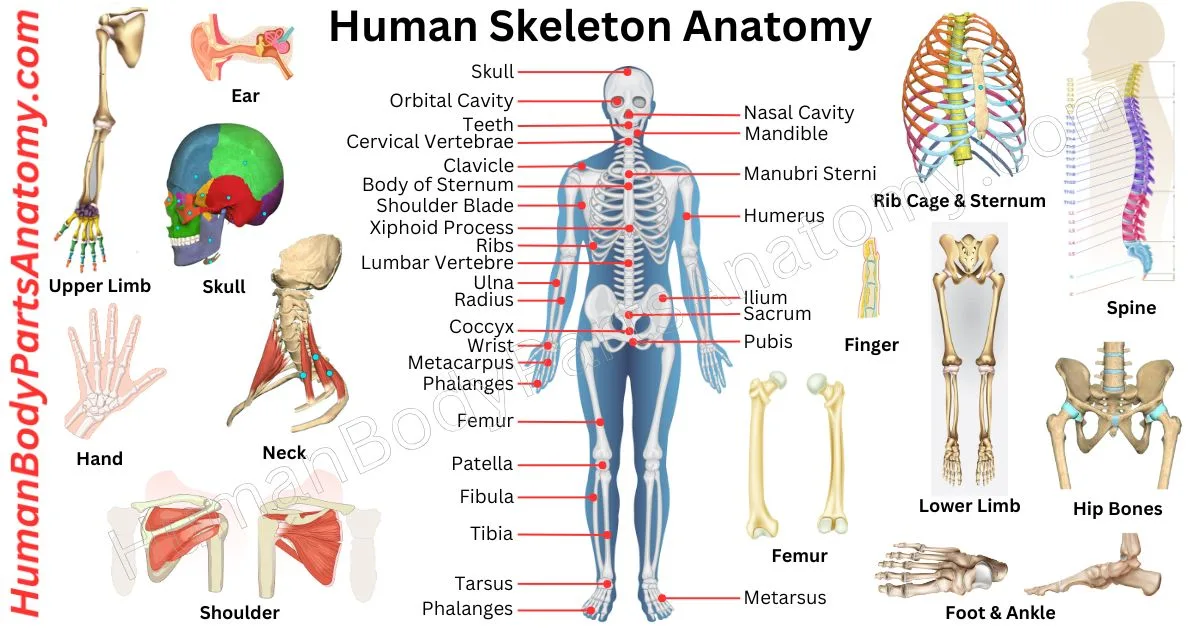
Human skeleton anatomy describes the bones inside our body that give us shape and support. The skeleton helps us stand, sit, walk, and move. It also protects important organs like the brain, heart, and lungs.[1][3]
Overview of Human Bone Anatomy
At birth, a human baby has roughly 270 to 300 bones.[2] As we grow, some bones join together. By the time we reach adulthood, the total number of bones in the human body becomes around 206.[1] The adult skeleton accounts for an estimated 12–15% of total body weight, although published figures vary with sex, body size, and the measurement method used.[1] Bones also have other important jobs. They produce blood cells in the bone marrow and store minerals such as calcium and phosphorus that keep bones strong.[3][4]
Bones grow and get stronger while we are children and young adults.[7] In most people, the amount of bone tissue in the skeleton peaks by the mid- to late 20s, when bones reach their maximum strength and density.[7] After about age 30, bone removal can begin to outpace bone formation, and adequate calcium, vitamin D, and weight-bearing activity become important for keeping the skeleton healthy.[8][7] Learning about the human skeleton helps us see how our bodies move and stay strong.
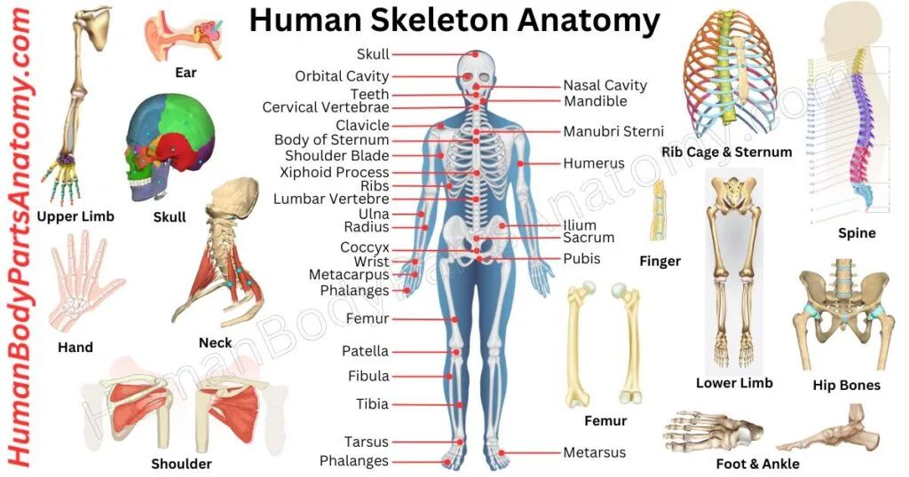
Human Skeleton Diagram Labeled
Bones of the Human Body
Skull (Head) Bones – 22 Bones
A. Cranial Bones (Protect the Brain) – 8 Bones
- Frontal (1)
- Parietal (2)
- Temporal (2)
- Occipital (1)
- Sphenoid (1)
- Ethmoid (1)
B. Facial Bones (Form the Face) – 14 Bones
- Nasal (2)
- Maxilla (2)
- Zygomatic (2)
- Palatine (2)
- Lacrimal (2)
- Inferior nasal concha (2)
- Vomer (1)
- Mandible (1)
Auditory Ossicles (Middle Ear) – 6 Bones
- Malleus (2)
- Incus (2)
- Stapes (2)
Hyoid Bone – 1 Bone
- Hyoid
Thoracic Cage (Chest) – 25 Bones
A. Sternum – 1 Bone
B. Ribs – 24 Bones (12 Pairs)
- True ribs (1–7)
- False ribs (8–10)
- Floating ribs (11–12)
Vertebral Column (Spine) – 33 Bones
A. Cervical Spine (Neck) – 7 Bones
- C1 – Atlas
- C2 – Axis
- C3 to C7 (5)
B. Thoracic Spine (Upper Back) – 12 Bones
- T1 to T12
C. Lumbar Spine (Lower Back) – 5 Bones
- L1 to L5
D. Sacrum – 5 fused bones
- S1 to S5 (fused)
E. Coccyx – 4 fused bones
- Tailbone
Upper Limb Bones – 64 Bones
A. Shoulder Girdle – 4 Bones
- Clavicle (2)
- Scapula (2)
- Humerus (2)
- Radius (2)
- Ulna (2)
C. Wrist (Carpal Bones) – 16 Bones (8 per hand)
- Scaphoid
- Lunate
- Triquetrum
- Pisiform
- Trapezium
- Trapezoid
- Capitate
- Hamate
D. Hand Bones – 38 Bones
- Metacarpals (10)
- Phalanges (28)
- Proximal
- Middle
- Distal
Pelvic Girdle – 2 Bones
- Hip bones (2)
- Ilium
- Ischium
- Pubis (fused in adults)
Lower Limb Bones – 62 Bones
- Femur (2)
- Patella (2)
- Tibia (2)
- Fibula (2)
B. Ankle (Tarsal Bones) – 14 Bones (7 per foot)
- Talus
- Calcaneus
- Navicular
- Cuboid
- Medial cuneiform
- Intermediate cuneiform
- Lateral cuneiform
C. Foot Bones – 38 Bones
- Metatarsals (10)
- Phalanges (28)
Sesamoid Bones (Variable)
- Patella (largest sesamoid bone)
- Small sesamoid bones in the hands and feet
Human Skeleton Anatomy
Skull
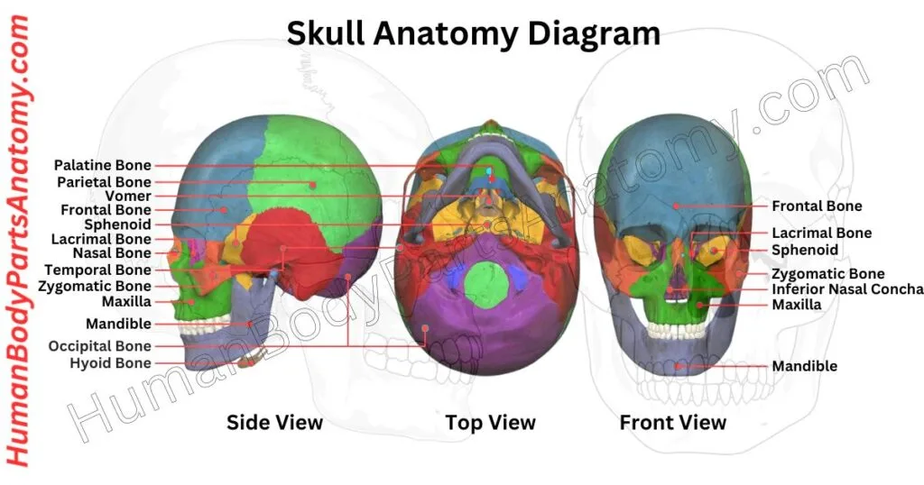
The human skull is one of the most important structures in the body. It’s a strong, bony framework that does far more than just protect the brain — it also shapes the face, houses vital sense organs, and supports many of the functions we rely on every day, from seeing and hearing to breathing and chewing.[9]
What Is the Skull Made Of?
The human skull is made up of 22 bones.[9] When the six tiny ear bones (called ossicles — three in each ear) and the hyoid bone in the throat are included, that number rises to 29.[10][11] Nearly all of these bones are held tightly together by fibrous joints called sutures, which interlock like puzzle pieces to form a rigid, protective shell.[9]
The Two Main Parts of the Skull
The skull is divided into two key sections. The first is the neurocranium, which wraps around and protects the brain. The second is the viscerocranium, which forms the face and includes the lower jaw, known as the mandible.[9] Together, these two sections give the skull its distinctive shape.
Three Types of Skull Bones
Skull bones fall into three main categories: cranial bones, of which there are eight (which form the braincase), facial bones, of which there are 14 (which shape the face and support the eyes, nose, and mouth), and the ossicles (three tiny bones in each ear that play a critical role in transmitting sound).[10]
Why Is the Skull Located at the Top of the Body?
The skull sits at the very top and front of the body — and that’s no accident. The brain and all major sense organs are concentrated in the head, a biological arrangement known as cephalization. The cranium is the most cephalad part of the axial skeleton, positioning the brain for rapid access to sensory information from the environment, which is essential for fast reflexes and decision-making.[9]
What Does the Skull Protect?
Inside and around the skull, you’ll find the brain, eyes, ears, nose, and mouth. These organs work together to give us the ability to see, hear, smell, taste, and think.[9] The skull is uniquely designed to support all of them: it keeps the eyes properly spaced for depth perception, positions the ears on opposite sides of the head so we can detect the direction of sounds, and provides openings for the nose and mouth to function freely.
Key Functions of the Human Skull
Beyond protection, the skull plays several important roles. It provides attachment points for the muscles used in chewing and facial expression, and it allows neurovascular structures to pass between the inside and outside of the cranial cavity.[9]
It also supports the top of the spine, balancing the head and allowing it to move in multiple directions. The nasal cavity within the skull warms, humidifies, and filters inspired air before it reaches the lower airways.[12]
Understanding the skull’s structure helps explain how the human body is built for both survival and sensory awareness — making it one of the most fascinating and functional structures in anatomy.
Read More – Skull Anatomy: Parts of the Skull, Structure, Cranial, Facial Bones & Functions
Vertebral Column or Spine
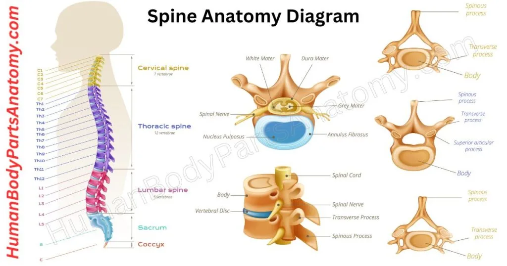
The spine — also called the vertebral column or backbone — is one of the most essential structures in the human body. It serves three critical roles: protecting your spinal cord, supporting your body’s upright posture, and enabling everyday movement.[13][14]
How Is the Spine Structured?
Your spine is made up of 33 small bones called vertebrae, stacked neatly on top of each other from the base of your skull to your tailbone.[13] Between each vertebra sits a soft, rubbery pad called an intervertebral disc. These discs work like shock absorbers — cushioning impact, preventing bone-on-bone friction, and giving your spine the flexibility to bend, twist, and rotate without sacrificing its supportive strength.[14][15]
Together, the vertebrae form a hollow channel called the vertebral (spinal) canal, which acts as a protective tunnel surrounding and shielding the delicate spinal cord and its branching nerve roots.[14]
The Five Regions of the Spine
Vertebrae are organized into five distinct regions, each serving a specific function:[13][15]
- Cervical (neck) — 7 vertebrae that support the head and enable neck movement.
- Thoracic (mid-back) — 12 vertebrae that anchor the rib cage and protect chest organs.
- Lumbar (lower back) — 5 large vertebrae that bear most of the body’s weight.[14]
- Sacral (pelvic area) — 5 fused vertebrae that connect the spine to the hips.[15]
- Coccygeal (tailbone) — 4 small fused vertebrae that provide a base for pelvic floor muscles.[13][15]
What Does the Spine Actually Do?
Beyond structure, the spine plays a direct role in your quality of life. It keeps your body balanced while standing, walking, or running.[13]
The spine allows you to bend forward to tie your shoes, twist to look over your shoulder, and carry heavy loads — the vertebral bodies bear the majority of the axial load, increasing in size toward the lower back.[14] It also houses the spinal cord, through which nerve signals travel between the brain and the rest of the body — making it critical for sensation and muscle control.[13][14]
When the spine is healthy, most people never think about it. But injuries, poor posture, or degenerative conditions like herniated discs or spinal stenosis can quickly impact mobility and overall well-being.[13]
Read More – Spine Anatomy: Parts of the Spine, Vertebrae, Curves, Spinal Cord & Diagram
Hip Bone
The hip — the bone of which is medically called the os coxae, or coxa — is one of the most important joints in the human body. It supports your body weight, keeps you balanced, and makes everyday movements like walking, running, and sitting possible.[17]
Where Is the Hip Located?
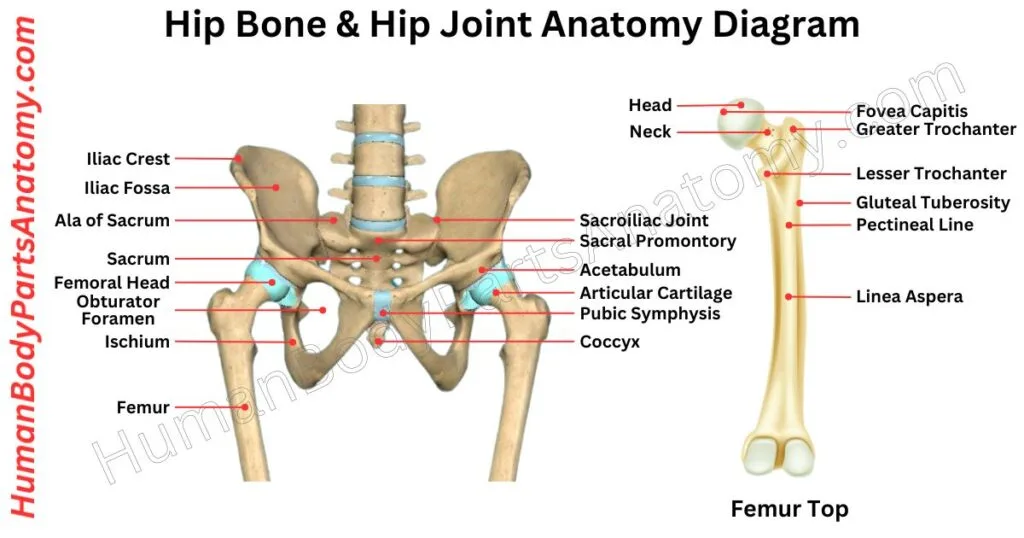
Your hip sits on the outer side of your pelvis, slightly toward the front and side of your buttocks. It rests just below the iliac crest — the curved upper edge of your pelvic bone — and near a large pelvic opening called the obturator foramen, formed by the superior and inferior rami of the ischium and pubis.[16]
What Makes Up the Hip?
The hip is built for both strength and movement. It is surrounded by powerful muscles, ligaments, tendons, and soft tissues that protect a bony landmark on your upper thigh bone called the greater trochanter.[18] These structures work together to keep the joint stable while allowing a wide range of motion, with three major ligaments — iliofemoral, ischiofemoral, and pubofemoral — anchoring the femur in its socket.[16]
In adults, the hip bone is formed by three bones — the ilium, ischium, and pubis — that fuse together over time. They form a deep, cup-shaped socket known as the acetabulum, which cradles the rounded head of the femur (thigh bone) to create the hip joint.[16] The acetabular labrum, a rim of connective tissue, deepens this socket to increase stability and resist dislocation.[16]
Why Is Hip Health Important?
Your hips carry your entire upper body weight with every step you take.[16] When something goes wrong — whether it is hip pain, arthritis, a stress fracture, or a muscle strain — it can quickly affect your mobility and quality of life.
Understanding basic hip anatomy makes it easier to recognize early warning signs, communicate with your doctor, and take steps to protect your long-term joint health.
Read More – Hip Bone Anatomy: Parts of Hip Bone, Ilium, Pubis, Functions & Diagram
Auditory Ossicles (Middle Ear) – 6 Bones
Deep inside each middle ear sit three of the tiniest bones in the human body — the malleus, incus, and stapes, giving six in total across both ears.[10][11] Together, they form a miniature chain that links your eardrum to the inner ear.[11]
Despite their incredibly small size, these bones — known as the auditory ossicles — are essential to how you hear every sound around you.[11]
The Malleus (Hammer) — First in Line
The malleus is the first bone in the chain and the one directly attached to the eardrum (tympanic membrane).[11] Its nickname, “the hammer,” comes from its shape.
Key facts about the malleus:
- Its long handle is connected to the eardrum.
- Its rounded head sits in a small cavity called the epitympanic recess.
- It links directly to the incus at the incudomalleolar joint, passing vibrations forward.
When your eardrum moves, the malleus moves with it — kicking off the entire hearing process.[11]
The Incus (Anvil) — The Middle Link
Positioned between the malleus and stapes, the incus is called “the anvil” because of its shape. It acts as the essential connector between the two other bones.[11]
What makes up the incus:[11]
- A central body that attaches to the malleus
- One limb that anchors to the back wall of the middle ear for stability
- Another limb that connects to the stapes, passing the vibrations along
Without the incus, the sound signal would have no bridge to cross.
The Stapes (Stirrup) — The Smallest Bone in the Human Body
The stapes holds a remarkable record: it is the smallest bone in the human body.[11] Shaped like a tiny riding stirrup, it completes the ossicle chain with precision.
Structure of the stapes:[11]
- A head that connects to the incus
- Two thin arms that form the stirrup shape
- A flat base called the footplate, which rests against the oval window
When the stapes vibrates, its footplate presses on the oval window — transferring mechanical energy into the fluid-filled inner ear, where hearing signals are generated and sent to the brain.[11]

Read More – Ear Anatomy: Parts of the Ear, Outer, Middle & Inner Ear & Structures
Hyoid Bone
The hyoid bone is a small, U- or horseshoe-shaped bone located in the midline at the front of your neck. It sits just below your lower jaw (inferior to the base of the mandible) and above your larynx, at roughly the level of the fourth cervical vertebra.[19] You can actually feel it yourself — simply place your fingers where your chin meets your neck and swallow. That subtle movement you feel? That’s your hyoid bone.
Why Is the Hyoid Bone Unique?
The hyoid is unlike any other bone in the human body. It does not articulate directly with any other bone. Instead, it is held in place by a network of muscles, ligaments, and cartilage.[19][20] This is why it is often called the body’s only “floating bone.”
What Does the Hyoid Bone Do?
The hyoid bone plays a key role in three essential functions: speaking, swallowing, and breathing — the patency of the airway between the oropharynx and the tracheal rings depends in part on its position.[19] It acts as an anchor point and provides support for several important structures in your head and neck, including your:[19][20]
- Tongue
- Throat
- Voice box (larynx)
- Epiglottis (the flap that keeps food out of your airway)
- Muscles in the floor of your mouth
Without the hyoid, these everyday actions would not be possible.
Parts of the Hyoid Bone
The hyoid bone has three main parts:[19]
- Body (Central Section). This is the central, quadrilateral-shaped broad segment of the bone. The front surface is slightly rounded and curves outward, while the back surface curves inward.
- Greater Horns (cornua majora). Two long extensions that project backward and outward from each side of the body, forming the limbs of the “U.” They serve as attachment points for many important neck muscles, making them critical for movement and stability.[19]
- Lesser Horns (cornua minora). Two smaller projections located near the top of the bone, close to where the greater horns begin. They point upward and slightly backward toward the base of the skull. A ligament called the stylohyoid ligament connects at the tip of each lesser horn, linking the hyoid to the styloid process of the temporal bone of the skull.[21]
Rib Cage
The rib cage, also known as the thoracic cage, is one of the most important structures in the human body. This bony framework sits at the center of your upper body, and it does far more than most people realize — from shielding your vital organs to helping you breathe with every single breath you take.[22]
What Makes Up the Rib Cage?
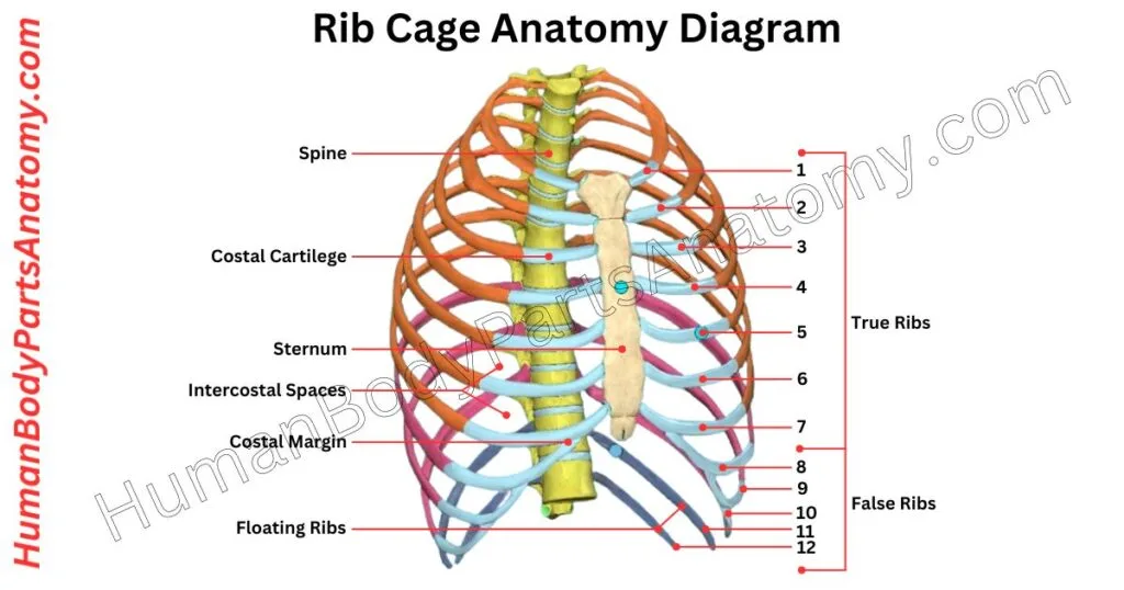
The rib cage is made up of three main components:[22][23]
- 12 pairs of ribs — curved bones that form the walls of the chest
- The sternum — the flat bone running down the center of your chest
- 12 thoracic vertebrae — the section of the spine the ribs attach to at the back
Each rib articulates posteriorly with the thoracic vertebrae at the costovertebral joints; the first rib is an exception, articulating with the first thoracic vertebra only.[22]
Anteriorly, the ribs are classified into three groups. The first seven pairs (true ribs) attach directly to the sternum through their own costal cartilage — a flexible, rubbery tissue that gives the rib cage just enough give to expand and compress during breathing.
Ribs 8 to 10 (false ribs) attach indirectly, joining the seventh costal cartilage, and ribs 11 and 12 (floating ribs) do not reach the sternum at all.[22]
What Does the Rib Cage Do?
The rib cage serves several critical functions:
- Protects Vital Organs. It shields the heart, lungs, and major blood vessels from injury — acting like a natural armor for the body’s most essential systems.[22]
- Supports Breathing. The ribs expand outward when you inhale and contract when you exhale, helping the lungs draw in oxygen and release carbon dioxide efficiently.[22]
- Supports the Shoulder Girdle. The rib cage provides structural support for the shoulders through the scapulothoracic articulation, enabling a full range of arm movement.[26]
- Anchors Key Muscles. Muscles in the neck, back, chest, and abdomen all attach to the rib cage, making it a central hub for movement, stability, and posture.[22]
Why Is the Rib Cage Important?
The rib cage is uniquely designed to be both strong and flexible. This combination enables it to absorb impact and protect delicate organs while still allowing for free movement with every breath you take.[22] Without it, normal functions like breathing, lifting, and even standing upright would not be possible.
Read More – Rib Cage Anatomy: Ribs, Sternum, Thoracic, Vertebrae & Functions Explained
Sternum
The sternum, commonly called the breastbone, is a flat, partially T-shaped bone running vertically down the center of your chest. It forms the anterior portion of the chest wall, connecting with the ribs via the costal cartilages and with the collarbone to form a protective shield around your vital organs.[23]
Understanding the sternum’s structure can help you better recognize chest pain, injuries, or medical procedures that involve this area.
The Three Parts of the Sternum
The sternum is made up of three distinct sections, each with a specific role:[23]
1. Manubrium (Top Section) – The manubrium is the broad, quadrangular, upper portion of the sternum. At the very top, you can feel a small indentation called the suprasternal (jugular) notch — that slight hollow at the base of your throat.[23]
On each side, the clavicular notches of the manubrium articulate with the medial end of each collarbone (clavicle), forming the sternoclavicular joints — the only bony articulation linking your arms to the rest of your skeleton.[23][28] The manubrium also articulates with the costal cartilages of the first pair of ribs.[23]
2. Body (Middle Section) – Also called the mesosternum or gladiolus, the body is the longest part of the sternum. It runs down the center of the chest and provides attachment points for the cartilages of ribs 3 through 7, inferior to the sternal angle.[23]
Where the manubrium and body meet, you’ll find a slight outward ridge known as the sternal angle, or angle of Louis — a landmark doctors and nurses use to locate the second rib and count down to other ribs during physical exams.[23]
3. Xiphoid Process (Bottom Section) – The xiphoid process is the small, triangular tip at the bottom of the sternum. It is the softest and most flexible part: it remains largely cartilaginous until around age 40 and is usually completely calcified by age 60, although these age-related changes vary considerably between individuals.[23]
Its size and shape vary from person to person.[23] This section serves as an attachment point for the diaphragm and some abdominal muscles, playing a quiet but important role in breathing and core stability.[23]
Why Is the Sternum Important?
The sternum’s primary job is protection. It forms the front wall of the thoracic cavity, shielding the heart, lungs, and major blood vessels from external impact.[23] It also plays a structural role in breathing, moving slightly with each breath as the rib cage expands and contracts.[22]
In medical settings, the sternum is central to procedures like CPR chest compressions and open-heart surgery, where surgeons split it (a procedure called a sternotomy) to access the chest cavity.[23]

Shoulder
Your shoulder is one of the most complex and mobile joints in the human body — the glenohumeral joint exhibits the highest degree of mobility of any joint in the body.[24] The shoulder region is built from three bones: the scapula (shoulder blade), the clavicle (collarbone), and the humerus (upper arm bone).[27][28] Three further structures described below — the acromion, the coracoid process, and the glenoid cavity — are not separate bones but distinct anatomical parts of the scapula, each with its own clinical importance.[24][25]
Understanding these bones can help you recognize common shoulder problems, including injuries from falls, accidents, and conditions such as arthritis.
1. Scapula (Shoulder Blade)
The scapula, commonly referred to as the shoulder blade, is a sturdy, flat, triangular bone situated on the upper back that connects the clavicle to the humerus and forms the posterior part of the shoulder girdle.[25] It serves as an anchor point for 17 muscles, making it one of the most heavily muscled bones in the body.[26]
A substantial share of overall shoulder motion occurs between the scapula and the chest wall at the scapulothoracic articulation, which slides across ribs 2 through 7 and works in coordination with the glenohumeral joint to position the socket during arm movement.[26] The scapula itself performs six motions — protraction, retraction, elevation, depression, upward rotation, and downward rotation — that together allow full functional movement of the upper limb.[25]
2. Clavicle (Collarbone)
The clavicle, or collarbone, is a sigmoid-shaped long bone that runs between the shoulder and the chest. It is the structural link connecting the axial and appendicular skeletons and, with the scapula, forms the pectoral girdle.[27]
The clavicle has a joint at each end — the sternoclavicular joint medially and the acromioclavicular joint laterally — and both are prone to developing arthritis over time.[27] It is also one of the most commonly fractured bones in the human body, whether from direct impact or from force transmitted by a fall onto an outstretched hand.[27]
3. Acromion
The acromion is a flat, bony extension that projects from the top of the scapula. It forms the highest point of the shoulder and gives it that characteristic square, rounded shape.[24]
The acromion, together with the coracoid process and the coracoacromial ligament, forms the roof of the shoulder joint, protecting the rotator cuff tendons beneath it.[24][29] When the space under the acromion narrows — for example with a downward-sloping, curved, or hooked acromion, or during repetitive overhead activity — it can lead to subacromial impingement and bursitis.[30]
4. Coracoid Process
The coracoid process is another bony projection that extends from the scapula, pointing forward toward the front of the body. While it may be small, it plays a critical role in shoulder stability.[24]
Several important muscles and ligaments attach to the coracoid process. The coracoclavicular ligament (its conoid and trapezoid components) anchors the clavicle to the coracoid, while the coracohumeral and coracoacromial ligaments provide further peripheral reinforcement.[24] The coracoid also serves as the attachment point for the short head of the biceps brachii, the coracobrachialis, and the pectoralis minor.[26][29]
5. Glenoid Cavity (Shoulder Socket)
The glenoid cavity, or glenoid fossa, is the shallow, cup-shaped socket at the lateral angle of the scapula. The rounded head of the humerus fits into this socket to create the glenohumeral joint.[24][25]
Because the socket is relatively shallow, the joint is inherently unstable and relies heavily on surrounding muscles, tendons, ligaments, and the glenoid labrum for stability.[24] Any irregularity in the glenoid cavity — such as damage from injury, a bony Bankart lesion, or wear — can therefore cause joint instability, dislocation, or degenerative change.[24]
Frozen shoulder (adhesive capsulitis) is a separate condition and is not caused by glenoid irregularity. It results from fibrotic thickening of the glenohumeral joint capsule, producing progressive stiffness and loss of both active and passive motion, most typically external rotation. It affects roughly 2% to 5% of the general population, has a mean age of onset around 55, and shows a slight female predominance of about 1.4 to 1. Diabetes, thyroid disorders, prolonged immobilisation, and previous shoulder injury are recognised risk factors, and people with diabetes or thyroid disease tend to have longer, more severe courses.[31] The outlook is generally good: with early treatment about 80% of patients regain near-normal shoulder function, though 10% to 20% have some residual stiffness or discomfort.[31]

Read More – Shoulder Anatomy: Parts of the Shoulder, Bones, Joint Structure & Diagram
Arm & Forearm Bones
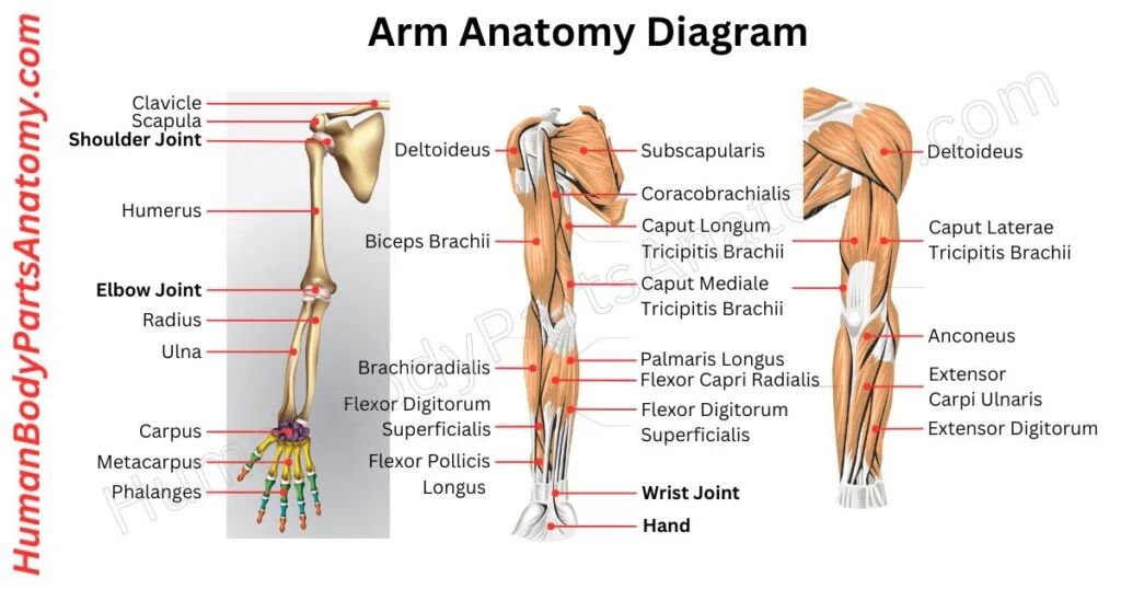
Your arm is made up of three major bones that work together to give you strength, flexibility, and a wide range of motion. These bones are the humerus (upper arm), the radius, and the ulna (both in the forearm).[32][35] Understanding what each bone does can help you make sense of arm injuries, pain, and treatment options.
1. Humerus: The Upper Arm Bone
The humerus is the long bone that runs from your shoulder down to your elbow. At the top, its head articulates with the glenoid fossa of the scapula to form the glenohumeral joint — a synovial ball-and-socket joint.[32] This design allows your arm to move along multiple planes, including flexion, extension, abduction, adduction, and internal and external rotation.[32]
At the lower end, the humerus widens into the medial and lateral epicondyles and the condyle, whose capitulum articulates with the head of the radius and whose trochlea articulates with the trochlear notch of the ulna, forming the elbow joint.[32] Many important muscles and ligaments attach along the length of the humerus, including the common flexor and extensor origins at the epicondyles — muscles that extend all the way down into your hand and power your grip and finger movements.[35]
In most cases, the humerus only becomes a medical concern when it breaks. Humerus fractures can happen in several different locations along the bone — near the shoulder, in the middle of the shaft, or near the elbow. Each type may require a different treatment approach, ranging from a sling or cast to surgery.[32]
Read More – Arm Anatomy: Parts of Arm, Bones, Muscles & Joints with Functions & Diagram
2. Radius
The radius is one of the two long bones of the forearm (antebrachium).[33] It lies on the thumb side of the forearm and, in anatomical position, is the lateral bone at both the elbow and the wrist.[33]
One of the radius’s most unique features is that it rotates. When you turn your palm up or down (supination and pronation), the radius rotates around the ulna, with the distal radius crossing over the distal ulna during pronation.[33]
At the elbow, the radius forms part of a complex, three-part joint with the humerus and ulna. Its proximal head is cylindrical with a shallow concave upper surface that cups the rounded capitulum of the humerus, forming the radiocapitellar articulation — a pivot joint stabilised against the radial notch of the ulna by the annular ligament.[33]
At the other end, the radius plays a major role in forming the wrist joint, articulating with the scaphoid and lunate carpal bones.[33] It carries the larger share of the force travelling from the hand up through the forearm: in a wrist with neutral ulnar variance, roughly 80% of the load crossing the wrist passes through the radius.[38]
The radius and ulna articulate with each other at both the proximal and distal radioulnar joints and are further connected along their length by the interosseous membrane, a fibrous layer running obliquely between the two bones.[35]
3. Ulna
The ulna is the second forearm bone, running along the pinky-finger side (medial side) of your arm.[34] Unlike the radius, the ulna does not rotate about its own axis during pronation and supination — it stays in a relatively fixed position regardless of how you turn your hand, and is generally considered the stabilising bone of the forearm.[35]
At the elbow, the trochlear notch of the ulna forms a hinge joint with the trochlea of the humerus.[34][35] You can feel this joint working every time you bend and straighten your arm. The bony point you feel at the back of your elbow is the olecranon, a proximal extension of the ulnar shaft that sits within the olecranon fossa of the humerus when the elbow is fully extended and helps prevent hyperextension.[34][35]
At the wrist, the head of the ulna articulates with the articular disc of the distal radioulnar joint and has no direct contact with the carpal bones.[35] It therefore bears less of the load from hand and wrist movements than the radius does — about 20% of the load crossing the wrist is carried by the ulna together with the triangular fibrocartilage complex — though it remains important for wrist stability.[35][38]
Like the radius, the ulna is connected to its neighbour at both ends and along the full length of the forearm by the interosseous membrane and associated ligaments.[35]
Ulnar fractures are a common injury, often occurring alongside radius fractures, particularly from falls or direct impacts.[34]
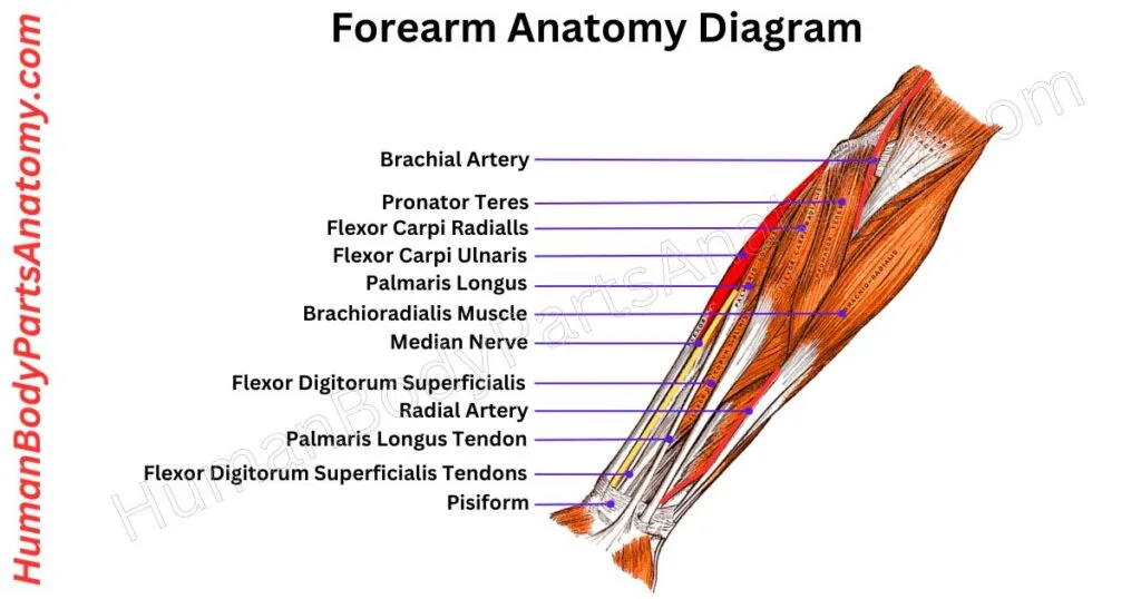
Read More – Forearm Anatomy: Parts of the Forearm, Radius, Ulna, Muscles & Diagram
Wrist
The wrist is made up of eight small bones called carpal bones, arranged in two rows: a proximal row (scaphoid, lunate, triquetrum, and pisiform, moving from the thumb side toward the little finger) and a distal row (trapezium, trapezoid, capitate, and hamate).[36] Each bone plays a specific role in movement, stability, and load distribution. Understanding these bones helps explain why wrist injuries can be complex — and why proper diagnosis matters.
1. Scaphoid
The scaphoid is one of the most important bones in the wrist. It sits in the proximal row of carpal bones but bridges both rows, helping coordinate movement throughout the entire wrist.[36]
Its name comes from its boat-like appearance. Most of its surface is covered by articular cartilage, which is what allows it to bridge the joint between the two rows of carpal bones; proximally it articulates with the radius.[33][36]
The small area of bone not covered by cartilage is where ligaments attach and where blood vessels — supplied by the radial artery — enter the bone.[36]
This blood supply is critical. The scaphoid has a single blood supply that enters distally and runs backward (retrograde) to supply the proximal portion, which makes the proximal pole particularly vulnerable to avascular necrosis when the bone is fractured.[36]
The scaphoid is the most commonly fractured carpal bone. On falling onto an outstretched hand, it receives most of the force transmitted from the radius because of its size and position.[36] Fractures typically cause radial-sided wrist pain and tenderness in the anatomical snuffbox; non-displaced fractures can be missed on plain films and may need CT or MRI to confirm.[36] Because the scaphoid is so central to wrist mechanics, a fracture that does not heal properly (a nonunion) can cause long-term pain, stiffness, and a predictable pattern of arthritis known as scaphoid nonunion advanced collapse.[37]
For context, carpal fractures are the least common hand fracture overall, accounting for about 12% of adult hand fractures, compared with 34% for metacarpal and 54% for phalangeal fractures.[36]
2. Lunate
The lunate sits in the center of the proximal row of wrist bones and has a crescent shape when viewed from the side.[36]
Like most carpal bones, the lunate is largely covered in cartilage, which allows for a wide range of wrist motion.[36] Isolated lunate fractures are uncommon, but the bone is frequently involved in dislocations, and about 20% of lunates have only a single intraosseous blood supply, placing them at higher risk of avascular necrosis.[35][36] Because the lunate lies close to the median nerve, anterior dislocation of the bone can compress the nerve inside the carpal tunnel, producing numbness in the thumb, index, middle and radial half of the ring finger and thenar weakness.[36]
It can also cause problems when the ulna is longer than the radius — a configuration called positive ulnar variance. In a wrist with neutral variance, roughly 80% of the load crossing the wrist passes through the radius and about 20% through the ulna and triangular fibrocartilage complex (TFCC); when positive ulnar variance exceeds about 2.5 mm, the share carried on the ulnar side can rise from 20% to 40%.[38] That sustained overload allows the ulnar head to repeatedly abut the ulnar-sided carpus, degenerating the TFCC and the cartilage of the lunate and triquetrum and disrupting the lunotriquetral ligament — a condition called ulnar impaction (ulnocarpal abutment) syndrome.[38]
3. Triquetrum
The triquetrum is a pyramid-shaped bone on the pinky (small finger) side of the wrist, in the proximal row of carpal bones. It articulates with the pisiform, the lunate, and the hamate.[36]
Though less commonly discussed, the triquetrum plays an important role in the overall balance and strength of the wrist.
4. Trapezoid
The trapezoid is the smallest carpal bone, sitting in the distal row between the trapezium and the capitate.[36] It anchors the index finger metacarpal — the long bone running to your index finger — firmly in place.[41]
The trapezoid has two areas of blood supply but no intraosseous anastomoses between them.[36]
5. Trapezium
The trapezium is a saddle-shaped bone in the distal row of carpal bones. It forms the first carpometacarpal joint with the base of the thumb metacarpal.[36][41] Its unique saddle shape is what gives the thumb its remarkable range of motion — allowing it to rotate, flex, extend, and oppose the other fingers.[41]
Two problems are commonly seen with the trapezium. Fractures can occur, but the most frequent issue is osteoarthritis at the base of the thumb — a condition called basal joint arthritis or CMC (carpometacarpal) arthritis.
This condition is markedly more common in women than in men. In a large Finnish population study the age-adjusted radiographic prevalence was 7% in men and 15% in women, obesity was a strong determinant in both sexes, and restricted thumb mobility with local tenderness and swelling frequently accompanied the radiographic changes.[40]
6. Capitate
The capitate is the largest bone in the wrist and sits at the center of the distal row of carpal bones.[36] It articulates with multiple bones in both the wrist and hand and sits beneath the middle finger metacarpal.[36][41]
Because of its central location, the capitate contributes significantly to overall wrist motion and serves as a hub for transmitting forces across the wrist. Like the scaphoid, it has a single intraosseous blood supply, which raises its risk of avascular necrosis after fracture.[36]
7. Hamate
The hamate is a large, wedge-shaped bone in the distal row of carpal bones. It supports the ring and little finger metacarpals and articulates with the triquetrum and capitate.[36] It also serves as an attachment point for several ligaments, including the transverse carpal ligament, which forms the roof of the carpal tunnel; its hook additionally forms the ulnar margin of the carpal tunnel and the radial wall of Guyon’s canal.[39]
The hamate can be injured in two distinct ways. The body of the hamate may fracture from direct trauma.[39]
More distinctively, the hook of the hamate — a bony projection on the palm side — can fracture from repeated impact, classically from “grounding” a golf club or “checking” a baseball bat, and also from racket sports.[36][39] It typically presents with pain over the hypothenar eminence and pins-and-needles in the ulnar nerve distribution.[36] Plain X-rays are frequently insufficient; special views or, more often, CT scanning are needed to make the diagnosis.[39][36] The current standard of care is excision of the fractured hook fragment, after which return to sport at around six weeks is usual.[36]
8. Pisiform
The pisiform is a small bone that sits on the pinky side of the wrist in the proximal carpal row, embedded within the flexor carpi ulnaris tendon.[36] It is classified as a sesamoid bone — a bone that forms inside a tendon, similar to the kneecap in the knee.[4][36]
Like other sesamoid bones, the pisiform changes the direction of pull of the tendon it sits in, improving the mechanical efficiency of wrist and finger flexion.[4] It articulates with the triquetrum on its dorsal surface and serves as an attachment site for tendons and ligaments — the flexor carpi ulnaris inserts onto it, and the hypothenar muscles take origin from it and from the hook of the hamate.[36]
While injuries to the pisiform are uncommon, it can fracture from a fall onto an outstretched hand or develop arthritis in its joint with the triquetrum.[36]

Read More – Wrist Anatomy: Parts of the Wrist, 8 Carpal Bones, Tendons & Diagram
Hand Bones
Each hand contains 27 bones in total: 8 carpals, 5 metacarpals, and 14 phalanges, along with several consistently present sesamoid bones.[41] The metacarpals sit in the palm, connecting your wrist to your fingers.
1. Metacarpal Bones
The five metacarpal bones form the core structure of the palm. Each one bridges two important joints: on the wrist side, it connects to a carpal (wrist) bone at the carpometacarpal (CMC) joint.[41]
On the finger side, it links to the finger bones at the metacarpophalangeal (MCP) joint — the joint you know as your knuckle.[41]
Doctors number the metacarpals 1 through 5, starting from the thumb. Like other long bones in the body, each metacarpal has three sections: the base (closest to the wrist), the shaft or body (the long middle portion), and the head (nearest the fingers).[41]
Those rounded heads are what form the bumps you see across your knuckles when you make a fist. The first metacarpal — the one connected to your thumb — is set apart from the rest and articulates with the trapezium at the first CMC joint.[41]
This separate positioning, together with the saddle shape of the trapezium, is exactly what gives your thumb its wide range of motion, including the ability to rotate and oppose the other fingers.[41] That’s what lets you grip, pinch, and pick things up.
Metacarpals 2 through 5, on the other hand, are tightly bound together at the wrist end by strong ligaments.[41] This close arrangement adds stability to the hand and also has a practical benefit: if one of these four bones fractures, the neighbouring bones and their interconnecting ligaments act like natural splints, often keeping the break from shifting out of place.[41]
2. Phalanges
The phalanges are the small bones that make up your fingers and thumbs. Each hand contains 14 phalanges — 28 across both hands— and their arrangement differs depending on which digit you’re looking at.[41]
How Many Phalanges Does Each Finger Have?
Your thumb has just two phalanges: a proximal phalanx (closer to the palm) and a distal phalanx (at the fingertip). Each of your other four fingers has three phalanges: proximal, middle, and distal — moving from the knuckle outward to the tip.[41]
Just like the metacarpals, each phalanx has a base, a body, and a head. The base of each proximal phalanx sits against the rounded head of the metacarpal below it, forming the metacarpophalangeal (knuckle) joint where finger movement begins.[41]
Femur
The femur, commonly known as the thigh bone, is the longest, heaviest, and strongest bone in the human body.[18] It is located in the upper leg and connects the hip joint above to the knee joint below, playing a central role in nearly every movement you make.[18]
At its upper end, the femur forms the hip joint by fitting into a cup-shaped socket in the pelvis called the acetabulum.[16][17] This ball-and-socket design allows a wide range of motion, including walking, running, sitting, and rotating the leg.[16]
At its lower end, the femoral condyles meet the tibia (shinbone), and the anterior surface articulates with the patella (kneecap), together forming the knee joint, which supports bending and straightening of the leg.[42]
One of the femur’s most important jobs is weight-bearing. Whether you are standing still or moving, this bone transmits the load of your upper body down to the lower limbs.[18] Its compact bone is thickest in the middle third of the shaft, where mechanical stresses are highest.[18]
The femur also serves as a major attachment point for some of the body’s most powerful muscles, including the quadriceps, hamstrings, and glutes, which are organised into anterior, medial, and posterior compartments around the bone.[18]
These muscles, along with key tendons and ligaments, attach directly to the femur — including at the greater and lesser trochanters — and work together to control hip and knee movement.[18]
Because of its size and central location, the femur is critical to mobility, stability, and overall musculoskeletal health. Injuries to the femur, such as fractures or stress injuries, can significantly impact a person’s ability to walk and perform daily activities, making proper bone health essential at every age.[18]

Read More – Femur Anatomy: Parts of Femur, Structure, Functions, Location & Diagram
What is Patella?
The patella, commonly known as the kneecap, is the largest sesamoid bone in the human body.[43] A sesamoid bone is a small, rounded bone that forms within a tendon, and the patella sits at the front of the knee within the tendon of the quadriceps femoris.[4][43]
Shape and Structure of the Patella
The patella has a roughly triangular shape with three distinct borders. The pointed lower tip is called the apex, and it faces downward toward the shin.[43]
The flat upper border is known as the base, which is where the quadriceps tendon attaches. The two side edges make up the medial (inner) and lateral (outer) borders.[43]
The bone has two main surfaces:[43]
- Front surface (anterior): This side lies just deep to the fascia lata and the skin, which is why you can easily feel your kneecap when you press on the front of your knee.
- Back surface (posterior): This side faces inward toward the knee joint. It contains two smooth, cartilage-covered areas called articular facets — one medial and one lateral. These facets articulate with the condyles of the femur, forming the patellofemoral joint.
The back surface of the patella is covered by some of the thickest articular cartilage in the body, which helps it handle the significant pressure placed on the knee during everyday activities like walking, climbing stairs, and squatting.[43]
What Does the Patella Do?
The patella plays a critical role in improving the efficiency of the knee. It acts as a pivot point that displaces the quadriceps tendon–patellar ligament linkage away from the knee’s axis of rotation.[43] This increases the effective moment arm — the mechanical distance over which the muscle force acts.[43]
By increasing this distance, the patella amplifies the torque your thigh muscles generate when straightening the leg. Displacing the extensor mechanism in this way produces roughly 60% additional torque during the last 15° of knee extension.[43] Without the kneecap, your quadriceps would need to work considerably harder to extend the knee.
The patella also protects the anterior articular surface of the distal femur and the deeper structures of the knee from direct impact, and it shields the quadriceps tendon from friction by minimising tendon contact with the femur as the knee bends and straightens.[43]
Why the Patella Matters for Your Health
Because the patella is under constant stress, it is a common site for knee problems. Conditions like patellar tendinitis (jumper’s knee), chondromalacia patellae (softening of the cartilage behind the kneecap), and patellar instability or dislocation are among the most frequent knee complaints seen by orthopedic doctors.[43]
Keeping the quadriceps and surrounding muscles strong is one of the best ways to support the extensor mechanism and maintain healthy knee function long-term.[43]
What is Tibia?
The tibia, commonly known as the shin bone, is one of the most important bones in the human body. It plays a central role in how you stand, walk, run, and move every day.[44]
The tibia is the second-largest bone in the body, after the femur.[44] In most adults it measures roughly 36–38 cm (about 14–15 inches), varying with overall stature. Despite being a weight-bearing bone that handles significant daily stress, it is also one of the strongest bones in your body — significantly larger and stronger than its neighbour, the fibula.[44]
What Does the Tibia Do?
The tibia serves several critical functions:[44]
- Weight-bearing: It supports your entire body weight whenever you’re standing or moving.
- Stabilization: It helps keep your body balanced and stable during physical activity.
- Joint connection: It bridges your knee and ankle joints, enabling coordinated leg movement.
- Muscle attachment: Muscles, tendons, and ligaments in your leg, knee, and ankle all anchor to the tibia.
Where Is the Tibia Located?
You have one tibia in each leg. It’s the larger of the two lower-leg bones, positioned toward the front and inner (medial) side of your leg. The other bone, the fibula (calf bone), runs alongside it on the outer edge and is connected to it by the interosseous membrane.[44]
The tibia extends from just below your knee all the way down to your ankle joint.[44]
What Does the Tibia Look Like?
The tibia has three distinct sections, each with a specific role:
1. Proximal Aspect (Upper End) – This is the top portion of the tibia, located just beneath the knee. It forms a wide, flat surface — almost like a shelf — that supports the knee joint. It includes the medial condyle, lateral condyle, and intercondylar eminence.
2. Shaft (Middle Section) – The long, triangular midsection of the bone. This is what forms the visible ridge of your shin and carries most of your body weight. It includes structures like the anterior border, posterior surface, soleal line, and lateral border.
3. Distal Aspect (Lower End) – The bottom portion of the tibia connects to the fibula and the talus bone, forming the ankle joint. It also sits above the heel bone (calcaneus). This section includes the medial malleolus — the small bony bump you can feel on the inner side of your ankle — and the fibular notch.
What Is the Fibula?
The fibula is the calf bone — the slender, smaller bone running along the outer (lateral) side of your lower leg.[45] It works alongside the tibia (shinbone), the larger bone beside it, to give your lower leg its shape and strength. Together, these two bones form the structural foundation of everything below your knee.[45]
While the fibula may be smaller, it plays a surprisingly important role. It anchors key muscles and tendons, stabilizes the ankle joint, and connects critical ligaments from the knee down through the lower leg.[45][46]
The fibular shaft is essentially non-weight-bearing.[46] The fibula as a whole carries only a modest share of the axial load; figures reported in the biomechanical literature and summarised in a 2024 finite-element analysis range from about 7% to 17% of the compressive load in the weight-bearing lower extremity.[47] Because it is thin, superficial, and lightly loaded compared with the tibia, fibular injuries are common — most often as part of ankle fractures, where the distal fibula (lateral malleolus) is involved — while isolated shaft fractures are frequently managed non-operatively.[46]
What Does the Fibula Do?
The fibula serves several essential functions in your lower body:[45]
- Provides structure to the outer calf and lower leg
- Stabilizes the ankle joint, forming the bony bump you feel on the outside of your ankle (called the lateral malleolus)
- Supports muscles and tendons that control foot and ankle movement
- Anchors knee ligaments to the lower leg, helping maintain joint stability
- Assists with weight distribution during walking, running, and other activities [47]
Where Is the Fibula Located?
The fibula is positioned on the lateral side of your lower leg, running from just behind and below the tibial head at the knee all the way down to the ankle. It sits parallel to and slightly behind the tibia, joined to it by the interosseous membrane, which forms a syndesmotic joint with very little mobility.[45] While the tibia is the primary weight-bearing bone, the fibula provides crucial support and balance to the entire lower leg.[45]
What Does the Fibula Look Like?
The fibula has three main parts:[45]
- The head — a rounded upper end bearing a circular facet that articulates with the lateral condyle of the tibia just below the knee
- The neck — the narrowed segment just distal to the head
- The shaft — a long, narrow middle section with lateral, medial, and posterior surfaces, whose shape is determined by its muscle attachments
- The lateral malleolus — the lower end that forms the outer part of your ankle joint
How Big Is the Fibula?
The fibula is one of the longest bones in the human body and is conventionally ranked third by length, after the femur and the tibia — although average fibular and humeral lengths are close, and rankings differ between reference sources and populations. In most adults, the fibula measures roughly 34–36 cm (about 14 inches). The plural form of fibula is fibulae.
Foot Bones
The foot is the part of your lower limb that sits below the ankle joint. It is one of the most complex structures in the human body — built specifically to support your body weight, absorb impact, and help you move efficiently. Its structural organisation enables upright stance and forms the foundation of bipedal gait mechanics.[48]
The skin on top of the foot (the dorsal side) is soft and flexible, while the skin on the bottom (the plantar side) is thick, tough, and tightly attached to a strong band of tissue called the plantar aponeurosis, which supports the medial longitudinal arch of the foot.[50]
How Many Bones Are in the Foot?
Each human foot contains 26 bones — so the two feet together contain 52 bones, roughly 25% of all the bones in the adult body.[48][1] These bones are organized into three main groups: tarsal bones, metatarsals, and phalanges.[49]
1. Metatarsals (The Forefoot)
The 5 metatarsals are long, slender bones that make up the middle section of the foot, connecting the tarsal bones to the toes.[48] They are numbered 1 through 5, starting from the big toe side. These bones play a key role in weight distribution when you walk or stand.[48]
2. Phalanges (The Toe Bones)
The toes contain 14 phalanges in each foot. Most toes have three phalanges each — a proximal (base), middle, and distal (tip) bone. The big toe (hallux) is the exception, with only two phalanges: a proximal and a distal bone.[48]
- Sesamoid Bones — Two small, pea-shaped bones sit beneath the head of the first metatarsal at the big toe joint, embedded within a tendon and separated by an intersesamoidal ridge and ligament as part of the plantar plate complex.[48] Though tiny, they redirect tendon forces and help tendons move smoothly, making push-off during walking and running more efficient.[4][48]
Ankle Bones (Tarsal Bones)
The tarsus is a group of 7 bones that make up the back portion of the foot.[49] Together, these tarsal bones give your foot its structure, support the arches that help you walk and run, and serve as key anchoring points for the muscles of your leg and foot.[49][50]
The seven tarsal bones are the calcaneus, talus, navicular, cuboid, and three cuneiform bones (medial, intermediate, and lateral).[49]
1. Talus
The talus is the bone that links your leg to your foot. On its upper and outer sides, it articulates with the tibia and fibula to form the talocrural (tibiotalar) joint, which is your ankle joint.[49][51]
Underneath, it connects to the calcaneus to form the subtalar (talocalcaneal) joint and to the navicular and calcaneus together to form the talocalcaneonavicular joint.[51] The talus also plays a role in supporting the medial longitudinal arch — one of the primary weight-bearing arches of the foot — where the head of the talus functions as the keystone.[50]
2. Calcaneus
The calcaneus is the largest tarsal bone and is commonly known as the heel bone.[48][52] It connects to the talus and cuboid bones, forming the subtalar and calcaneocuboid joints, and participates with the talus and navicular in the talocalcaneonavicular articulation.[52][54]
Because of its position, the calcaneus helps support both the medial and lateral longitudinal arches of the foot, making it essential for balance and shock absorption during movement.[50]
3. Navicular Bone
Shaped like a small boat, the navicular bone sits between the talus behind it and the three cuneiform bones in front of it.[53] It also sits on the inner (medial) side, next to the cuboid.[53]
This placement allows it to form several joints, including the cuneonavicular, cuboideonavicular, and talocalcaneonavicular joints.[54] The navicular is a key building block of the medial longitudinal arch of the foot.[50]
4. Cuboid Bone
The cuboid bone is found on the outer (lateral) edge of the foot, in the distal row of tarsal bones. It sits just behind the 4th and 5th metatarsal bones, in front of the calcaneus, and to the outer side of the navicular and lateral cuneiform bones.[49]
As a result, it forms four joints: the tarsometatarsal, calcaneocuboid, cuboideonavicular, and cuneocuboid joints.[54] Because of its position on the outer edge, the cuboid contributes to the rigid lateral longitudinal arch of the foot, together with the calcaneus and the 4th and 5th rays.[48][50]
5. Three Cuneiform Bones
The cuneiform bones are three wedge-shaped bones named for where they sit: medial (inner), intermediate (middle), and lateral (outer). All three articulate with the navicular bone behind and with their corresponding metatarsal bones in front.[49]
They also connect to each other, forming the tarsometatarsal, cuneonavicular, cuneocuboid, and intercuneiform joints.[54] The cuneiform bones support both the medial longitudinal arch and the transverse arch — the arch that runs obliquely across the tarsometatarsal joints.[48][50]
FAQ’s-
The adult human body contains 206 bones, organized into the axial skeleton (80 bones) and the appendicular skeleton (126 bones).[1] Newborns have roughly 270–300 bones that gradually fuse into the adult count by early adulthood.[2] Minor variations — such as extra sesamoid or sutural bones — can occur in a small percentage of people but do not alter the established baseline of 206.[1]
The human skeleton is divided into two main divisions: the axial skeleton (80 bones), which forms the central vertical axis of the body and includes the bones of the head, the vertebral column, the ribs, and the sternum; and the appendicular skeleton (126 bones), which comprises the free upper and lower limbs along with the girdles that attach them to the axial skeleton.[1] Together, these two divisions account for all 206 bones in the adult body.[1]
The axial skeleton (80 bones) runs along the body’s central axis — the skull, spine, and rib cage — and primarily protects vital organs and supports the head and trunk.[1][10] The appendicular skeleton (126 bones) includes the limbs and their connecting girdles, and is built for movement and locomotion.[1] While the axial skeleton is largely protective and relatively fixed, the appendicular skeleton is highly mobile: the hip, for example, joins the limb to the axial skeleton through a relatively free ball-and-socket joint, while the sacroiliac joints binding the pelvis to the spine are comparatively stiff.[16]
Bones are classified into five types based on shape: long bones (e.g., femur, humerus), short bones (e.g., carpal and tarsal bones), flat bones (e.g., skull, sternum, ribs), irregular bones (e.g., vertebrae, sphenoid), and sesamoid bones (e.g., patella).[4] Each type is structurally adapted to its specific mechanical role — from bearing weight and enabling movement to protecting organs and reducing tendon friction.[3][4]
The skeletal system performs several major functions: structural support, organ protection, enabling movement via muscle–bone leverage, mineral storage, hematopoiesis (blood cell production in red bone marrow), and energy storage in the form of fat within marrow.[3][4] Bone is the body’s principal mineral reservoir: more than 99% of the body’s calcium is stored in bone and teeth as hydroxyapatite,[5] and about 85% of the body’s phosphorus is likewise held in bones and teeth.[6] These roles make the skeleton a metabolically active, continuously remodeling organ system — not merely a passive framework, since remodeling continues throughout life in response to physiologic demand.[3]
A newborn has approximately 275 to 300 bones, compared to 206 in a fully developed adult.[2] The higher count in infants reflects separate bone segments and cartilage that gradually harden and fuse through a process called ossification, which continues into early adulthood.[2] Notable examples include the skull’s fontanelles and sutures,[9] the sacrum’s five separate vertebrae,[15] and the three-part hip bone — the ilium, ischium, and pubis, whose secondary ossification is not complete until adulthood.[16]
The axial skeleton consists of 80 bones along the body’s central vertical axis, divided into three regions: the skull (28 bones — 8 cranial, 14 facial, 6 ear ossicles), the vertebral column (26 bones in the adult — 7 cervical, 12 thoracic, 5 lumbar, plus the fused sacrum and coccyx), and the thoracic cage (25 bones — 1 sternum and 24 ribs), plus the hyoid bone (1).[10] This framework protects the brain, spinal cord, heart, and lungs, and anchors the muscles of the neck, back, and chest.[9][22]
References –
- National Cancer Institute (NIH). Divisions of the Skeleton. SEER Training Modules: Anatomy & Physiology — Skeletal System. Cited 2026. https://training.seer.cancer.gov/anatomy/skeletal/divisions/
- Cleveland Clinic. Here’s How Many Bones Babies Have. Reviewed February 2024. https://health.clevelandclinic.org/how-many-bones-does-a-baby-have
- Cowan PT, Launico MV, Kahai P. Anatomy, Bones. StatPearls [Internet]. Treasure Island (FL): StatPearls Publishing; last update 21 April 2024. NCBI Bookshelf ID: NBK537199. https://www.ncbi.nlm.nih.gov/books/NBK537199/
- Baig MA, Bacha D. Histology, Bone. StatPearls; last update 1 May 2023. NCBI Bookshelf ID: NBK541132. https://www.ncbi.nlm.nih.gov/books/NBK541132/
- National Institutes of Health, Office of Dietary Supplements. Calcium — Fact Sheet for Health Professionals. Updated 2026. https://ods.od.nih.gov/factsheets/Calcium-HealthProfessional/
- National Institutes of Health, Office of Dietary Supplements. Phosphorus — Fact Sheet for Health Professionals. Updated 2026. https://ods.od.nih.gov/factsheets/Phosphorus-HealthProfessional/
- National Institute of Arthritis and Musculoskeletal and Skin Diseases (NIH). Kids and Their Bones: A Guide for Parents. Reviewed May 2023. https://www.niams.nih.gov/health-topics/kids-and-their-bones
- MedlinePlus Medical Encyclopedia (U.S. National Library of Medicine, NIH). Aging changes in the bones — muscles — joints. https://medlineplus.gov/ency/article/004015.htm
- Anderson BW, Kortz MW, Black AC, Al Kharazi KA. Anatomy, Head and Neck, Skull. StatPearls [Internet]; last update 9 November 2023. NBK499834. PMID: 29763009. https://www.ncbi.nlm.nih.gov/books/NBK499834/
- National Cancer Institute (NIH). Axial Skeleton (80 bones). SEER Training Modules. Cited 2026. https://training.seer.cancer.gov/anatomy/skeletal/divisions/axial.html
- George T, Fakoya AO, Bordoni B. Anatomy, Head and Neck, Ear Ossicles. StatPearls [Internet]; last update 24 February 2024. NBK570549. https://www.ncbi.nlm.nih.gov/books/NBK570549/
- Freeman SC, Karp DA, Kahwaji CI. Physiology, Nasal. StatPearls; last update 1 May 2023. NBK526086. https://www.ncbi.nlm.nih.gov/books/NBK526086/
- DeSai C, Jozsa F, Agarwal A. Neuroanatomy, Spine (previously Anatomy, Back, Vertebral Column). StatPearls; last update 4 May 2025. NBK526133. PMID: 30247844. https://www.ncbi.nlm.nih.gov/books/NBK526133/
- Waxenbaum JA, Reddy V, Williams C, Futterman B. Anatomy, Back, Lumbar Vertebrae. StatPearls; last update 31 January 2026. NBK459278. https://www.ncbi.nlm.nih.gov/books/NBK459278/
- Sattar MH, Guthrie ST. Anatomy, Back, Sacral Vertebrae. StatPearls; last update 30 July 2023. NBK551653. https://www.ncbi.nlm.nih.gov/books/NBK551653/
- Wobser AM, Adkins Z, Wobser RW. Anatomy, Abdomen and Pelvis: Bones (Ilium, Ischium, and Pubis). StatPearls; last update 24 July 2023. NBK519524. PMID: 30137809. https://www.ncbi.nlm.nih.gov/books/NBK519524/
- Gold M, Munjal A, Varacallo MA. Anatomy, Bony Pelvis and Lower Limb, Hip Joint. StatPearls; last update 25 July 2023. NBK470555. https://www.ncbi.nlm.nih.gov/books/NBK470555/
- Chang A, Breeland G, Black AC, Hubbard JB. Anatomy, Bony Pelvis and Lower Limb: Femur. StatPearls; last update 17 November 2023. NBK532982. PMID: 30422577. https://www.ncbi.nlm.nih.gov/books/NBK532982/
- Garg R, Fakoya AO, Menezes RG. Anatomy, Head and Neck: Hyoid Bone. StatPearls; last update 3 May 2025. NBK539726. https://www.ncbi.nlm.nih.gov/books/NBK539726/
- Khan YS, Fakoya AO, Bordoni B. Anatomy, Head and Neck: Suprahyoid Muscle. StatPearls; last update 25 March 2025. NBK546710. https://www.ncbi.nlm.nih.gov/books/NBK546710/
- Abuhaimed AK, Alvarez R, Menezes RG. Anatomy, Head and Neck, Styloid Process. StatPearls; last update 9 January 2023. NBK540975. https://www.ncbi.nlm.nih.gov/books/NBK540975/
- Safarini OA, Bordoni B. Anatomy, Thorax, Ribs. StatPearls; last update 10 July 2023. NBK538328. PMID: 30855912. https://www.ncbi.nlm.nih.gov/books/NBK538328/
- Altalib AA, Miao KH, Menezes RG. Anatomy, Thorax, Sternum. StatPearls: last update 24 July 2023. NBK541141. https://www.ncbi.nlm.nih.gov/books/NBK541141/
- Chang LR, Anand P, Varacallo MA. Anatomy, Shoulder and Upper Limb, Glenohumeral Joint. StatPearls; last update 3 March 2025. NBK537018. https://www.ncbi.nlm.nih.gov/books/NBK537018/
- Cowan PT, Mudreac A, Varacallo MA. Anatomy, Back, Scapula. StatPearls; last update 8 August 2023. NBK531475. https://www.ncbi.nlm.nih.gov/books/NBK531475/
- Frank RM, Ramirez J, Chalmers PN, McCormick FM, Romeo AA. Scapulothoracic Anatomy and Snapping Scapula Syndrome. Anat Res Int. 2013;2013:635628. doi:10.1155/2013/635628. PMID: 24369502; PMCID: PMC3863500.
- La Rose J, Charlick M, Varacallo MA. Anatomy, Shoulder and Upper Limb, Clavicle. StatPearls; last update 2026. NBK525990. PMID: 30252246. https://www.ncbi.nlm.nih.gov/books/NBK525990/
- Anatomy, Shoulder and Upper Limb, Shoulder. StatPearls; last update 24 July 2023. NBK536933. https://www.ncbi.nlm.nih.gov/books/NBK536933/
- McCausland C, Sawyer E, Eovaldi BJ, Varacallo MA. Anatomy, Shoulder and Upper Limb, Shoulder Muscles. StatPearls; last update 8 August 2023. NBK534836. PMID: 30521257. https://www.ncbi.nlm.nih.gov/books/NBK534836/
- Subacromial Bursitis. StatPearls; last update 26 June 2023. NBK541096. https://www.ncbi.nlm.nih.gov/books/NBK541096/
- Li D, St Angelo JM, Taqi M. Adhesive Capsulitis (Frozen Shoulder). StatPearls; last update 28 March 2025. NBK532955. PMID: 30422550. https://www.ncbi.nlm.nih.gov/books/NBK532955/
- Anatomy, Shoulder and Upper Limb, Humerus. StatPearls; last update 7 August 2023. NBK534821. https://www.ncbi.nlm.nih.gov/books/NBK534821/
- Bair MM, Gondal AZ. Anatomy, Shoulder and Upper Limb, Forearm Radius. StatPearls; last update 31 July 2023. NBK544512. https://www.ncbi.nlm.nih.gov/books/NBK544512/
- Anatomy, Shoulder and Upper Limb, Forearm Ulna. StatPearls; last update 24 July 2023. NBK547749. https://www.ncbi.nlm.nih.gov/books/NBK547749/
- Anatomy, Shoulder and Upper Limb, Forearm Bones. StatPearls; last update 20 June 2023. NBK545260. https://www.ncbi.nlm.nih.gov/books/NBK545260/
- Tang A, Varacallo MA. Anatomy, Shoulder and Upper Limb, Hand Carpal Bones. StatPearls; last update 28 November 2022. NBK535382. PMID: 30571003. https://www.ncbi.nlm.nih.gov/books/NBK535382/
- Scaphoid Wrist Fracture. StatPearls; last update 4 August 2023. NBK536907. https://www.ncbi.nlm.nih.gov/books/NBK536907/
- Xu W, Ho PC, Nakamura T, Ecker JO, Fujio K, Lee JY, et al. Guidelines for the Diagnosis and Treatment of Ulnar Impaction Syndrome (2024). J Wrist Surg. 2024;14(1):2–13. doi:10.1055/s-0044-1787156. PMID: 39896902; PMCID: PMC11781854.
- Abrego MO, Ahmed AS, De Cicco FL. Hamate Fractures. StatPearls; last update 20 July 2023. NBK544314. https://www.ncbi.nlm.nih.gov/books/NBK544314/
- Haara MM, Heliövaara M, Kröger H, Arokoski JPA, Manninen P, Kärkkäinen A, et al. Osteoarthritis in the carpometacarpal joint of the thumb: prevalence and associations with disability and mortality. J Bone Joint Surg Am. 2004 Jul;86(7):1452–7. doi:10.2106/00004623-200407000-00013. PMID: 15252092.
- Arias DG, Black AC, Varacallo MA. Anatomy, Shoulder and Upper Limb, Hand Bones. StatPearls; last update 30 July 2023. NBK547684. https://www.ncbi.nlm.nih.gov/books/NBK547684/
- Anatomy, Bony Pelvis and Lower Limb, Knee. StatPearls; last update 5 November 2023. NBK500017. https://www.ncbi.nlm.nih.gov/books/NBK500017/
- Cox CF, Sinkler MA, Black AC, Launico MV, Hubbard JB. Anatomy, Bony Pelvis and Lower Limb, Knee Patella. StatPearls; last update 27 October 2023. NBK519534. PMID: 30137819. https://www.ncbi.nlm.nih.gov/books/NBK519534/
- Bourne M, Sinkler MA, Murphy PB. Anatomy, Bony Pelvis and Lower Limb: Tibia. StatPearls; last update 8 August 2023. NBK526053. PMID: 30252309. https://www.ncbi.nlm.nih.gov/books/NBK526053/
- Gupton M, Munjal A, Kang M. Anatomy, Bony Pelvis and Lower Limb: Fibula. StatPearls; last update 23 May 2023. NBK470591. PMID: 29261984. https://www.ncbi.nlm.nih.gov/books/NBK470591/
- Fibula Fractures. StatPearls; last update 7 August 2023. NBK556139. https://www.ncbi.nlm.nih.gov/books/NBK556139/
- Alito A, Vitali A, Bruschetta A, Mantineo EM, Filardi V. Finite element analysis of the fibula’s contribution to lower extremity torsional stiffness. J Orthop. 2024. doi:10.1016/j.jor.2024.10.007. PMCID: PMC11882339.
- MacGregor R, Byerly DW. Anatomy, Bony Pelvis and Lower Limb: Foot Bones. StatPearls; last update 24 May 2026. NBK557447. https://www.ncbi.nlm.nih.gov/books/NBK557447/
- Ficke J, Fakoya AO, Byerly DW. Anatomy, Bony Pelvis and Lower Limb: Foot and Ankle. StatPearls; last update 24 May 2026. NBK546698. https://www.ncbi.nlm.nih.gov/books/NBK546698/
- Anatomy, Bony Pelvis and Lower Limb: Arches of the Foot. StatPearls; last update 27 August 2025. NBK587361. https://www.ncbi.nlm.nih.gov/books/NBK587361/
- Anatomy, Bony Pelvis and Lower Limb, Foot Talus. StatPearls; last update 8 August 2023. NBK541086. https://www.ncbi.nlm.nih.gov/books/NBK541086/
- Anatomy, Bony Pelvis and Lower Limb: Calcaneus. StatPearls; last update 23 May 2023. NBK519544. https://www.ncbi.nlm.nih.gov/books/NBK519544/
- Anatomy, Bony Pelvis and Lower Limb: Navicular Bone. StatPearls; last update 21 August 2023. NBK547675. https://www.ncbi.nlm.nih.gov/books/NBK547675/
- Manganaro D, Dollinger B, Nezwek TA, Sadiq NM. Anatomy, Bony Pelvis and Lower Limb, Foot Joints. StatPearls; last update 21 August 2023. NBK536941. https://www.ncbi.nlm.nih.gov/books/NBK536941/
Read More-
Human Body-
- Human Anatomy: Guide to Bones, Muscles, Organs, Systems, Functions & Diagram
- Human Muscle Anatomy: Validated Guide to Every Major Muscles & Functions
Head, Face & Senses-
- Nose Anatomy: Parts of the Nose, Structure, Nasal Cavity & Sinuses Explained
- Skull Anatomy: Parts of the Skull, Structure, Cranial, Facial Bones & Functions
- Mouth Anatomy: Guide on Parts of Mouth, Lips, Palate, Gums & Oral Cavity
- Eye Anatomy: Parts of the Eye, Cornea, Lens, Retina, Optic Nerve & Diagram
- Ear Anatomy: Parts of the Ear, Outer, Middle & Inner Ear & Structures
Brain & Nervous System-
- Brain Anatomy: Parts of the Brain, Structure, Functions & Regions Explained
- The 4 Lobes of the Brain: Complete Guide with Locations & Functions
Spine & Back-
- Cervical Spine Anatomy: C1–C7 Vertebrae, Muscles & Nerves Explained
- Spine Anatomy: Parts of the Spine, Vertebrae, Curves, Spinal Cord & Diagram
- Neck Muscle Anatomy: Guide with Key Muscles, Groups, Functions & Diagrams
- Rib Cage Anatomy: Ribs, Sternum, Thoracic, Vertebrae & Functions Explained
Organs-
- Pancreas Anatomy: Parts of Pancreas, Structure, Location, Functions & Role
- Thyroid Anatomy: Guide on Key Parts, Location, Structure & Functions
- Stomach Anatomy: Parts of Stomach, Regions, Layers & Digestive Function
- Heart Anatomy: Guide on Parts of Heart, Chambers, Valves & Blood Flow
- Liver Anatomy: Key Parts of Liver, Functions, Lobes, Segments & Diagram
- Kidney Anatomy: Guide on Parts of Kidney, Structure, Functions & Diagram
Upper Limb-
- Shoulder Anatomy: Parts of the Shoulder, Bones, Joint Structure & Diagram
- Hand Anatomy: Parts of the Hand, Bones, Muscles with Functions & Diagram
- Arm Anatomy: Parts of Arm, Bones, Muscles & Joints with Functions & Diagram
Lower Limb-
- Hip Muscle Anatomy: Guide on Key Muscle Groups, Names, Functions & Diagram
- Hip Bone Anatomy: Parts of Hip Bone, Ilium, Pubis, Functions & Diagram
- Leg Anatomy: Parts of the Leg, Bones, Muscles & Lower Leg with Functions
- Knee Anatomy: Parts of Knee, Bones, Ligaments, Cartilage & Joint Structure
- Thigh Muscle Anatomy: Key Muscle Groups, Names, Functions & Diagram
Medical Disclaimer
All content on HumanBodyPartsAnatomy.com is educational and based on verified, peer-reviewed medical sources. Articles are authored or reviewed by qualified medical or biomedical professionals to ensure accuracy.
This website does not provide medical advice, diagnosis, or treatment. Always consult a licensed healthcare professional for personal medical guidance.
No commercial or promotional interests influence the medical content published on this site.