📅 Published on July 11, 2025 | 🕒 Last updated on July 6, 2026
Overview of Cerebrum Anatomy
The cerebrum, also known as the telencephalon or forebrain, is the largest and most superior part of the human brain.[1] It plays a central role in voluntary movement, thinking, learning, emotions, and memory.[2][1] Structurally, cerebrum anatomy is divided into two C-shaped hemispheres—left and right—separated by a deep groove called the longitudinal fissure.[1] The parts of the cerebrum hemisphere have an outer layer of grey matter called the cerebral cortex.[2] It is responsible for higher brain functions,[2] and an inner core of white matter that connects different brain regions.[1] Beneath the cortex lie important subcortical structures such as the hippocampus (involved in memory),[1] basal ganglia (which help regulate movement),[1][7] and the olfactory bulb (responsible for smell).[1][8]
During fetal development, the cerebrum forms from the forebrain or prosencephalon.[3] The upper part, called the pallium, becomes the cerebral cortex, while the lower part, the subpallium, forms the basal ganglia.[3][7]
Working alongside the cerebellum, the cerebrum governs all conscious and voluntary actions, making it essential for nearly every function that defines human behavior and intelligence.[1]
In this article, we will explore the different parts of the cerebrum with their structure, locations in the brain, what it does, and how they work as part of the brain.
Parts of the Brain Cerebrum

Different Parts of the Cerebrum Anatomy
- Cerebral Hemispheres
- Cerebral Cortex
- Lobes of the Cerebrum
- White Matter
- Corpus Callosum
Parts of the Cerebrum Anatomy
1. Cerebral Hemispheres
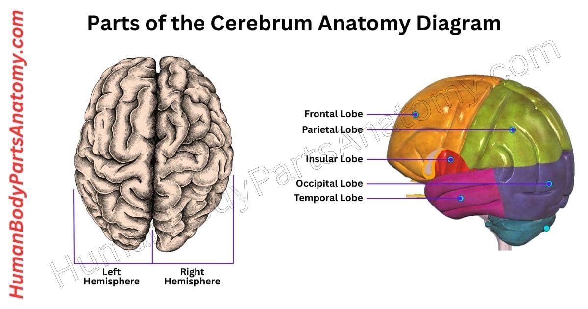
The cerebrum is split into two halves—right and left—by a deep groove called the medial longitudinal fissure. Each half is called a cerebral hemisphere.[1]
Interestingly, the brain works in a cross-wired way: the right hemisphere mainly controls the left side of the body, and the left hemisphere mainly controls the right.[1]
Scientists believe this crisscross setup happens due to a twisting of the body axis during early embryo development.[1]
Although the two hemispheres look similar and share many functions, they are not the same. Some tasks are handled more by one side than the other. This feature is known as brain lateralization.[1]
2. Cerebral Cortex
The cerebral cortex is the outermost layer of the brain and plays a major role in how we think, feel, and act. Although it is only 2 to 4 millimeters thick—about as thick as a coin.[2]
It covers the entire surface of the brain and accounts for nearly half its weight. This is because it is packed with billions of nerve cells, organized into six distinct layers.[2]
The cortex appears wrinkled due to its many folds. These folds include grooves called sulci and raised areas called gyri.[2]
This folded structure increases surface area, allowing more neurons to fit in a small space. More neurons mean a better ability to process complex information.[2]
3. Lobes of the Cerebrum
The cerebral cortex is divided into four main regions, called lobes, each with its own specialized tasks:[2]
- Frontal lobe – Involved in planning, decision-making, movement, and problem-solving.[2]
- Parietal lobe – Handles touch, pressure, and understanding of spatial relationships.[2]
- Temporal lobe – Important for hearing, language, and forming memories.[2]
- Occipital lobe – Processes visual signals and helps you make sense of what you see.[2]
These regions work together to control advanced brain functions such as language, memory, emotions, learning, and personality. Despite its thinness, the cerebral cortex is essential for everything that makes us uniquely human.[2]

Read More – Lobes of the Brain: Complete Guide with Names, Functions & Diagram
4. White Matter
White matter is a key part of the central nervous system, made mostly of myelinated axons—long nerve fibers wrapped in a fatty layer called myelin.[5]
This myelin gives white matter its pale color and plays a crucial role in speeding up the transmission of electrical signals across thebrain and spinal cord.[5]
Although once thought to be support tissue, white matter is now recognized as essential for brain function. It helps different parts of the brain talk to each other by carrying signals between regions of grey matter, where processing and decision-making happen.[5]
This relay system helps the brain work smoothly and efficiently, influencing how we learn, think, and respond. The whitish appearance of this tissue in preserved samples comes from myelin’shigh-fat content, which reflects light.[5]
In fresh tissue, it looks more pinkish-white due to blood vessels running through it. By insulatingnerve fibers, myelin ensures messages travel quickly and with less energy, making communication across the nervous system faster and more reliable.[5]
5. Corpus Callosum
The corpus callosum also called the callosal commissure, is a thick, curved band of nerve fibers located deep inside the brain, just beneath the cerebral cortex.[6]
It serves as a vital bridge between the left and right cerebral hemispheres, allowing them to share information and coordinate activities. This structure is unique to placental mammals.[6]
Measuring around 10 centimeters in length, the corpus callosum is the brain’s largest white matter structure and contains 200 to 300 million axons, which are the long, thread-like parts of neurons that carry signals.[6]
The corpus callosum is divided into several key regions, each linking different parts of the brain:[6]
- The rostrum connects the front underside of the hemispheres.[6]
- The genu curves forward and connects the front lobes.[6]
- The body or trunk links the middle regions.[6]
- The splenium connects the back parts of the brain.[6]
Together, these sections form a communication highway that keeps both sides of the brain working together smoothly.[6]
FAQ’s
The cerebrum is the largest and most developed part of the brain, located in the upper front portion of the skull. It’s responsible for higher brain functions like thinking, memory, learning, reasoning, and voluntary movement.[1]
The cerebrum is divided into four main lobes:
Frontal lobe – controls movement, speech, planning, and decision-making.[2]
Parietal lobe – processes touch, pressure, and spatial awareness.[2]
Temporal lobe – handles hearing, memory, and emotion.[1][2]
Occipital lobe – processes visual information.[2]
Each lobe works together to coordinate complex mental and physical actions.[2]
The cerebrum governs conscious thought and voluntary actions. The cerebellum, located beneath it, fine-tunes movement and balance. The brainstem, at the base of the brain, controls essential automatic functions like breathing, heartbeat, and sleep cycles.[1]
The cerebrum is divided into left and right hemispheres connected by the corpus callosum.[1]
The left hemisphere focuses on logic, math, language, and analytical tasks.[1]
The right hemisphere manages creativity, spatial ability, and emotion.[2][1]
Both sides work together for balanced brain activity.[1]
The cerebrum has an outer layer of gray matter (cerebral cortex) made of neuron cell bodies, and inner white matter that carries signals between brain regions.[1] Its surface folds into gyri (ridges) and sulci (grooves) to increase surface area for more neural connections.[2]
The cerebrum manages conscious activities like movement, speech, memory, problem-solving, learning, emotions, and sensory perception. It integrates data from all senses to help you think, act, and respond to your environment.[1]
Conditions like stroke, traumatic brain injury, brain tumors, epilepsy, and Alzheimer’s disease can damage parts of the cerebrum, leading to problems with speech, movement, memory, or behavior depending on which region is affected.[1]
Medical Disclaimer
All content on HumanBodyPartsAnatomy.com is educational and based on verified, peer-reviewed medical sources. Articles are authored or reviewed by qualified medical or biomedical professionals to ensure accuracy.
This website does not provide medical advice, diagnosis, or treatment. Always consult a licensed healthcare professional for personal medical guidance.
No commercial or promotional interests influence the medical content published on this site.