📅 Published on April 11, 2024 | 🕒 Last updated on July 15, 2026
Overview of Tooth Anatomy
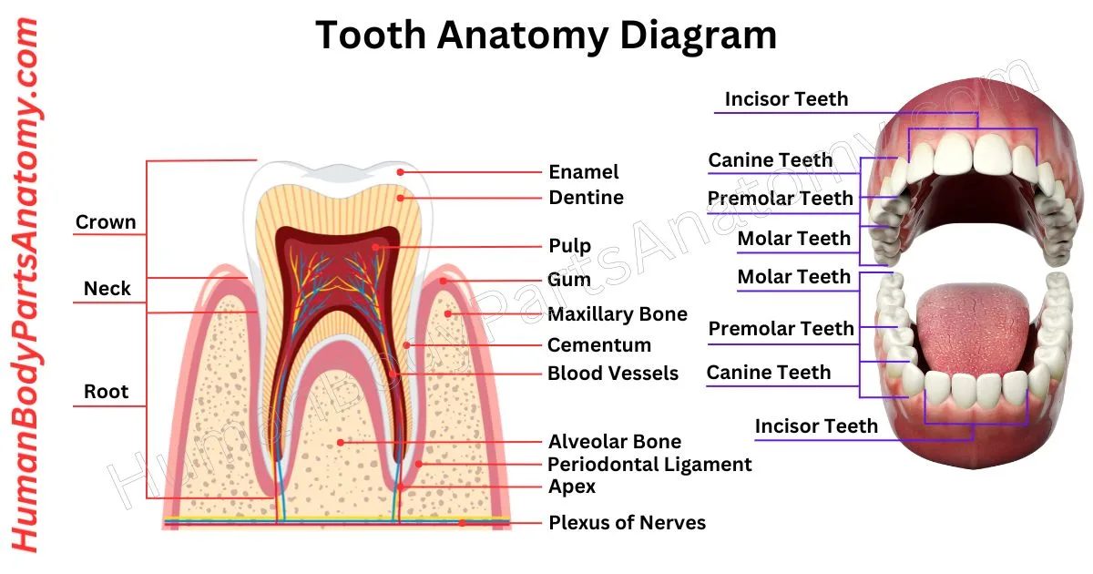
Teeth are important for breaking down food before we swallow it.[1] In the tooth anatomy, we can find four types of teeth, each with a different job.[2] Incisors cut food, canines tear it, and molars and premolars crush it.[2] Most people have 32 teeth,[3] but that can vary. The outside layer, called enamel, is the hardest part of our bodies.[4][5] Teeth are stuck in either the upper or lower jaw and are kept safe by gums.[6] Like many animals, humans get two sets of teeth in their lives.[7] The first set, baby teeth, usually has 20 teeth.[8] They start showing up around six months old,[9][10] which might make babies a bit fussy. Sometimes, babies are even born with teeth called neonatal teeth.[5][6][11]
Teeth anatomy studies tooth structure, such as how they grow and look.[2] It helps dentists figure out teeth during treatments. While how teeth fit together isn’t part of dental anatomy, naming teeth and their parts is super important in this field.[2]
Anatomy of Tooth Diagram

Parts of Tooth
- Crown
- Enamel
- Dentin
- Pulp
- Cementum
- Root
- Root canal
- Periodontal Ligament
- Apical Foramen
- Gingiva (Gum)
- Alveolar Bone
- Incisors
- Canines
- Premolars
- Molars
- Occlusal Surface
- Cusp
- Fissure
- Ridge
- Enamel rod
- Enamel tuft
- Cementoenamel Junction (CEJ)
- Interdental Papilla
- Apex
- Dentin Tubules
- Dentinoenamel Junction (DEJ)
- Pulp Chamber
Teeth Anatomy
Crown
In dentistry, the crown is the top part of a tooth covered by enamel, visible when you smile.[6] Accidents or decay can cause it to chip or break. Dentists use artificial crowns to cover damaged teeth or implants.[18]
Bridges are used to fill the gaps when a tooth is missing. They can be attached to nearby natural teeth or implants. It is made from cement or stainless steel; cement bridges resemble regular teeth, while stainless steel can be silver or gold.[18]
Enamel
Tooth enamel is a protective cover for your teeth.[4] It is the hardest part of your body, even tougher than your bones.[5][4] Enamel protects teeth from cavities, wear and tear, infections, and sensitivity to hot, cold, and sweet foods.[4]
It is mostly made of calcium and phosphorus, which form super-strong crystals.[5][12] The layer underneath (called dentin) the enamel can be different colors, like white or yellow.[13]
Dentin
Dentine is a crucial part of teeth that provides support just under the enamel.[13] It is made mainly of minerals, like hydroxyapatite, forming 70% of its composition. The rest includes water, collagen, and other organic stuff.[12][13]
Inside the dentine are tiny channels called dentinal tubules, which carry sensations from the tooth’s surface to the inner pulp.[13]
There are three types of dentine: primary, secondary, and tertiary. Primary dentine forms before the tooth comes out, while secondary dentine adds layers after erupting.[19]
Tertiary dentine forms in response to damage, like from heat, chemicals, bacteria, or injury. It is denser and darker than the other types and has fewer tubules.[19]
Pulp
The dental pulp is like the heart of a tooth. It has important nerves, blood vessels, and special cells that keep the tooth healthy.[14]
It is protected by layers of hard stuff called dentin and enamel. But if you get holes in your teeth, cracks, or grind your teeth, the pulp can get exposed and need fixing.[14]
The functions of dental pulp are diverse:
- Immune cells fight germs to keep your mouth healthy.[14]
- Nerves feel hot/cold, and pressure changes to warn them of problems.[14]
- Dental pulp makes dentin to protect your teeth.[14]
- Makes important proteins to keep dentin strong.[14]
- Blood vessels in the pulp keep teeth moist to make them stronger.[14]
Cementum
Cementum is a tough tissue, which is important for securing teeth in place.[15][16] It covers the tooth’s root and connects to the periodontal ligament and dentin.[16] Along with the ligament, bone, and gum, cementum forms a good support system for teeth.[15]
This tissue develops gradually from cementoblast cells. Cementum reaches bone but lacks blood vessels, making it avascular.[16]
It has a yellowish color and softer texture, compared to dentin, giving strength and flexibility, which is crucial for tooth stability.[16]
Root
The root is the component of the tooth that is underneath the gums. The upper layer of the crown is enamel. Dentin is located beneath the enamel and surrounds the pulp.[6]
The pulp contains the tooth’s blood vessels and nerves.[14] Some teeth have one or two roots, whereas molars might have four.[2]
Root Canal
A root canal is a tunnel inside your tooth. It has a big part at the top called the pulp chamber and smaller tunnels called root canals that go down into the roots. The dental pulp, like the tooth’s nerve, lives in these tunnels.[14]
Sometimes, there are even smaller branches called accessory canals, mostly near the tips of the roots.[14] The number of root canals varies depending on the tooth.[2][14] Some have one, two, three, or even more; sometimes, one root can have more than one canal.[2]
It is like a little network of tunnels inside your tooth; some teeth have more complicated tunnels than others.[14]
Periodontal Ligament
The periodontal ligament (PDL) is a soft, stretchy band that connects your teeth to the bone around them. It is not the bone or gums that keep your teeth in place, but this special ligament.[15]
It comprises different types of collagen and has tiny blood vessels and nerves running through it.[15]
When you chew or grind your teeth, the PDL helps absorb the pressure and protects your teeth from damage. Instead of being stuck in a place like a nail, your teeth can be gently moved because of the PDL.[15]
If you grind your teeth too much, the ligament can swell and make your teeth feel loose. But once the pressure stops, the PDL heals, and your tooth returns to normal.[15]
Apical Foramen
In dental anatomy, the apical foramen is a tiny hole at the end of a tooth’s root. It is important because it is where blood vessels, nerves, and other tissues enter the tooth’s soft inner part, called the pulp.[2][14]
This little opening also shows where the pulp and the tissues supporting the tooth meet. Usually, it is about 0.5mm to 1.5mm away from the tip of the root.[2]
Gingiva (Gums)
The gums, called gingiva, are the pink tissue around your teeth. They are important for protecting your teeth and supporting their health.[17]
Blood vessels from the carotid artery bring blood to the gums, while nerves from the trigeminal nerve give them feeling.[17]
The gums are part of the periodontium, which includes structures that support your teeth. It consists of the periodontal ligament, alveolar bone, and cementum.[15][17]
One important feature of the gums is the junctional epithelium, a protective barrier at the base of the gum line. It helps shield against damage and bacteria. The gums also help you sense things in your mouth and absorb nutrients.[17]
Furthermore, the gum tissue plays a role in fighting off infections that can lead to gum disease.[17]
Alveolar Bone
The alveolar bone is like a strong foundation for your teeth. It holds them in place and supports them while you eat. It comprises two main layers: dense bone on the outside and trabecular bone on the inside.[2]
The dense bone is tough, like a protective shell, while the trabecular bone is softer and more flexible, providing cushioning.[2]
The main job of the alveolar bone is to keep your teeth firmly anchored in your mouth and to absorb the pressure when you bite and chew.[2]
It comprises two parts: the alveolar bone proper, which surrounds the tooth roots, and the supporting alveolar bone, which helps hold everything together.[2]
The alveolar bone is essential for maintaining healthy teeth and a stable bite.[2]
Incisors
Incisors are those front teeth that look like little chisels.[2] They help us cut up our food.[2][3] We have four on the top and four on the bottom.[3]
In the upper jaw, they are called teeth 7 through 10; in the lower jaw, they are teeth 23 through 26.[3]
Babies usually start getting these teeth between 6 and 16 months old.[9][10] In each jaw, you have two types: central and lateral incisors.[2][3] They are some of the earliest teeth to show up in your mouth![9][10]
Canines
Canines are the sharp teeth in mammal mouths.[2][3] It is also known as fangs, cuspids, or dog teeth.[3][2] Most mammals have four canines, two on top and two on bottom,[2][3] but they vary in size and sharpness.[2]
Some species, like sheep and deer, only have them on top. In some animals, like gorillas and pigs, males have larger canines than females. Rodents don’t have canines.[2]
Humans have small canines with big roots.[2][3] We get baby canines around 16-23 months and lose them around 9-12 years when adult teeth come in.[9][10]
Premolars
Premolars, called bicuspids, are important teeth between canines and molars.[2][3] Two premolars are in each section in adult human mouths, making eight in total.[3]
They usually have two pointed parts and help transition food from front to back during chewing. Their mix of canine and molar traits helps grind food for digestion.[2]
Molars
Molars are big, flat teeth in the back of your mouth.[2][3] They help you grind food when you chew. In people, molars usually have four or five bumps on top.[2]
Grown-ups have 12 molars in sets of three at the back.[3] The last molar in each set is called a wisdom tooth.[3] It usually shows up last, around age 20,[3] but that can be different for everyone. Sometimes, people don’t get a wisdom tooth at all![3]
Occlusal Surface
The top surface of a tooth touches or comes close to touching the top surface of another tooth.[2]
Each tooth has five surfaces:
- Occlusal – the top surface that bites or chews.[2]
- Mesial – the front side.[2]
- Distal – the back side.[2]
- Buccal – the side facing the cheeks.[2]
- Lingual – the side facing the tongue.[2]
Cusp
A cusp is like a little bump on your tooth, either on the top or the front. Canines, or “eye teeth,” have one bump each. Premolars, also called bicuspids, have two bumps each.[2]
Molars usually have either four or five bumps. Sometimes, especially in certain groups of people, the big molars at the top of your mouth.[2] The first ones especially have an extra bump on the inside called the Cusp of Carabelli.[7]
Fissure
Fissures are tiny grooves and pits in teeth. Most tooth decay starts here. There are two types: external and internal.[20]
- External fissures only affect the outer layer of the tooth, called enamel. They usually don’t hurt but can still let in bacteria. Sealants can help prevent this.[20]
- Internal fissures go deeper into the tooth. They can be painful and may reach the sensitive parts inside. In serious cases, they might need special treatment to save the tooth.[20]
Sealants can be put on any tooth with fissures as long as it is healthy and doesn’t already have fillings. They are often used on back teeth and are good for kids, teens, and adults who might get fissures.[20]
Ridge
Mamelons are rounded bumps at the top of a tooth, which gives it a wavy look. They are common in kids’ teeth as they grow, but can also be seen in adults.[21]
While they usually wear down naturally as we chew, they might stick around if teeth don’t align properly. If they are still there, then they can be permanent.[21]
Enamel Tuft
Enamel tufts are tiny branches found where dentin and enamel meet. They stick out into the enamel. Sometimes, people mix them with enamel lamellae, straight lines instead of branches.[22]
Enamel lamellae mostly go from the enamel surface toward the junction. In contrast, tufts go the other way, from the intersection toward the surface.[22]
Cementoenamel Junction
The Cementoenamel Junction (CEJ) is where the tooth’s crown’s enamel meets the root’s cementum.[2][16] It forms a kind of “neck” for the tooth.[2]
It is important because it is the portion where the gums attach to the tooth, keeping it in place. Tooth loss can happen if there is resorption (loss of tooth structure) at this junction, which affects about 5 to 10 percent of people.[2]
Dentists use the CEJ as a reference point for measuring gum health. It can vary between people and even between teeth in the same person.[2]
Interdental Papilia
The interdental papilla, or gum tissue between teeth, sits above the gum line on the sides of your teeth. It is shaped like a cone for the front teeth and more flattened for the back teeth.[2]
You might see a small space called a ‘black triangle’ between your teeth if missing. Dentists can sometimes fix this with braces.[2]
Whether the papilla forms depends on how close the bone is to the space between your teeth; you may not have a papilla if there is more than 8mm of space. But if it is 5mm or less, you will have papilla.[2]
Apex
The apex is like the root tip of a tooth. It is a little entrance known as the apical foramen, through which blood vessels and nerves enter.[2]
Sometimes, there are many smaller doors around the main entrance, similar to those on the side entrance. These smaller ones, called auxiliary canals, are separated by hard tissue.[2]
The main entryway, known as the foramen, links the tooth to the surrounding tissue. If the blood and nerve connections are lost, the tooth loses vitality. Typically, the primary door measures around 0.3 mm thick.[2]
Dentin Tubules
Dentinal tubules are tiny channels in teeth that connect the pulp inside to the outer layers. They act like highways for substances to move in and out, which could potentially harm the tooth.[13]
Researchers examined eight upper premolar teeth under a special microscope. They found more tubules near the top of the tooth, closer to the pulp.[13]
These tubules vary in size, with larger ones near the pulp and smaller ones near the root tip. Inside the tooth, there are more tubules than outside.[13]
The shape of the tubule openings changes: they are mostly round near the top but become irregular toward the root bottom.[13]
Dentinoenamel Junction (DEJ)
The dentinoenamel junction (DEJ) is a vital boundary in your tooth where the outer enamel meets the inner dentin.[2][13] It acts as a protective barrier, preventing cracks from spreading between these layers.[13]
It is composed of tough materials like collagen, proteins (such as SIBLINGs), and molecules from your blood serum.[24]
Its scalloped shape enhances its resilience,[24] while enzymes, metalloproteinases, and lipoproteins play crucial roles in its formation and maintenance, ensuring the strength and health of your tooth.[24]
Pulp Chamber
Inside a tooth, there’s a crucial part called the pulp chamber. It holds a jelly-like material called pulp. This chamber sits in the top part of the tooth, called the crown. It turns into the root canal as it goes down into the roots.[14] The shape and how many canals there are depend on the tooth’s shape.[2][14]
A tooth has three main parts: enamel, dentin, and pulp.[6][13][14] Enamel is the hard outer layer,[4][5] dentin lies beneath it and supports the structure,[13] and the pulp contains nerves and blood vessels that keep the tooth alive.[14]
Humans have four types: incisors (front teeth for cutting food), canines (pointed for tearing), premolars (for crushing), and molars (back teeth for grinding).[2][3] Each type supports specific eating functions, with adults typically having 8 incisors, 4 canines, 8 premolars, and 12 molars.[3]
Most adults have 32 permanent teeth,[3][7] though some may have fewer if wisdom teeth are removed or missing.[3] This includes 20 on top and 12 on bottom,[3][7] replacing the 20 primary (baby) teeth from childhood.[8]
Dentin is the yellowish layer beneath enamel, forming the bulk of the tooth.[13] It’s sensitive due to tiny tubules connected to nerves and provides structural support, though it’s softer than enamel and prone to decay if exposed.[13]
It varies: incisors and canines typically have 1 root, premolars have 1-2, and molars have 2-3 for stability.[2] Roots anchor teeth in the jaw via periodontal ligaments[15] and are covered by cementum.[16]
Tooth sensitivity occurs when enamel wears down or gums recede, exposing dentin.[4][13] Dentin has tiny tubules that allow temperature changes to reach the nerve,[13] causing discomfort or pain.[4]
References-
- National Library of Medicine. (2023). Physiology, Tooth. StatPearls Publishing, Treasure Island (FL). Available from NCBI Bookshelf: https://www.ncbi.nlm.nih.gov/books/NBK538475/ — Publisher: StatPearls (NIH/NCBI indexed clinical resource).
- Morris A, Tadi P. (2023). Anatomy, Head and Neck, Teeth. StatPearls Publishing, Treasure Island (FL). Available from NCBI Bookshelf: https://www.ncbi.nlm.nih.gov/books/NBK557543/ — Peer-reviewed clinical reference via NIH/NCBI Bookshelf.
- Cleveland Clinic. (2025). Teeth: Anatomy, Types, Function & Care. Health Library. Last reviewed November 17, 2025. Available at: https://my.clevelandclinic.org/health/body/24655-teeth — Medically reviewed patient education resource.
- Cleveland Clinic. (2025). Tooth Enamel: What It Is, Function & Care. Health Library. Last reviewed October 7, 2025. Available at: https://my.clevelandclinic.org/health/body/24798-tooth-enamel — Clinically reviewed educational content.
- Lacruz RS, Habelitz S, Wright JT, Paine ML. (2017). Dental Enamel Formation and Implications for Oral Health and Disease. Physiological Reviews, 97(3):939-993. PMCID: PMC6151498. DOI: 10.1152/physrev.00030.2016. Available at: https://www.ncbi.nlm.nih.gov/pmc/articles/PMC6151498/ — High-impact physiology journal review article.
- National Library of Medicine. (2025). Tooth Anatomy. MedlinePlus Medical Encyclopedia. Last reviewed by Linda J. Vorvick, MD. Available at: https://medlineplus.gov/ency/article/002214.htm — NIH MedlinePlus peer-reviewed medical encyclopedia.
- Hambridge H, et al. (2023). Anatomy, Permanent Dentition. StatPearls Publishing, Treasure Island (FL). Last updated May 22, 2023. Available at: https://www.ncbi.nlm.nih.gov/books/NBK570590/ — NIH/NCBI indexed reference.
- Anatomy, Head and Neck, Primary Dentition. (2023). StatPearls Publishing, Treasure Island (FL). Last updated July 31, 2023. Available at: https://www.ncbi.nlm.nih.gov/books/NBK573074/ — Evidence-based clinical reference.
- American Dental Association. Eruption Charts. MouthHealthy – Oral Health Information from the ADA. Available at: https://www.mouthhealthy.org/all-topics-a-z/eruption-charts — Official U.S. professional dental association guidance.
- Cleveland Clinic. (2025). Teething (Teething Syndrome): Symptoms & Tooth Eruption Chart. Health Library. Last reviewed December 5, 2025. Available at: https://my.clevelandclinic.org/health/articles/11179-teething-teething-syndrome — Physician-reviewed content.
- DeSeta F, et al. (2022). Natal and Neonatal Teeth: A Review and Case Series. British Dental Journal, 232(7):503-508. PMID: 35396420. DOI: 10.1038/s41415-022-4091-3. Available at: https://pubmed.ncbi.nlm.nih.gov/35396420 — Peer-reviewed clinical dental journal.
- McGuire T, et al. (2023). Histology, Tooth. StatPearls Publishing (NIH/NCBI). Available at: https://www.ncbi.nlm.nih.gov/books/NBK572055/ — Indexed biomedical reference.
- Arola D, et al. (2017). The Tooth: Its Structure and Properties. Dental Clinics of North America, 61(4):651-668. PMCID: PMC5774624. DOI: 10.1016/j.cden.2017.05.001. Available at: https://www.ncbi.nlm.nih.gov/pmc/articles/PMC5774624/ — Authoritative specialty review journal.
- Davis A, Roberson A, Paranjpe A. (2025). Anatomy, Head and Neck, Dental Pulp. StatPearls Publishing. Last updated December 9, 2025. Available at: https://www.ncbi.nlm.nih.gov/books/NBK537112/ — NIH/NCBI clinical reference.
- Torabi S, Soni A. (2023). Histology, Periodontium. StatPearls Publishing. Last updated March 27, 2023. Available at: https://www.ncbi.nlm.nih.gov/books/NBK570604/ — Peer-reviewed academic source.
- Janik-Skucha-Nowak A, et al. (2025). Root Cementum Molecular Structure and Its Role in Maintaining Oral Health—Systematic Review. PMCID: PMC12652686. Available at: https://pmc.ncbi.nlm.nih.gov/articles/PMC12652686/ — Systematic review indexed in PubMed Central.
- Potturi R, Reddy K, Chintalapani S. (2023). Anatomy, Head and Neck, Oral Gingiva. StatPearls Publishing. Available at: https://www.ncbi.nlm.nih.gov/books/NBK560662/ — Evidence-based NIH reference.
- Cleveland Clinic. (2023). Dental Crowns: Purpose, Procedure, Complications, Care. Available at: https://my.clevelandclinic.org/health/treatments/10923-dental-crowns — Clinically reviewed patient resource.
- Zhao H, et al. (2023). Physiologic Dentin Regeneration: Past, Present, and Future Perspectives. PMCID: PMC10750396. Available at: https://pmc.ncbi.nlm.nih.gov/articles/PMC10750396/ — Indexed biomedical research article.
- Fejerskov O, et al. (2023). Pit and Fissure Sealants. StatPearls Publishing (NIH/NCBI). Available at: https://www.ncbi.nlm.nih.gov/books/NBK448116/ — Preventive dentistry clinical reference.
- Bhardwaj A, et al. (2024). A Cross-Sectional Study on the Presence of Mamelons in Northeastern India. PMCID: PMC11685784. Available via PubMed Central — Peer-reviewed indexed research.
- Desoutter A, et al. (2022). Human Tooth Enamel Tuft Drapes Revealed by Microtomography. Archives of Oral Biology, 141:105487. PMID: 35738023. DOI: 10.1016/j.archoralbio.2022.105487. Available at: https://pubmed.ncbi.nlm.nih.gov/35738023 — International peer-reviewed oral biology journal.
- Arola D, et al. (2017). The Tooth: Its Structure and Properties. Dental Clinics of North America, 61(4):651-668. PMCID: PMC5774624. DOI: 10.1016/j.cden.2017.05.001 — High-authority dental materials review article.
- Vaddi A, et al. (2025). Multi-Modal Comparison of Murine and Human Incisal Dentin-Enamel Junctions. Acta Biochimica Polonica, 72:14642. DOI: 10.3389/abp.2025.14642. Available at: https://www.frontierspartnerships.org/journals/acta-biochimica-polonica/articles/10.3389/abp.2025.14642/full — Peer-reviewed biochemical research journal.
Read More-
Human Body-
- Human Anatomy: Guide to Bones, Muscles, Organs, Systems, Functions & Diagram
- Human Skeleton Anatomy: All 206 Bones Explained with Functions & Diagrams
- Human Muscle Anatomy: Validated Guide to Every Major Muscles & Functions
Head, Face & Senses-
- Nose Anatomy: Parts of the Nose, Structure, Nasal Cavity & Sinuses Explained
- Skull Anatomy: Parts of the Skull, Structure, Cranial, Facial Bones & Functions
- Tongue Anatomy: Muscles, Papillae, Taste Buds, Nerves & Taste Function
- Mouth Anatomy: Guide on Parts of Mouth, Lips, Palate, Gums & Oral Cavity
- Eye Anatomy: Parts of the Eye, Cornea, Lens, Retina, Optic Nerve & Diagram
- Ear Anatomy: Parts of the Ear, Outer, Middle & Inner Ear & Structures
Brain & Nervous System-
- Brain Anatomy: Parts of the Brain, Structure, Functions & Regions Explained
- The 4 Lobes of the Brain: Complete Guide with Locations & Functions
Spine & Back-
- Cervical Spine Anatomy: C1–C7 Vertebrae, Muscles & Nerves Explained
- Spine Anatomy: Parts of the Spine, Vertebrae, Curves, Spinal Cord & Diagram
- Neck Muscle Anatomy: Guide with Key Muscles, Groups, Functions & Diagrams
- Rib Cage Anatomy: Ribs, Sternum, Thoracic, Vertebrae & Functions Explained
Organs-
- Pancreas Anatomy: Parts of Pancreas, Structure, Location, Functions & Role
- Thyroid Anatomy: Guide on Key Parts, Location, Structure & Functions
- Stomach Anatomy: Parts of Stomach, Regions, Layers & Digestive Function
- Heart Anatomy: Guide on Parts of Heart, Chambers, Valves & Blood Flow
- Liver Anatomy: Key Parts of Liver, Functions, Lobes, Segments & Diagram
- Kidney Anatomy: Guide on Parts of Kidney, Structure, Functions & Diagram
Upper Limb-
- Shoulder Anatomy: Parts of the Shoulder, Bones, Joint Structure & Diagram
- Wrist Anatomy: Parts of the Wrist, 8 Carpal Bones, Tendons & Diagram
- Hand Anatomy: Parts of the Hand, Bones, Muscles with Functions & Diagram
- Arm Anatomy: Parts of Arm, Bones, Muscles & Joints with Functions & Diagram
Lower Limb-
- Hip Muscle Anatomy: Guide on Key Muscle Groups, Names, Functions & Diagram
- Hip Bone Anatomy: Parts of Hip Bone, Ilium, Pubis, Functions & Diagram
- Femur Anatomy: Parts of Femur, Structure, Functions, Location & Diagram
- Leg Anatomy: Parts of the Leg, Bones, Muscles & Lower Leg with Functions
- Knee Anatomy: Parts of Knee, Bones, Ligaments, Cartilage & Joint Structure
- Thigh Muscle Anatomy: Key Muscle Groups, Names, Functions & Diagram
Medical Disclaimer
All content on HumanBodyPartsAnatomy.com is educational and based on verified, peer-reviewed medical sources. Articles are authored or reviewed by qualified medical or biomedical professionals to ensure accuracy.
This website does not provide medical advice, diagnosis, or treatment. Always consult a licensed healthcare professional for personal medical guidance.
No commercial or promotional interests influence the medical content published on this site.