📅 Published on March 30, 2024 | 🕒 Last updated on July 6, 2026
Overview of Wrist Anatomy
The wrist joint, or the radiocarpal joint, is a crucial connection between the forearm and hand.[1] It allows various movements like bending, straightening, side-to-side, and twisting.[1][2] This joint is like a modified ball and socket, allowing flexibility while maintaining stability.[2][1] The wrist anatomy (joint) is made up of bones that form two surfaces: one is like a cup (formed by the end of the radius bone and a cartilage disk), and the other is like a ball (formed by the scaphoid, lunate, and triquetrum bones).[1] These bones allow movement in two main directions: up and down (like when you wave your hand) and side to side (like when you move your hand towards your thumb or pinky finger).[1][2] The wrist joint helps us do everyday tasks with our hands, like writing, typing, or picking things up.[2]
It is important to note that the ulna bone isn’t part of this joint; it connects to the wrist differently.[1] In this article, we will see the details of wrist anatomy with their parts, functions & diagrams.
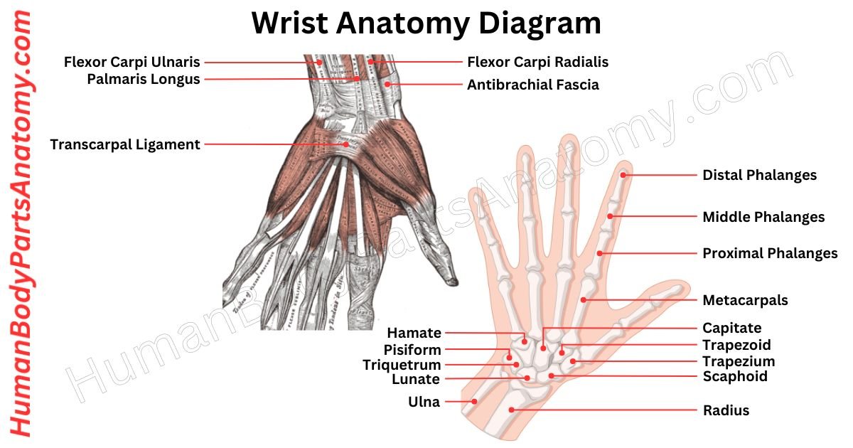
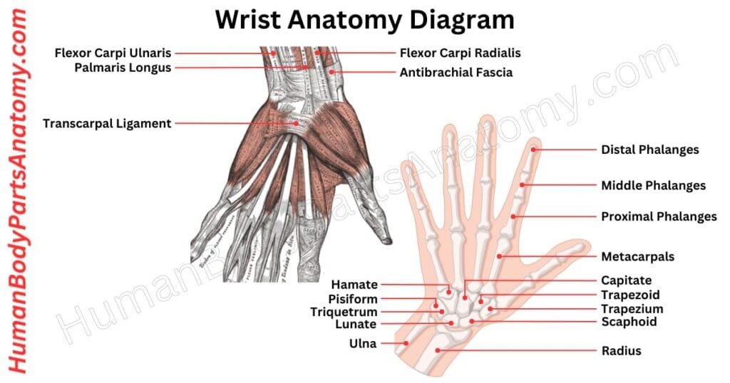
Wrist Anatomy Diagram
Parts of a Wrist
Bones
Carpal Bone Names
- Proximal row (closest to the forearm)
- Scaphoid
- Lunate
- Triquetrum
- Pisiform
- Distal row (closest to the hand)
- Trapezium
- Trapezoid
- Capitate
- Hamate
Joints
- Radiocarpal Joint
- Intercarpal Joints
- Midcarpal Joint
- Carpometacarpal Joints
- Intercarpometacarpal Joints
- Distal Radioulnar Joint
Muscles
- Flexor carpi radialis
- Palmaris longus
- Flexor carpi ulnaris
- Extensor carpi radialis longus
- Extensor carpi radialis brevis
- Extensor carpi ulnaris
Ligaments
- Radial collateral ligament
- Ulnar collateral ligament
- Palmar radiocarpal ligament
- Dorsal radiocarpal ligament
- Scapholunate ligament
- Triangular fibrocartilage complex (TFCC)
Tendons
- Flexor pollicis longus tendon
- Flexor digitorum superficialis tendons
- Flexor digitorum profundus tendons
- Extensor pollicis longus tendon
- Extensor digitorum tendons
- Extensor digiti minimi tendon
Wrist Anatomy: Bones
The carpal bones are eight tiny bones in your wrist that link it to your forearm. They are essential for wrist mobility. They connect with the bones in your forearm to facilitate smooth motion.[3]
These bones also support your hand muscles, such as those in your thumb and little finger. It helps them to function properly and boosts their capacity to understand things.[3]
They also form the carpal tunnel, which allows nerves and tendons to travel from your forearm to your hand.[4][5] It ensures everything works properly and you can feel things correctly.[4]
Proximal Row (closest to the forearm)
The proximal row of carpal bones is situated closest to the forearm. These are the 4 bones that contribute to wrist movement, stability, and overall hand function.[3]
1. Scaphoid
The scaphoid bone in your wrist is a boat shape, the biggest one in the row closest to your forearm.[3]
It is called “scaphoid” because it looks like a boat (“scaphos” in Greek means boat).[3] You can find it just below a spot on your wrist called the anatomical snuffbox.[1][6]
This bone connects to the radius on one end and the trapezium and trapezoid bones on the other. It also links up with the lunate and capitate bones nearby.[1][3]
If you feel the palm of your hand, you might notice a bony bump – that’s the scaphoid tubercle.[3]
2. Lunate
The lunate is a moon-shaped bone in your wrist that sits in the middle of the wrist bones. It connects with the radius on one side, the scaphoid bone on another, the triquetral bone on the other, and the capitate bone below. Its name comes from Latin, meaning “moon-shaped.”[3]
This bone helps your wrist move smoothly and supports your joints. It works together with the two bones in your forearm, the radius and ulna.[3]
Sometimes, “lunate” refers to a small stone tool with a straight, sharp edge and a curved back. It is called that because of its crescent shape, similar to the moon.[3]
3. Triquetrum
The triquetrum is a bone in your wrist that looks like a pyramid with three corners. It sits on the inner side of your wrist.[3]
It connects to the lunate bone on one side and the hamate bone on the other. Also, it has a small oval spot on its lower front side, connecting to the pisiform bone.[3]
4. Pisiform
The pisiform is a small bone shaped like a pea. It sits on the bottom of your wrist near a triquetrum bone. The pisiform bone has a smooth area on top where it connects to the triquetrum bone.[3]
It is special because a tendon surrounds it like a seed in a fruit. This tendon is from a muscle called the flexor carpi ulnaris.[3][7] You can feel the pisiform easily on the palm side of your hand.[3]
Distal Row (closest to the hand)
1. Trapezium
The trapezium bone is an important part of your wrist. It is on the outer side and is part of the lower row of wrist bones. It connects with other bones and helps make your wrist strong and stable.[3]
The trapezium has bumps and grooves where tendons and ligaments attach, which help with movement. Also, it is close to an artery that brings blood to your hand and fingers, keeping them healthy.[3]
2. Trapezoid
The trapezoid bone is like a small wedge in your wrist. Even though it looks tiny from the front, it is wider when you look at it from the back.[3]
It connects to other bones: the scaphoid on one side, the trapezium on another, and the capitate on another. Plus, it touches the second metacarpal bone at the bottom.[3]
3. Capitate
The capitate bone is the biggest of the eight wrist bones. It sits in the middle of the wrist and acts like a keystone. It has a round head, a narrow neck, and a body.[3]
The capitate bone connects with almost all other wrist bones except one.[3] It also links to several ligaments in the wrist.[1]
You can usually feel the capitate bone on the back of your wrist, close to the bone in the middle of your hand. It starts to harden into bone about two months after a baby is born.[3]
4. Hamate
The hamate bone is triangle-shaped in your wrist, near your pinky and ring fingers. It is one of the bones in the lower row of wrist bones.[3]
This bone has six sides, facing your palm, toward the middle of your body, and towards the outside. The palm-facing side has a little curved hook called the hamulus.[3]
Wrist Anatomy – Joint
Wrist anatomy consists of a Radiocarpal Joint, Intercarpal Joint, Midcarpal Joint, Carpometacarpal Joints, Inter Carpometacarpal Joints, and Distal Radioulnar Joint.[1][2]
1. Radiocarpal Joint
The wrist joint is where your forearm meets your hand.[1][2] It is special because it lets you move your hand in many directions.[1]
This joint allows you to bend your hand up and down, move it from side to side, and even twist it.[1][2] It is made by the end of your forearm bone connecting with a few small bones in your hand.[1]
2. Intercarpal Joints
The intercarpal joints in your wrist connect the small bones.[2][8] They are flexible, allowing smooth movements without a fixed pivot point. These joints work closely with the muscles in your wrist.[8]
When you bend your wrist forward (flexion), it mainly happens at the wrist joint itself. But when you straighten your wrist (extension), it mostly involves these intercarpal joints.[2]
They also help your wrist move sideways (abduction and adduction) and in circular motions (circumduction).[2]
3. Midcarpal Joint
The midcarpal joint is a pivot point between the carpal bones in your wrist.[2][8] It is made up of two special joints: one involving the capitate, hamate, and scaphoid bones and the other involving the trapezium, trapezoid, and scaphoid bones. These joints are shaped like saddles.[2]
Your wrist can bend and move because of these joints.[2] They absorb force when you do things like grip or lift objects.[2][3]
These bones also play a role in forming the arches of your hand, which are important for securely gripping objects.[3]
4. Carpometacarpal Joints
The carpometacarpal (CMC) joints connect the base of the metacarpal bones to the carpal bones in the hand.[2][9] They are found at the top part of the hand.[2]
These joints allow for a little sliding motion and are most flexible in the little finger.[2] Ligaments, like the anterior oblique ligament, help stabilize them.[1][2]
The thumb’s CMC joint allows various movements and adds strength for gripping things.[2][3] Cartilage covers and cushions the bones in these joints.[2]
5. Inter Carpometacarpal Joints
The wrist has intercarpal joints connecting the wrist bones to the bases of the finger bones. The wrist bones (carpals) link with the bases of the finger bones (metacarpals) through five joints called carpometacarpal joints.[2]
These joints have ligaments that provide strength.[1][2] They help optimize hand grip by allowing the finger bases to move smoothly.[2]
Among these joints, the thumb joint is the most flexible. It can move in different directions, allowing the thumb to grasp objects effectively.[2]
While some CMC joints are relatively fixed, others are more mobile. This mobility helps with gripping, particularly when the fourth and fifth finger bones move slightly toward the thumb.[2]
6. Distal Radioulnar Joint
The distal radioulnar joint is a pivot joint in your forearm, connecting the radius and ulna bones. It allows you to turn your palm up (supinate) or palm down (pronate).[1][10]
It is crucial to your forearm’s movement, working alongside the proximal radioulnar joint, forearm bones, and interosseous membrane.[1][10]
The distal radioulnar joint also helps your wrist handle weight by spreading forces across your forearm bones. Plus, it is closely linked to the wrist’s ulnocarpal joint.[1][10]
Wrist Anatomy – Muscle
The wrist anatomy consists of a Flexor Carpi Radialis, Palmaris Longus, Flexor Carpi Ulnaris, Extensor Carpi Radialis Longus, Extensor Carpi Radialis Brevis, and Extensor Carpi Ulnaris.[11][12][13]
1. Flexor Carpi Radialis
The flexor carpi radialis is a thin muscle in the forearm that helps bend the wrist and move the hand sideways.[11][12]
It is located on the palm side of the forearm and runs from the inner part of the upper arm bone to the base of the second metacarpal bone. The flexor carpi radialis is part of the muscles at the front of the forearm and sits just below the skin.[11][12]
Its main jobs are to bend the wrist, aid in sideways hand movement, and support the scaphoid bone. It starts from a bump on the inner side of the upper arm bone and attaches to the bases of the second and third metacarpal bones.[11][12]
The flexor carpi radialis is controlled by the median nerve (C6-C7) and gets blood supply from arteries around the forearm.[11]
2. Palmaris Longus
The palmaris longus (PL) muscle is a thin forearm muscle that helps to bend the wrist and smaller hand joints. It begins in the inner elbow and extends to the flexor retinaculum, which joins the palmar fascia.[12]
Despite its heterogeneity across people, it promotes hand stability by constricting palmar aponeurosis. Surgeons sometimes use their tendons in surgeries for the hand and arm.[12]
3. Flexor Carpi Ulnaris
The flexor carpi ulnaris is a powerful muscle in your forearm that bends and pulls your hand closer to the body.[7][11] It is unique because only the ulnar nerve controls it entirely.[7]
This muscle is divided into two parts: the humeral head, which originates on the inside of your elbow, and the ulnar head, which begins on the bony hump at the elbow. They adhere to the tiny bones of your hand.[7][11]
The flexor carpi ulnaris bends your wrist and slides it sideways toward your little finger.[7]
4. Extensor Carpi Radialis Longus
The extensor carpi radialis longus is a forearm muscle important for moving the wrist and hand. It works with other muscles to extend the wrist, move it sideways (radial deviation), bend the elbow, and make a fist. You can feel it easily just below and behind the elbow.[13]
The extensor carpi radialis longus starts from specific points on the humerus bone and grabs onto some fibers from the elbow’s outer bump. It then runs down to attach at the base of the second metacarpal bone in the hand.[13]
The radial nerve from the neck’s C6 and C7 roots tells the extensor carpi radialis longus when to contract. It gets blood from the radial artery.[13]
5. Extensor Carpi Radialis Brevis
The extensor carpi radialis brevis is a forearm muscle that helps straighten and move the wrist to the side.[13][14]
It is found at the back of the forearm and starts from a bony bump on the outer side of the upper arm called the lateral epicondyle.[13][14]
The extensor carpi radialis brevis tendon travels through a passage in the wrist and attaches to the base of the third finger bone on the backside of the hand.[13][14]
The extensor carpi radialis brevis muscle is shorter and thicker compared to another muscle nearby called the extensor carpi radialis longus. They both share a protective covering around their tendons.[14]
6. Extensor Carpi Ulnaris
The Extensor Carpi Ulnaris muscle is found along the outer edge of the forearm. It stretches from the elbow down to the base of the little finger.[13][15]
Its main job is to straighten and pull the wrist towards the pinky side. Keeping the wrist steady and aid movement towards the ulnar side is crucial.[13][15]
It originates from a bony bump on the outer part of the elbow called the lateral epicondyle. It ends by attaching to the base of the fifth metacarpal bone in the hand.[13][15]
The radial nerve supplies signals to the Extensor Carpi Ulnaris muscle. In contrast, its blood supply comes from the ulnar artery.[13]
Wrist Anatomy – Ligament
The wrist anatomy consists of a Radial Collateral Ligament, Ulnar Collateral Ligament, Palmar Radiocarpal Ligament, Dorsal Radiocarpal Ligament, and Scapholunate Ligament.[1]
1. Radial Collateral Ligament
The radial collateral ligament is a strong band that connects the upper arm bone (humerus) with the forearm bone (radius).[1]
It is a tough ligament that starts from the outer edge of the radius bone at the wrist and attaches to a wrist bone called the scaphoid.[1]
Its main job is to prevent too much wrist bending towards the thumb. Additionally, it helps keep the elbow joint stable by resisting forces that might push the joint inward.[1]
2. Ulnar Collateral Ligament
The ulnar collateral ligament is a strong band inside your elbow that connects your upper arm to your forearm bone. It helps keep your elbow stable when you move your arm around.[1]
It is made up of three parts. The main part of the ulnar collateral ligament, the anterior band, is important for keeping your elbow steady and supporting your arm.[1][10]
3. Palmar Radiocarpal Ligament
The palmar radiocarpal ligament connects the lower arm bone (radius) to the bones in your wrist. It is made up of different strands of fibers that crisscross each other.[1]
It allows your hand to move smoothly as your forearm twists. This ligament also helps to keep your wrist stable.[1]
It is positioned on the front side of your wrist, right where you can feel your pulse. This ligament sits in front of certain hand muscle tendons.[1]
4. Dorsal Radiocarpal Ligament
The dorsal radiocarpal ligament is an extrinsic ligament in your wrist that connects the back of your hand bones.[1]
It starts from the back of the long bone in your forearm (radius) and stretches downwards towards the outer side of your wrist.[1]
It attaches to two small bones in your wrist (lunate and triquetral) and ends at a bony bump on the triquetral bone.[1]
The dorsal radiocarpal ligament and other tissues, like cartilage and tendons, help keep your wrist steady from the back.[1]
5. Scapholunate Ligament
The scapholunate ligament (more precisely, the scapholunate interosseous ligament) is a key intrinsic ligament in the wrist that connects the scaphoid and lunate bones.[1][16]
It has three components: dorsal, proximal (membranous), and volar bands, with the dorsal band being the strongest.[16]
This ligament is crucial for maintaining proper alignment and coordinated movement between the scaphoid and lunate during wrist motion.[16]
It is frequently injured and injury can lead to scapholunate dissociation and eventually scapholunate advanced collapse (SLAC wrist).[16]
FAQ’s-
The wrist is made up of eight small carpal bones arranged in two rows between the forearm and the hand. These bones connect the radius and ulna (forearm bones) to the hand and allow flexible movement.[3]
The wrist contains multiple joints, mainly the radiocarpal joint and midcarpal joint, which work together to enable bending, straightening, and side-to-side motion of the hand.[1][2]
Wrist movement is controlled by forearm muscles, not muscles located directly in the wrist. The flexor muscles (flexor carpi radialis, flexor carpi ulnaris, palmaris longus) flex the wrist, while extensor muscles (extensor carpi radialis longus, extensor carpi radialis brevis, extensor carpi ulnaris) extend it. These muscles have their muscle bellies in the forearm and connect to the wrist and hand via long tendons.[11][12][13]
Common causes include acute injuries such as falls onto an outstretched hand leading to fractures (especially scaphoid and distal radius fractures) or ligament sprains.[1][6][17] Other causes include repetitive strain from activities, arthritis, carpal tunnel syndrome (compression of the median nerve), tendinitis, and ganglion cysts.[4][17]
The wrist joint allows the hand to flex, extend, rotate slightly, and move in radial and ulnar deviation. These movements make everyday activities like typing, gripping, lifting, and writing possible. The wrist serves as a crucial transition point between the forearm and hand, allowing for fine motor control and force transmission.[1][2]
Major nerves passing through the wrist region include the median nerve (through the carpal tunnel), ulnar nerve (through Guyon’s canal), and the superficial branch of the radial nerve (along the radial aspect).[4][5][7] These nerves provide sensation and motor control to the hand and fingers.[4][5]
Frequent injuries involve scaphoid or Colles’ fractures from falls, ligament sprains, tendinitis from overuse, or lunate dislocations, often requiring prompt medical attention to avoid complications.[1][4][6][17]
References-
- Erwin J, Varacallo M. (2023). Anatomy, Shoulder and Upper Limb, Wrist Joint. StatPearls [Internet]. Treasure Island (FL): StatPearls Publishing; Updated September 4, 2023.
Available from: https://www.ncbi.nlm.nih.gov/books/NBK534779/ — NCBI Bookshelf ID: NBK534779 - Cleveland Clinic. (2023). Anatomy of the Hand and Wrist. Cleveland Clinic Health Library. Published September 25, 2023.
Available from: https://my.clevelandclinic.org/health/body/25060-anatomy-of-the-hand-and-wrist — Peer-reviewed Academic Medical Center Resource - Arias DG, Varacallo M. (2022). Anatomy, Shoulder and Upper Limb, Hand Carpal Bones. StatPearls [Internet]. Treasure Island (FL): StatPearls Publishing; Updated November 28, 2022.
Available from: https://www.ncbi.nlm.nih.gov/books/NBK535382/ — NCBI Bookshelf ID: NBK535382 - Johns Hopkins Medicine. (2024). Carpal Tunnel Syndrome. Johns Hopkins Medicine Health Library.
Available from: https://www.hopkinsmedicine.org/health/conditions-and-diseases/carpal-tunnel-syndrome — Academic Institutional Clinical Reference - Mayo Clinic Staff. (2024). Carpal Tunnel Syndrome – Symptoms and Causes. Updated March 6, 2024.
Available from: https://www.mayoclinic.org/diseases-conditions/carpal-tunnel-syndrome/symptoms-causes/syc-20355603 — Evidence-Based Clinical Guideline Resource - Wagner ER, Elhassan BT, Kakar S. (2023). Scaphoid Fracture. StatPearls [Internet]. Treasure Island (FL): StatPearls Publishing.
Available from: https://www.ncbi.nlm.nih.gov/books/NBK536907/ — NCBI Bookshelf ID: NBK536907 - Alshami AM, Cairns CW, Souvlis T. (2024). Anatomy, Shoulder and Upper Limb, Forearm Flexor Carpi Ulnaris Muscle. StatPearls [Internet]. Treasure Island (FL): StatPearls Publishing; Updated January 30, 2024.
Available from: https://www.ncbi.nlm.nih.gov/books/NBK526051/ — NCBI Bookshelf ID: NBK526051 - Erne H, Akel A, Jungwirth-Weinberger A, Cerny MK, Landsiedl F, Giardini P. (2022). Anatomy, Biomechanics, and Loads of the Wrist Joint. Life (Basel). 2022;12(2):188.
doi:10.3390/life12020188 — PMCID: PMC8880601 - Arias DG, Black AC, Varacallo M. (2023). Anatomy, Shoulder and Upper Limb, Hand Carpometacarpal Joints. StatPearls [Internet]. Treasure Island (FL): StatPearls Publishing; Updated August 14, 2023.
Available from: https://www.ncbi.nlm.nih.gov/books/NBK547684/ — NCBI Bookshelf ID: NBK547684 - Arias DG, Varacallo M. (2023). Anatomy, Shoulder and Upper Limb, Distal Radio-Ulnar Joint. StatPearls [Internet]. Treasure Island (FL): StatPearls Publishing; Updated August 14, 2023.
Available from: https://www.ncbi.nlm.nih.gov/books/NBK547720/ — NCBI Bookshelf ID: NBK547720 - Bordoni B, Varacallo M. (2023). Anatomy, Shoulder and Upper Limb, Forearm Muscles. StatPearls [Internet]. Treasure Island (FL): StatPearls Publishing; Updated June 5, 2023.
Available from: https://www.ncbi.nlm.nih.gov/books/NBK536975/ — NCBI Bookshelf ID: NBK536975 - Walkowski AD, Gubbels AL. (2023). Anatomy, Shoulder and Upper Limb, Hand Palmaris Tendon. StatPearls [Internet]. Treasure Island (FL): StatPearls Publishing; Updated July 24, 2023.
Available from: https://www.ncbi.nlm.nih.gov/books/NBK519516/ — NCBI Bookshelf ID: NBK519516 - Goubier JN, Walkowski AD, Goldman EM. (2023). Anatomy, Shoulder and Upper Limb, Wrist Extensor Muscles. StatPearls [Internet]. Treasure Island (FL): StatPearls Publishing; Updated August 28, 2023.
Available from: https://www.ncbi.nlm.nih.gov/books/NBK534805/ — NCBI Bookshelf ID: NBK534805 - Walkowski AD, Goldman EM. (2023). Anatomy, Shoulder and Upper Limb, Forearm Extensor Carpi Radialis Brevis Muscle. StatPearls [Internet]. Treasure Island (FL): StatPearls Publishing; Updated August 28, 2023.
Available from: https://www.ncbi.nlm.nih.gov/books/NBK539719/ — NCBI Bookshelf ID: NBK539719 - Sawyer E, Sajjad H, Tadi P. (2023). Anatomy, Shoulder and Upper Limb, Forearm Extensor Carpi Ulnaris Muscle. StatPearls [Internet]. Treasure Island (FL): StatPearls Publishing; Updated August 28, 2023.
Available from: https://www.ncbi.nlm.nih.gov/books/NBK539760/ — NCBI Bookshelf ID: NBK539760 - Pappou IP, Basel J, Deal DN. (2024). Scapholunate Advanced Collapse. StatPearls [Internet]. Treasure Island (FL): StatPearls Publishing; Updated January 8, 2024.
Available from: https://www.ncbi.nlm.nih.gov/books/NBK537124/ — NCBI Bookshelf ID: NBK537124 - American Academy of Orthopaedic Surgeons (AAOS). (2020; 2023 endorsements). Clinical Practice Guideline for the Management of Distal Radius Fractures.
Available from: https://www.aaos.org/quality/quality-programs/distal-radius-fractures — Evidence-Based Orthopedic Clinical Practice Guideline
Medical Disclaimer
All content on HumanBodyPartsAnatomy.com is educational and based on verified, peer-reviewed medical sources. Articles are authored or reviewed by qualified medical or biomedical professionals to ensure accuracy.
This website does not provide medical advice, diagnosis, or treatment. Always consult a licensed healthcare professional for personal medical guidance.
No commercial or promotional interests influence the medical content published on this site.