
📅 Published on May 5, 2024 | 🕒 Last updated on May 20, 2026
Overview of Tongue Anatomy
The tongue is a muscle in your mouth that helps you eat, talk, and taste food.[1] It is covered with tiny bumps called taste buds, which enable you to perceive sweet, salty, sour, bitter, and umami flavors.[2][3] The tongue helps move food around so you can chew and swallow.[1] It also helps keep your teeth clean. Tongue anatomy consists of strong muscles and has a lot of nerves and blood vessels, which makes it sensitive to touch and temperature.[1] Saliva keeps our mouths moist. The tongue has two parts: the front, which you can see when you stick out your tongue, and the back, which is closer to your throat.[1] A line down the middle is called the median sulcus, showing where the left and right sides meet.[1]
The tongue is a combination of intrinsic & extrinsic muscles.[1] The intrinsic muscles are not connected to bones; they help change the shape of the tongue.[1] The extrinsic muscles are attached to bones. These muscles can move the tongue around.[1] This article will illustrate the anatomy of the tongue, including its various parts, names & functions.
Tongue Anatomy Diagram
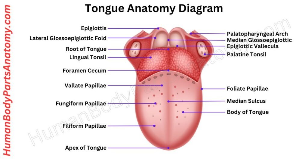
Parts of Tongue
- Apex
- Body
- Root
- Ventral Surface
- Papillae
- Taste buds
- Lingual frenulum
- Saliva Glands
- Sublingual Gland
- Submandibular glands
- Lingual Tonsils
Tongue Anatomy: Parts & Functions
Apex
The apex of the tongue is the tip that touches your front teeth. Linguists study how sounds are made by focusing on different tongue parts. The apex is the front end, while the blade is the part behind it facing your teeth.[4]
Body
The body of the tongue is the central part of your tongue. It is the portion between the tip at the front, and the base at the back, where the tongue connects to a bone in your throat called the hyoid bone.[1][4]
The tongue comprises muscles, connective tissue, and a soft covering called a mucous membrane. This part of the tongue is crucial because it helps you talk, swallow food, and sense different tastes.[1][2]
The surface of the body of the tongue has tiny bumps called papillae, which contain taste buds. These taste buds let you taste sweet, salty, sour, and bitter flavors.[2][3]
Root
The root of the tongue is at the back of the mouth and is less movable than the front part, which is more flexible. It is attached to the hyoid bone (a small bone in the throat) by muscles called Hyoglossi and Genioglossi and a hyoglossal membrane.[1][4]
The root of the tongue connects to other important areas. It links to the epiglottis (a flap that covers the windpipe during swallowing) by three folds of tissue known as the glossoepiglottic folds.[1]
It also connects to the soft palate through arches of tissue called glossopalatine arches and to the pharynx (part of the throat) through other muscles and membranes.[1]
Ventral Surface
The ventral surface is underneath the tongue, facing the mouth’s bottom. It is smoother and less bumpy compared to the top part (the dorsal surface), which has little papillae with taste buds.[1][3][4]
You can see some blood vessels on the ventral surface because the skin is thinner. This tongue part helps it move around easily, which is important for talking and eating.[1][4]
The ventral surface has a small piece of tissue called the frenulum, which connects the tongue to the floor of the mouth. This frenulum helps keep the tongue steady while it moves.[1][5]
Papillae
Lingual papillae are tiny bumps on the top of your tongue that have a rough texture. These papillae have four types: circumvallate, fungiform, filiform, and foliate. They all have different shapes and functions.[2][3]
The circumvallate, fungiform, and foliate types have taste buds, which help you taste food. The filiform type doesn’t have taste buds; instead, it enables you to grip food and gives your tongue its rough feel.[2][3]
Taste buds
Taste buds are small clusters of cells on the tongue that help us taste different flavors. They are mostly found on our tongue, but in a few other places, like the roof of our mouth, the throat, and the flap.[2][3]
Taste buds can detect five main flavors: salty, sour, bitter, sweet, and umami (a savory taste). Any part of the tongue with taste buds can detect the above flavors.[2][3]
When we eat something, food dissolves in saliva and comes into contact with these pores. These pores have small openings that allow the taste buds to send information to our brains.[2][3]
Most people have between 2,000 and 8,000 taste buds on their tongues. Each taste bud has a lifespan of about 10 days, and then it gets replaced by a new one.[6][7][8]
Lingual frenulum
The lingual frenulum is a thin piece of tissue that connects the bottom of your tongue to the floor of your mouth. It runs from near the base of the tongue to the tip, along the center. At the base, it joins the tissue that lines the floor of your mouth.[5]
Small bumps called sublingual caruncles are on each side of the frenulum’s base. These have openings for the submandibular ducts, from where saliva comes out. The blue lines on either side of the frenulum are the deep lingual veins, which drain blood from the tongue.[1]
The lingual frenulum helps keep your tongue in place and supports it while you talk, eat, or swallow.[5]
Saliva Glands
Saliva helps us digest food by breaking down starch into sugars. We have three main pairs of saliva-making glands and lots of smaller ones. These glands can be grouped based on what they produce.[9]
Some make a watery type of saliva (serous), others make a slimy kind (mucous), and some make a mix of both. The watery saliva contains an enzyme called alpha-amylase, which helps break down starch.[9]
The slimy saliva contains a protein called mucin, which helps keep things slippery. Every day, our glands make about 1200 to 1500 milliliters of saliva.[9]
Our body makes saliva when our parasympathetic nervous system gets active, usually when we eat. This system uses acetylcholine to tell our glands to produce more saliva.[9]
Sublingual Gland
The sublingual gland is a small gland under your tongue that makes saliva. It is one of three saliva-making glands in your mouth. This gland produces about 3-5% of your total saliva.[1][9]
It sits below your mouth’s floor, and you can feel it behind your bottom canine teeth. To find it, you can put one finger inside your mouth and press gently with the other hand’s fingers from the outside.[1][9]
This gland has one main tube and about 20 smaller tubes called ducts. The main tube joins with the tube of the submandibular gland to empty saliva through a tiny bump called the sublingual caruncle.[1][9]
The other small tubes open into the mouth along a raised line of tissue, the sublingual fold, on either side of the frenulum of the tongue.[1][9]
Submandibular Glands
The submandibular glands are one of three types of salivary glands in your mouth, and they are about the size of a walnut. They sit under your jawbone and above your tongue bone. These glands have two parts separated by a muscle in your mouth.[9]
Their duct, called Wharton’s duct, drains saliva from the base of your tongue. Blood from your facial and lingual arteries supplies these glands. Your nervous system controls them.[1][9]
These glands make saliva, which helps you chew, swallow, and keep your mouth clean. When you are not eating, they make most of your saliva. When you eat, the parotid gland makes more.[9]
Lingual Tonsils
The lingual tonsils are collections of lymphatic, immune tissue at the back of the tongue. These tonsils are made up of groups of cells from the immune system.[10][11]
They respond when they detect harmful bacteria, viruses, or parasites. When these germs come into contact with the lingual tonsils, the immune cells activate to start defending the body.[10][11]
They are positioned to catch germs before they get further into your body, like your stomach or lungs. When germs show up, the white blood cells in the tonsils kick into action to fight them off.[10][11]
Tongue Anatomy -Intrinsic Muscles
The tongue has four sets of tiny muscles that start and end in the tongue itself. These muscles are the superior longitudinal, inferior longitudinal, transverse, and vertical. They’re named based on how they run or move within the tongue.[1]
Superior Longitudinal Muscle
The superior longitudinal muscle of the tongue is found just under the top surface of the tongue. It helps give the tongue its shape and flexibility. It allows our tongues to move in different directions.[1]
This muscle starts near the back of the tongue, close to the epiglottis flap, and extends toward the front and sides of the tongue.[1]
As an intrinsic muscle, it is responsible for fine-tuning the movements of the tongue, like rolling or bending. These movements are important for speaking, swallowing, and other tongue functions.[1]
Inferior Longitudinal Muscle
The inferior longitudinal muscle is a muscle inside your tongue. It is found on the bottom side of the tongue, between two other muscles—the genioglossus and the hyoglossus.[1]
It helps to move your tongue by making it shorter and thicker. This muscle is controlled by a nerve called the hypoglossal nerve.[1][12]
In cross-section, this muscle looks thin and oval-shaped. It runs from the back to the front of your tongue. At the back, some of its fibers are attached to a bone in your neck called the hyoid bone.[1]
At the front, it connects with other tongue muscles like the styloglossus, hyoglossus, and genioglossus.[1]
Transverse Muscle
The transverse muscle of the tongue is a muscle inside your tongue that helps control its shape. It starts from a line in the middle of the tongue and stretches outward to the sides.[1]
This muscle is controlled by the hypoglossal nerve, which is responsible for tongue movements. When this muscle tightens, your tongue is narrower and longer.[1][12]
Vertical Muscle
The vertical muscle in the tongue is a muscle inside the tongue. When it tightens, it makes the tongue wider, flatter, and longer. This muscle helps with different tongue movements.[1]
It gets signals from the hypoglossal nerve, also called cranial nerve XII. You can find this muscle along the sides and near the front of the tongue.[1][12]
Tongue Anatomy -Extrinsic Muscles
Genioglossus Muscle
The genioglossus is a muscle in your tongue shaped like a fan. It has two parts—one on the left and one on the right—separated by a thin tissue wall.[13]
This muscle starts inside your lower jaw, near your chin, and connects to the hyoid bone in your neck and the bottom of your tongue.[13]
It plays a big role in sticking your tongue out, moving it side to side, and even helps with breathing. The hypoglossal nerve, or cranial nerve XII, controls this muscle.[1][12][13]
Hyoglossus
The hyoglossus muscle is a key extrinsic muscle of the tongue. It has two main parts: one at the front (shaped like a square) and one at the back (shaped like a triangle).[14]
They start from different points on the hyoid bone and join together in the tongue. The front part is thicker and has several smaller parts, while the back has three bundles of fibers.[14]
This muscle helps pull the tongue’s lower part downwards and backward when swallowing. This action is important for pushing food down the throat smoothly. It also plays a role in controlling tongue movements for clear speech.[1][14]
Styloglossus
The styloglossus muscle helps move your tongue in different ways. It is part of the muscle group that works together for actions like swallowing. This muscle starts at the styloid process in your skull and attaches to the side of your tongue.[15]
When you chew food, the styloglossus muscle helps your tongue form a groove to swallow the food. It also helps pull your tongue back and up after chewing.[15]
This muscle has two main parts: front and back. The front parts meet in the middle of the tongue’s bottom, making an arch. The back parts split into smaller bundles that go into the tongue, helping it move.[15]
Overall, the function of the styloglossus muscle is to pull your tongue back and up, helping with chewing and swallowing.[1][15]
Palatoglossus
The palatoglossus muscle is one of the external muscles of the tongue found near the soft palate. It helps shape the sides of the throat and forms the palatoglossal arches. These arches divide the mouth and throat.[1][16]
This muscle works against the levator veli palatini muscle and helps lift the back of the tongue. It also pulls down the soft palate, making the throat narrower.[1][16]
During swallowing, it pushes food down the throat and seals off the mouth to prevent food from going back up. It stops saliva from leaking into the throat by supporting the palatoglossal arch.[16]
The tongue is made of skeletal muscle fibers, connective tissue, blood vessels, and nerves, all covered by a mucous membrane. Unlike most muscles, tongue muscles are arranged in multiple directions, allowing precise movement for speech, chewing, and swallowing.[1]
Yes, the tongue is a muscular organ made up of intrinsic muscles (for shape changes) and extrinsic muscles (for movement), all covered by mucous membrane.[1]
Taste buds are nerve cell clusters in papillae that detect five basic tastes—sweet, salty, bitter, sour, and umami—and send sensory signals to the brain.[2][3]
Its flexibility comes from muscle fibers arranged in three directions (longitudinal, transverse, vertical) and extrinsic muscles allowing protrusion, retraction, and shaping.[1]
The bumps on the tongue are called papillae. They increase surface area, help grip food, and house taste buds. Most bumps are normal, but inflammation or infection can make them more noticeable or painful.[2][3]
Yes. Changes in tongue color, texture, or coating may signal conditions such as vitamin deficiencies, infections, dehydration, digestive disorders, or systemic diseases. Persistent changes should be evaluated by a healthcare professional.[3]
Motor control is mainly by the hypoglossal nerve (CN XII), with sensory input from trigeminal (touch, anterior), facial (taste, anterior), and glossopharyngeal (posterior).[1][12]
References-
- NCBI (StatPearls). (2023). Anatomy, Head and Neck, Tongue. National Library of Medicine.
PMID: Not listed — https://www.ncbi.nlm.nih.gov/books/NBK507782/ - Roper SD, Chaudhari N. (2017). Taste buds: cells, signals and synapses. Nature Reviews Neuroscience, 18, 485–497.
PMID: 28655883 — DOI: 10.1038/nrn.2017.68 — https://pubmed.ncbi.nlm.nih.gov/28655883/ - Cleveland Clinic. (2022). Tongue: Definition, Location, Anatomy & Function. Cleveland Clinic Health Library.
https://my.clevelandclinic.org/health/body/22845-tongue - Sanders I, Mu L. (2013). A three-dimensional atlas of human tongue muscles. The Anatomical Record, 296(7), 1102–1114.
PMCID: PMC3687025 — PMID: 23650264 — https://pmc.ncbi.nlm.nih.gov/articles/PMC3687025/ - Mills N, Pransky SM, Geddes DT, Mirjalili SA. (2019). What is a tongue tie? Defining the anatomy of the in-situ lingual frenulum. Clinical Anatomy, 32(6), 749–761.
PMCID: PMC6850428 — PMID: 30701608 - Taste receptor polymorphisms and longevity: A systematic review and meta-analysis. (2021).
PMCID: PMC8429150 — https://pmc.ncbi.nlm.nih.gov/articles/PMC8429150/ - IQWiG (InformedHealth.org). (2023). How does our sense of taste work? National Library of Medicine.
https://www.ncbi.nlm.nih.gov/books/NBK279408/ - Barlow LA, Klein OD. (2015). Developing and regenerating a sense of taste. Current Topics in Developmental Biology, 111, 401–419.
PMCID: PMC4647210 — PMID: 25662267 — DOI: 10.1016/bs.ctdb.2014.11.012 — https://www.ncbi.nlm.nih.gov/pmc/articles/PMC4647210/ - Sajjad A, Sajjad SS. (2024). Anatomy, Head and Neck: Salivary Glands. StatPearls, NCBI.
PMID: Not listed — https://www.ncbi.nlm.nih.gov/books/NBK538325/ - [Marra A, Khan M. (2024). Anatomy, Head and Neck: Tonsils. StatPearls, NCBI.
PMID: Not listed — https://www.ncbi.nlm.nih.gov/books/NBK539792/ - Arambula A, Brown JR, Neff L. (2021). Anatomy and physiology of the palatine tonsils, adenoids, and lingual tonsils. World Journal of Otorhinolaryngology – Head and Neck Surgery, 7(3), 155–160.
PMCID: PMC8356106 — https://pmc.ncbi.nlm.nih.gov/articles/PMC8356106/ - NCBI (StatPearls). (2022). Neuroanatomy, Cranial Nerve XII (Hypoglossal).
PMID: Not listed — https://www.ncbi.nlm.nih.gov/books/NBK532869/ - Sanders I, Mu L. (2013). Human tongue neuroanatomy and muscle innervation.
PMCID: PMC2955167; PMC3687025 — https://pmc.ncbi.nlm.nih.gov/articles/PMC2955167/ - NCBI (StatPearls). (2023). Anatomy, Head and Neck: Hyoglossus Muscle.
PMID: Not listed — https://www.ncbi.nlm.nih.gov/books/NBK574565/ - Akbar S, Hohman MH. (2023). Anatomy, Head and Neck: Styloglossus. StatPearls, NCBI.
https://www.ncbi.nlm.nih.gov/books/NBK574498/ - NCBI (StatPearls). (2023). Anatomy, Head and Neck: Palatoglossus Muscle.
PMID: Not listed — https://www.ncbi.nlm.nih.gov/books/NBK549823/
Read More-
Human Head
- Skull Anatomy: Complete Guide with Parts, Names, Functions & Diagram
- Ultimate Guide to Eye Anatomy: Parts, Structure, Functions & Diagram
- Mouth Anatomy: Complete Guide with Parts, Names, Functions & Diagram
- Complete Guide to Tooth Anatomy: Learn Parts, Names & Diagram
- Ultimate Guide to Ear Anatomy: Parts, Structure, Functions & Diagram
Organs
External Sources-
- Wikipedia
- KenHub
- Optometrists
- Cleveland Clinic
- American Academy of Ophthalmology
Medical Disclaimer
All content on HumanBodyPartsAnatomy.com is educational and based on verified, peer-reviewed medical sources. Articles are authored or reviewed by qualified medical or biomedical professionals to ensure accuracy.
This website does not provide medical advice, diagnosis, or treatment. Always consult a licensed healthcare professional for personal medical guidance.
No commercial or promotional interests influence the medical content published on this site.