
📅 Published on February 6, 2024 | 🕒 Last updated on July 17, 2026
Overview of Finger Anatomy
Fingers are unique parts of the hand found in humans and many other animals. Over millions of years, they have developed to handle both delicate and strong tasks with skill.[1] Each finger has three bones, called phalanges, which are joined together and surrounded by muscles, tendons, and ligaments that help them move precisely.[2][3] Tendons link the muscles to the bones, allowing the fingers to bend, grip, and release objects easily.[4] Ligaments stabilize the joints and keep them in position,[5] while nerves provide sensory feedback like pressure, temperature, and texture.[6] Additionally, blood vessels supply essential nutrients and oxygen to keep the finger tissues healthy and functioning properly.[7]
All these parts work together to help us do many things, from simple actions like writing and typing to stronger movements like lifting and holding tools.[8] Learning about finger anatomy shows how these movements happen and reveals how complex our hands really are.[2]
In this article, we will explore the structure of the fingers, their key parts, and how they work together to perform various tasks, offering you a detailed overview of this essential part of the human body.
Finger Anatomy Diagram
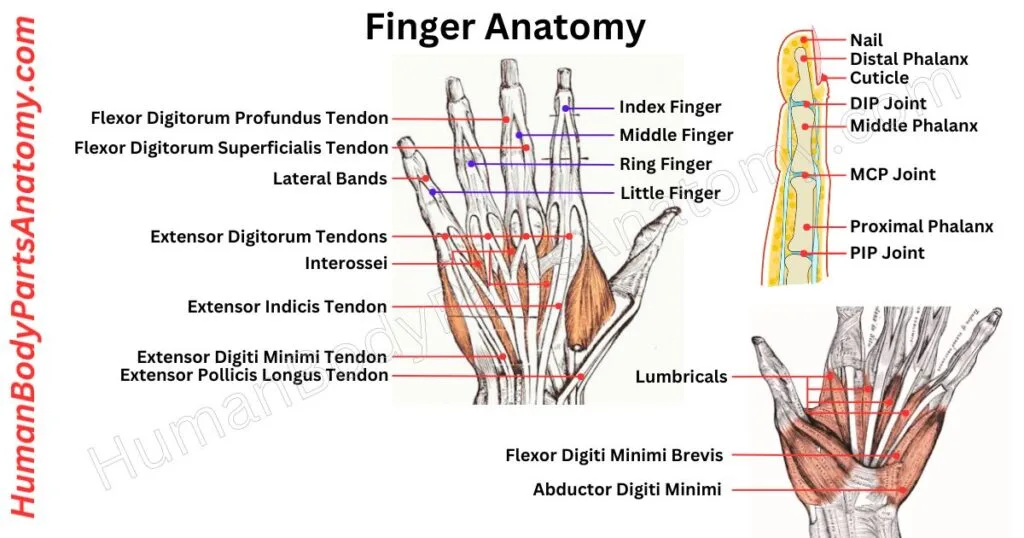
Parts of the Finger
Muscles
- Flexor Digitorum Profundus
- Flexor Digitorum Superficialis
- Extensor Digitorum
- Lumbricals
- Interossei
Joints
- Metacarpophalangeal (MCP) Joint
- Proximal Interphalangeal (PIP) Joint
- Distal Interphalangeal (DIP) Joint
Tendons
- Flexor Digitorum Profundus
- Flexor Digitorum Superficialis
- Flexor Pollicis Longus
- Extensor Digitorum
Ligaments
- Collateral ligaments
- Volara plate
- Palmar ligaments
- Dorsal ligaments
- Sagittal bands
Finger Parts
- Nail
- Phalanx bones
- Blood vessels
- Nerves
- Pulp
- Sesamoid bones
Finger Anatomy: Joint
The finger joints help us move our fingers with control and precision.[3] They let us bend, straighten, rotate, and grip things easily.[9] We can write, hold tools, and do other tasks that require fine hand movements.[8]
Metacarpophalangeal (MCP) Joint
The bottom part of the pinky finger connects to the head of the fifth metacarpal bone, forming the metacarpophalangeal (MCP) joint.[3] It can bend, flex, twist, turn, and move in various directions.[9]
This joint is kept together by some tough ligaments and lubricated by a smooth substance called synovial fluid.[5][3]
Proximal Interphalangeal (PIP) Joint
This joint is located in the middle of your finger and can only move in one direction – bending or straightening.[3]
The joint plays a crucial role in gripping and grasping objects.[8] The surrounding tough ligaments provide stability and prevent injury.[5]
Distal Interphalangeal (DIP) Joint
It is a tiny joint at the tip of your finger.[3] This joint might be small and move in one direction.[9]
This joint is essential for all intricate finger movements, from playing musical instruments to typing on a keyboard.[8]
Finger Anatomy: Tendon
Flexor Digitorum Profundus
FDP tendons help bend the fingers at the fingertip joint.[4] The same muscles regulate them as the rest of the fingers.[10] This muscle is divided into four tendons that go down the forearm via the carpal tunnel and finally join to the fingertip bone.[4]
In contrast to other flexors in the hand, these tendons travel within protective sheaths along the hand and fingers near the bone.[12]
Flexor Digitorum Superficialis
FDS tendons play a vital role in bending the middle joints of the index, middle, ring, and little fingers.[4] A shared muscle powers them, split into four tendons from the forearm into the carpal tunnel.[11] They smoothly slide in sheaths along the fingers and hands.[12]
At the finger level, each tendon separates into two cords.[10] Independently enter the middle bone on either side of the flexor digitorum profundus tendon and continue down to the finger.[4] This intricate system ensures coordinated and efficient finger movement.[8]
Extensor Digitorum Communis
The EDC (Extensor Digitorum Communis) tendons extend from the forearm to your fingertips.[4] Their primary responsibility is straightening the index, middle, ring, and little fingers.[10]
They run down the forearm, passing through the retinaculum, which keeps them in place.[4] These tendons work together to expand the fingers’ joints, providing for smooth and coordinated movement.[8]
Extensor Digiti Minimi Tendon
The EDM muscle is crucial for straightening the little finger.[4] Located in the forearm, it works with another muscle to control the small finger’s movement.[10] Its tendon passes through a wristband (retinaculum) for support, allowing smooth movement.[10]
Surprisingly, fewer than half of people have this tendon.[4] The EDM and other tendons ensure the straightening of the three joints in the small finger.[9]
Finger Anatomy: Ligaments
Collateral Ligaments
The collateral ligaments on each side of the finger joint are made of tough, highly organized collagen fibers.[5] This structure gives them strength and flexibility to handle the various activities of the fingers.[5] These ligaments serve a dual purpose:
- Support precise finger movements to enable us to perform fine motor tasks easily.[8]
- Protect the joint by stabilizing it against unwanted forces.[5]
Volara Plate
The volar plate ligament is found on the palm side of the finger joints.[5] It is made up of specialized collagen fibers and glycosaminoglycans.[5] This ligament has unique properties that provide strength and flexibility.[3]
Its primary role is stabilizing the finger joint and protecting the flexor tendons responsible for bending the fingers.[4]
The volar plate absorbs and distributes forces during movement, acting as a cushion to prevent excessive joint extension, which helps safeguard the joint from injury.[5] Its strength and elasticity make it crucial for maintaining normal finger function.[3]
Palmar & Dorsal Ligaments
The palmar and dorsal ligaments play a crucial role in finger movement. The palmar ligaments are located on the palm side of the finger and are essential in preventing joint hyperextension.[5]
These ligaments are highly organized collagen fibers that provide strength and stability to the joint.[3] On the other hand, the dorsal ligaments are located on the backside of the finger. They are crucial in preventing excessive joint movement.[5]
Sagittal Bands
The sagittal bands are another remarkable set of ligaments found in the fingers.[5] These bands are located on the top of the finger and are crucial in facilitating finger movement.[3]
Finger Anatomy: Parts
Nail
Nails and toenails are like skin accessories, made of tough keratin about half a millimeter thick.[13] They’re firmly attached to the nail bed but have a slight gap at the tip for practical uses like scratching.[14] Nails are essential for our sense of touch.[13]
The lateral folds on each side of the nail give a neat framing effect.[13] The proximal fold is at the base of the nail, acting as a skin border.[14]
A thin layer of skin called the cuticle covers this area. Altogether, our nails are fascinating structures with unique features.[13]
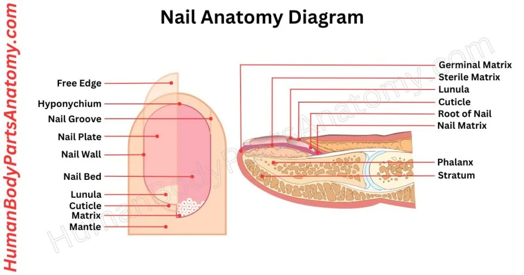
Read More – Nail Anatomy: Parts of the Nail, Plate, Bed, Matrix, Lunula & Structure
Phalanx Bones
The phalanges are small bones that make up the fingers.[2] Even though they are tiny, they are classified as long bones because of their structure.[15] Each phalange has a central shaft, a base at one end, and a head at the other.[2]
In total, there are fourteen phalanges in each hand. The four fingers (index, middle, ring, and little fingers) have three phalanges each—called the proximal, middle, and distal phalanges. The thumb is slightly different, as it only has the proximal and distal phalanges.[2]
These bones are joined by interphalangeal joints, which allow the fingers to move.[3] Blood is supplied to the phalanges through the palmar digital arteries, which send nutrients to the bones via small branches called nutrient rami.[7]
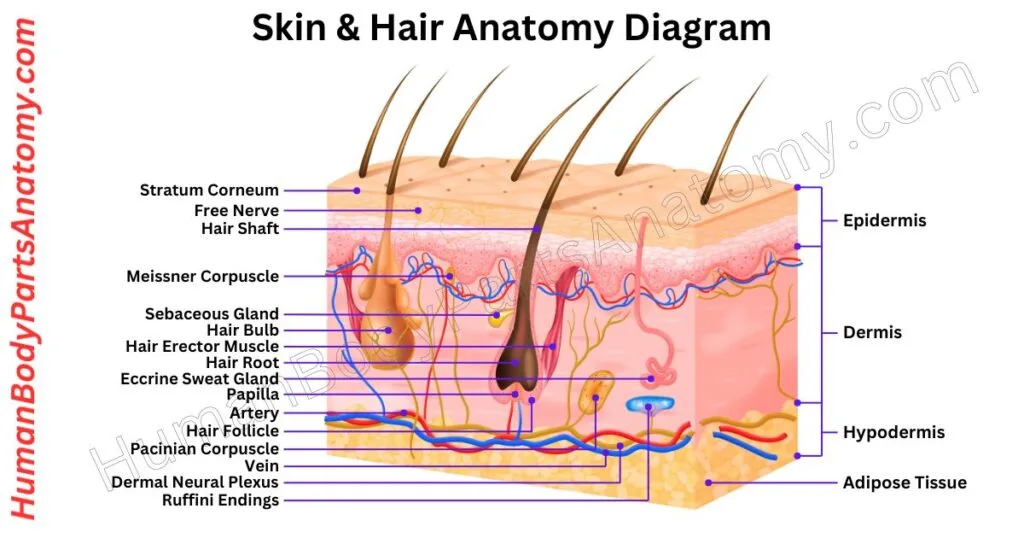
Skin
The function of the skin is to protect, regulate temperature, and feel sensations. The outer layer (epidermis) shields and gives color. In contrast, the inner layer (dermis) has fibers for strength, glands, hair follicles, and nerves.[16]
Below is the fatty layer (subcutis), providing insulation and energy storage. Together, they defend against harm, control body temperature, and allow us to feel pain and pleasure. The skin stays functional by constantly replacing old cells with new ones.[16]
FAQ’s
Each finger has three bones (phalanges) – the proximal, middle, and distal phalanges – except for the thumb, which has two bones. Altogether, the fingers contain 14 phalanges in one hand.[2]
Fingers have hinge joints that allow bending and straightening. These include the metacarpophalangeal (MCP) joints at the knuckles, proximal interphalangeal (PIP) joints, and distal interphalangeal (DIP) joints.[3] The thumb has only the MCP and interphalangeal (IP) joints.[9]
Finger movement is controlled by intrinsic hand muscles (such as lumbricals and interossei) and extrinsic forearm muscles (like flexor digitorum and extensor digitorum).[8][10] Together, they enable grip, precision, and fine motor skills.[8]
Fingers allow grasping, holding, and manipulating objects.[8] They are essential for fine motor skills, such as writing, typing, and playing musical instruments, as well as for sensory feedback due to their high concentration of nerve endings.[6][8]
Finger cracking happens due to gas bubbles forming and collapsing in the joint fluid (synovial fluid).[20] It is usually harmless, but frequent or painful popping may indicate joint issues, such as arthritis or ligament problems.[3]
Finger pain can result from injuries, fractures, sprains, or tendon issues.[16] Chronic pain or stiffness is often linked to arthritis, carpal tunnel syndrome, or repetitive strain injury.[19] Medical evaluation is recommended for persistent discomfort.[19]
Numbness or tingling in the fingers may be caused by nerve compression, commonly seen in carpal tunnel syndrome, cervical spine issues, or poor circulation. It’s important to identify the underlying cause for proper treatment.[17]
Yes. Bone alignment, joint flexibility, tendon function, and muscle strength all influence grip. Conditions such as arthritis, tendon injuries, or nerve damage can weaken grip strength and limit hand function.[18]
References-
- Alba DM, et al. (2015). The evolution of human and ape hand proportions. Proc Natl Acad Sci USA. PMCID: PMC4510966. https://pmc.ncbi.nlm.nih.gov/articles/PMC4510966/. DOI: 10.1073/pnas.1502746112.
- Arias DG, et al. (2023). Anatomy, Shoulder and Upper Limb, Hand Bones. StatPearls [Internet]. NBK547684. U.S. National Library of Medicine. https://www.ncbi.nlm.nih.gov/books/NBK547684/.
- Muppidi V, et al. (2025). Finger Dislocation. StatPearls [Internet]. NBK551508. PMID: 31855352. U.S. National Library of Medicine. https://www.ncbi.nlm.nih.gov/books/NBK551508/.
- Austin GJ, et al. (2023). Anatomy, Shoulder and Upper Limb, Hand Long Flexor Tendons. StatPearls [Internet]. NBK546607. U.S. National Library of Medicine. https://www.ncbi.nlm.nih.gov/books/NBK546607/.
- Varacallo M, et al. (2023). Anatomy, Shoulder and Upper Limb, Metacarpophalangeal Joints. StatPearls [Internet]. NBK538428. U.S. National Library of Medicine. https://www.ncbi.nlm.nih.gov/books/NBK538428/.
- Bordoni B, et al. (2023). Physiology, Sensory System. StatPearls [Internet]. NBK547656. U.S. National Library of Medicine. https://www.ncbi.nlm.nih.gov/books/NBK547656/.
- Sharma S, et al. (2023). Anatomy, Bones. StatPearls [Internet]. NBK537199. U.S. National Library of Medicine. https://www.ncbi.nlm.nih.gov/books/NBK537199/.
- InformedHealth.org. (2025). In brief: How do hands work? NCBI Bookshelf. https://www.ncbi.nlm.nih.gov/books/NBK279362/.
- Varacallo M, et al. (2023). Anatomy, Shoulder and Upper Limb, Hand Metacarpal Phalangeal Joint. StatPearls [Internet]. NBK538343. U.S. National Library of Medicine. https://www.ncbi.nlm.nih.gov/books/NBK538343/.
- Kozin SH, et al. (2023). Hand Tendon Transfers. StatPearls [Internet]. NBK459359. U.S. National Library of Medicine. https://www.ncbi.nlm.nih.gov/books/NBK459359/.
- Varacallo M, et al. (2022). Anatomy, Shoulder and Upper Limb, Flexor Digitorum Superficialis. StatPearls [Internet]. NBK539723. U.S. National Library of Medicine. https://www.ncbi.nlm.nih.gov/books/NBK539723/.
- Klauser AS, et al. (2021). Sonography of Tendon Pathology. J Ultrason. PMCID: PMC8678645. PMID: 34970442. https://pmc.ncbi.nlm.nih.gov/articles/PMC8678645/. DOI: 10.15557/JoU.2021.0052.
- Zargari O, et al. (2023). Anatomy, Shoulder and Upper Limb, Nails. StatPearls [Internet]. NBK534769. PMID: 30521190. U.S. National Library of Medicine. https://www.ncbi.nlm.nih.gov/books/NBK534769/.
- Hanly JG, et al. (2024). Nails. MedlinePlus Medical Encyclopedia. https://medlineplus.gov/ency/article/003247.htm.
- MedlinePlus Medical Encyclopedia. (2023). Long Bones. U.S. National Library of Medicine. https://medlineplus.gov/ency/article/002249.htm.
- MedlinePlus. (2025). Skin layers. U.S. National Library of Medicine. https://medlineplus.gov/ency/imagepages/8912.htm.
- MedlinePlus Medical Encyclopedia. (2024). Numbness and Tingling. U.S. National Library of Medicine. https://medlineplus.gov/ency/article/003206.htm.
- Li K, et al. (2022). Predicting handgrip power of young adult population among major ethnicities using artificial neural networks. PLoS One, 17(6):e0269166. PMCID: PMC9166194. https://pmc.ncbi.nlm.nih.gov/articles/PMC9166194/. (Peer-reviewed; U.S. National Library of Medicine)
- MedlinePlus Medical Encyclopedia. (2024). Finger pain. U.S. National Library of Medicine. https://medlineplus.gov/ency/article/003248.htm.
- Mayo Clinic Staff. (2017). Mayo Clinic Minute: A hand surgeon’s advice about knuckle cracking. https://newsnetwork.mayoclinic.org/discussion/mayo-clinic-minute-a-hand-surgeons-advice-about-knuckle-cracking/.
Read More-
Human Body-
- Human Anatomy: Guide to Bones, Muscles, Organs, Systems, Functions & Diagram
- Human Skeleton Anatomy: All 206 Bones Explained with Functions & Diagrams
- Human Muscle Anatomy: Validated Guide to Every Major Muscles & Functions
Head, Face & Senses-
- Nose Anatomy: Parts of the Nose, Structure, Nasal Cavity & Sinuses Explained
- Skull Anatomy: Parts of the Skull, Structure, Cranial, Facial Bones & Functions
- Mouth Anatomy: Guide on Parts of Mouth, Lips, Palate, Gums & Oral Cavity
- Eye Anatomy: Parts of the Eye, Cornea, Lens, Retina, Optic Nerve & Diagram
- Ear Anatomy: Parts of the Ear, Outer, Middle & Inner Ear & Structures
Brain & Nervous System-
- Brain Anatomy: Parts of the Brain, Structure, Functions & Regions Explained
- The 4 Lobes of the Brain: Complete Guide with Locations & Functions
Spine & Back-
- Cervical Spine Anatomy: C1–C7 Vertebrae, Muscles & Nerves Explained
- Spine Anatomy: Parts of the Spine, Vertebrae, Curves, Spinal Cord & Diagram
- Neck Muscle Anatomy: Guide with Key Muscles, Groups, Functions & Diagrams
- Rib Cage Anatomy: Ribs, Sternum, Thoracic, Vertebrae & Functions Explained
Organs-
- Pancreas Anatomy: Parts of Pancreas, Structure, Location, Functions & Role
- Stomach Anatomy: Parts of Stomach, Regions, Layers & Digestive Function
- Heart Anatomy: Guide on Parts of Heart, Chambers, Valves & Blood Flow
- Liver Anatomy: Key Parts of Liver, Functions, Lobes, Segments & Diagram
- Kidney Anatomy: Guide on Parts of Kidney, Structure, Functions & Diagram
Upper Limb-
- Forearm Anatomy: Parts of the Forearm, Radius, Ulna, Muscles & Diagram
- Shoulder Anatomy: Parts of the Shoulder, Bones, Joint Structure & Diagram
- Wrist Anatomy: Parts of the Wrist, 8 Carpal Bones, Tendons & Diagram
- Hand Anatomy: Parts of the Hand, Bones, Muscles with Functions & Diagram
- Biceps Brachii Anatomy: Parts of Bicep, Structure, Functions & Diagram
- Arm Anatomy: Parts of Arm, Bones, Muscles & Joints with Functions & Diagram
- Nail Anatomy: Parts of the Nail, Plate, Bed, Matrix, Lunula & Structure
Lower Limb-
- Hip Muscle Anatomy: Guide on Key Muscle Groups, Names, Functions & Diagram
- Hip Bone Anatomy: Parts of Hip Bone, Ilium, Pubis, Functions & Diagram
- Femur Anatomy: Parts of Femur, Structure, Functions, Location & Diagram
- Leg Anatomy: Parts of the Leg, Bones, Muscles & Lower Leg with Functions
- Knee Anatomy: Parts of Knee, Bones, Ligaments, Cartilage & Joint Structure
- Thigh Muscle Anatomy: Key Muscle Groups, Names, Functions & Diagram
Medical Disclaimer
All content on HumanBodyPartsAnatomy.com is educational and based on verified, peer-reviewed medical sources. Articles are authored or reviewed by qualified medical or biomedical professionals to ensure accuracy.
This website does not provide medical advice, diagnosis, or treatment. Always consult a licensed healthcare professional for personal medical guidance.
No commercial or promotional interests influence the medical content published on this site.