
📅 Published on January 27, 2024 | 🕒 Last updated on July 17, 2026
Overview of Biceps Anatomy
The biceps brachii — commonly referred to as the “biceps” — is a large, thick, fusiform muscle on the ventral (anterior) surface of the upper arm.[1] It consists of two proximal heads: the short head (caput breve) and the long head (caput longum).[1] The short head originates at the coracoid process of the scapula[1], while the long head originates from the supraglenoid tubercle above the glenoid socket.[1] Both heads share innervation via the musculocutaneous nerve with the coracobrachialis and brachialis muscles — all three forming the anterior compartment of the upper arm.[1][3] A defining anatomical feature is the biceps’ dual proximal attachment to the shoulder blade at both the coracoid process and supraglenoid tubercle.[1] It enables it to span two joints — the glenohumeral (shoulder) joint and the elbow — a property that distinguishes it from single-joint muscles of the upper arm.[1][5]
Anatomy of Bicep Diagram
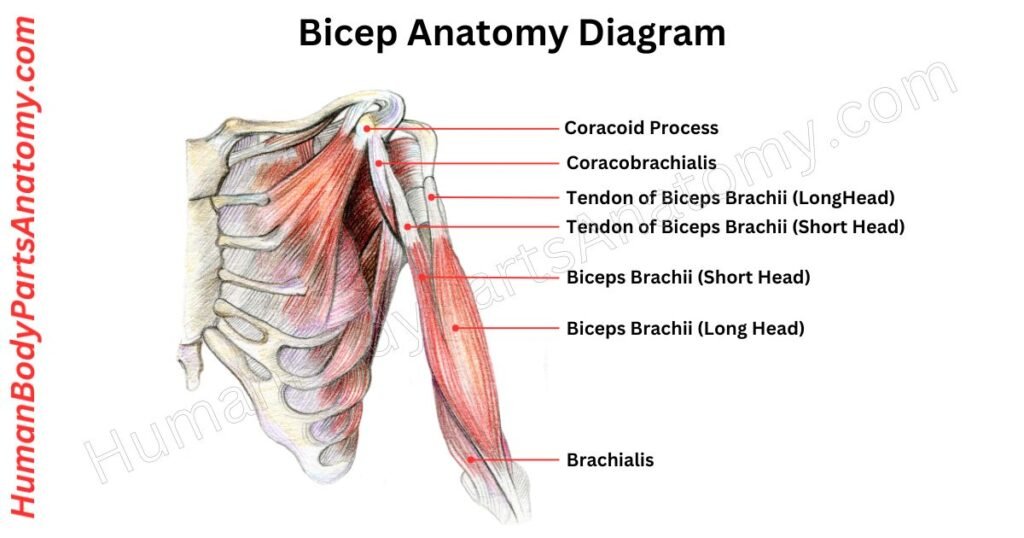
Parts of Bicep
- Short head
- Long head
- Brachialis
- Muscle belly
- Innervation
Bicep Anatomy: Parts & Functions
Short Head
The short head originates via a short, flat tendon from the apex of the coracoid process of the scapula.[1] This origin lies anterosuperior to the attachment of the coracobrachialis muscle, with which the short head shares a common proximal region.[3]
Long Head
The long head originates at the supraglenoid tubercle of the scapula and is anchored additionally by the superior glenoid labrum.[1]
The long head tendon is intra-articular — it passes through the interior of the glenohumeral joint capsule — but extrasynovial, lying outside the synovial membrane.[1]
After exiting the joint capsule, the long biceps tendon turns sharply at the proximal humerus and descends through the intertubercular (bicipital) groove — a bony channel between the greater and lesser tuberosities.[1]
This position is stabilized by the biceps pulley: a capsuloligamentous sling formed by the superior glenohumeral ligament, the coracohumeral ligament, and adjacent rotator cuff tendon fibers.[1]
- Long head of the biceps tendon pain: differential diagnosis and treatment.
- Diagnosis and treatment of distal biceps and anterior elbow pain in throwing athletes.
Brachialis
The brachialis muscle lies deep to the biceps brachii within the anterior compartment of the upper arm.[2][3] It originates from the distal anterior surface of the humerus and inserts onto the tuberosity of the ulna.[2]
Functionally, the brachialis is the primary flexor of the forearm at the elbow, providing elbow flexion at all physiologic positions — a role that has led to its classification as a “pure flexor.”[2][5]
It generates substantially greater flexion torque than the biceps brachii and does not participate in forearm supination or pronation.[2]
Muscle Belly
When the arm is flexed under resistance, the muscle belly becomes visibly prominent as the characteristic biceps bulge. The long and short heads converge to form a single belly on the anterior surface of the humerus before uniting distally into a common tendon that inserts on the radial tuberosity of the radius.[1]
The belly contains thousands of individual muscle fibers organized into bundles called fasciculi.[6] These fibers generate force in response to neural signals carried by the musculocutaneous nerve.
The entire muscle is enclosed by the epimysium. It is the outermost sheath of dense, irregular connective tissue — which protects the muscle, permits gliding against adjacent structures, and transmits contractile force to the skeleton through its continuity with the perimysium, endomysium, and distal tendon.[6]
Innervation
The biceps brachii receives both motor and sensory innervation from the musculocutaneous nerve.[1][3] This nerve arises from the C5 and C6 spinal roots and is a terminal branch of the lateral cord of the brachial plexus.[1][3]
As it passes through the arm, it sequentially innervates the coracobrachialis (first), then the biceps brachii and brachialis, supplying all muscles of the anterior compartment.[3]
FAQ’s-
The biceps attaches proximally to the scapula at the coracoid process (short head) and supraglenoid tubercle (long head), and inserts distally into the radial tuberosity of the radius via its tendon, with a secondary fibrous expansion through the bicipital aponeurosis into the forearm fascia.[1] This architecture enables the muscle to act on both the elbow and shoulder joints.[5]
The biceps act on the elbow joint (contributing to flexion) and the glenohumeral joint (where the long head tendon provides passive stabilization).[1][5] The brachialis — not the biceps — is the primary elbow flexor; the biceps is most accurately described as the forearm’s principal supinator, particularly when the elbow is flexed.[1][2]
The biceps brachii is primarily a powerful forearm supinator — rotating the palm upward — most effective when the elbow is flexed.[1] It also contributes to elbow flexion and plays a role in passive glenohumeral joint stabilization via the long head tendon.[1][5]
Post-exercise soreness is typically due to delayed onset muscle soreness (DOMS) — associated with microscopic disruption of muscle fibers and connective tissue following eccentric exercise.[8]
DOMS manifests 12–24 hours after strenuous or unaccustomed activity, peaks at 24–72 hours, and resolves spontaneously within 3–7 days.[8]
Sudden, sharp, or persistent pain with weakness in supination or flexion may indicate tendinopathy or tendon rupture and warrants clinical evaluation.[4][7]
Biceps tendinitis (inflammatory tenosynovitis of the long head tendon) is the most common type, typically arising from repetitive overhead activities.[7]
Biceps tendon rupture — avulsion of the tendon from its bony attachment — most often involves the proximal long head[4] and may produce an audible pop, acute pain, bruising, visible “Popeye” deformity, and greater than 50% loss of supination strength.[4][9]
Yes. Because the long head tendon is intra-articular,[1] biceps pain frequently co-exists with shoulder pathology, including rotator cuff tears, glenohumeral instability, and tenosynovial inflammation.[7][4] Persistent, mechanical, or weakness-associated biceps pain warrants clinician evaluation.
References-
- Tiwana MS, Charlick M, Varacallo MA. Anatomy, Shoulder and Upper Limb, Biceps Muscle. StatPearls [Internet]. Treasure Island (FL): StatPearls Publishing; 2025 Jan–. Last Update: January 30, 2024. NCBI NBK519538. PMID: 29763025. https://www.ncbi.nlm.nih.gov/books/NBK519538/
- Plantz MA, Bordoni B. Anatomy, Shoulder and Upper Limb, Brachialis Muscle. StatPearls [Internet]. Treasure Island (FL): StatPearls Publishing; 2026 Jan–. Last Update: February 21, 2023. NCBI NBK551630. https://www.ncbi.nlm.nih.gov/books/NBK551630/
- Tuck JM, Bhimji SS. Anatomy, Shoulder and Upper Limb, Musculocutaneous Nerve. StatPearls [Internet]. Treasure Island (FL): StatPearls Publishing; 2026 Jan–. Last Update: September 4, 2023. NCBI NBK534199. https://www.ncbi.nlm.nih.gov/books/NBK534199/
- Hsu D, Anand P, Mabrouk A, Chang KV. Biceps Tendon Rupture. StatPearls [Internet]. Treasure Island (FL): StatPearls Publishing; 2026 Jan–. Last Update: July 15, 2023. NCBI NBK513235. https://www.ncbi.nlm.nih.gov/books/NBK513235/
- Anatomy, Shoulder and Upper Limb, Elbow Joint. StatPearls [Internet]. Treasure Island (FL): StatPearls Publishing; 2026 Jan–. Last Update: December 9, 2025. NCBI NBK532948. https://www.ncbi.nlm.nih.gov/books/NBK532948/
- Dave HD, Shook M, Varacallo MA. Anatomy, Skeletal Muscle. StatPearls [Internet]. Treasure Island (FL): StatPearls Publishing; 2026 Jan–. Last Update: August 28, 2023. NCBI NBK537236. https://www.ncbi.nlm.nih.gov/books/NBK537236/
- Varacallo MA, Mair SD. Proximal Biceps Tendinitis and Tendinopathy. StatPearls [Internet]. Treasure Island (FL): StatPearls Publishing; 2026 Jan–. Last Update: August 4, 2023. NCBI NBK533002. PMID: 30422594. https://www.ncbi.nlm.nih.gov/books/NBK533002/
- Wilke J, Behringer M. Is “Delayed Onset Muscle Soreness” a False Friend? The Potential Implication of the Fascial Connective Tissue in Post-Exercise Discomfort. Int J Mol Sci. 2021 Aug 31;22(17):9482. PMID: 34502387. PMC8431437. DOI: 10.3390/ijms22179482. https://pmc.ncbi.nlm.nih.gov/articles/PMC8431437/
- Durrani S, Moradi A, Asmar A, et al. Distal biceps tendon rupture: a comprehensive overview. Front Surg. 2023 Oct 27;10:1258019. PMC10646517. DOI: 10.3389/fsurg.2023.1258019. https://pmc.ncbi.nlm.nih.gov/articles/PMC10646517/
Read More-
Human Body-
- Human Anatomy: Guide to Bones, Muscles, Organs, Systems, Functions & Diagram
- Human Skeleton Anatomy: All 206 Bones Explained with Functions & Diagrams
- Human Muscle Anatomy: Validated Guide to Every Major Muscles & Functions
Head, Face & Senses-
- Nose Anatomy: Parts of the Nose, Structure, Nasal Cavity & Sinuses Explained
- Skull Anatomy: Parts of the Skull, Structure, Cranial, Facial Bones & Functions
- Mouth Anatomy: Guide on Parts of Mouth, Lips, Palate, Gums & Oral Cavity
- Eye Anatomy: Parts of the Eye, Cornea, Lens, Retina, Optic Nerve & Diagram
- Ear Anatomy: Parts of the Ear, Outer, Middle & Inner Ear & Structures
Brain & Nervous System-
- Brain Anatomy: Parts of the Brain, Structure, Functions & Regions Explained
- The 4 Lobes of the Brain: Complete Guide with Locations & Functions
Spine & Back-
- Cervical Spine Anatomy: C1–C7 Vertebrae, Muscles & Nerves Explained
- Spine Anatomy: Parts of the Spine, Vertebrae, Curves, Spinal Cord & Diagram
- Neck Muscle Anatomy: Guide with Key Muscles, Groups, Functions & Diagrams
- Rib Cage Anatomy: Ribs, Sternum, Thoracic, Vertebrae & Functions Explained
Organs-
- Pancreas Anatomy: Parts of Pancreas, Structure, Location, Functions & Role
- Thyroid Anatomy: Guide on Key Parts, Location, Structure & Functions
- Stomach Anatomy: Parts of Stomach, Regions, Layers & Digestive Function
- Heart Anatomy: Guide on Parts of Heart, Chambers, Valves & Blood Flow
- Liver Anatomy: Key Parts of Liver, Functions, Lobes, Segments & Diagram
- Kidney Anatomy: Guide on Parts of Kidney, Structure, Functions & Diagram
Upper Limb-
- Forearm Anatomy: Parts of the Forearm, Radius, Ulna, Muscles & Diagram
- Shoulder Anatomy: Parts of the Shoulder, Bones, Joint Structure & Diagram
- Wrist Anatomy: Parts of the Wrist, 8 Carpal Bones, Tendons & Diagram
- Hand Anatomy: Parts of the Hand, Bones, Muscles with Functions & Diagram
- Finger Anatomy: Parts of Finger, Bones, Joints, Muscles, Tendons & Diagram
- Arm Anatomy: Parts of Arm, Bones, Muscles & Joints with Functions & Diagram
- Nail Anatomy: Parts of the Nail, Plate, Bed, Matrix, Lunula & Structure
Lower Limb-
- Hip Muscle Anatomy: Guide on Key Muscle Groups, Names, Functions & Diagram
- Hip Bone Anatomy: Parts of Hip Bone, Ilium, Pubis, Functions & Diagram
- Femur Anatomy: Parts of Femur, Structure, Functions, Location & Diagram
- Leg Anatomy: Parts of the Leg, Bones, Muscles & Lower Leg with Functions
- Knee Anatomy: Parts of Knee, Bones, Ligaments, Cartilage & Joint Structure
- Thigh Muscle Anatomy: Key Muscle Groups, Names, Functions & Diagram
Medical Disclaimer
All content on HumanBodyPartsAnatomy.com is educational and based on verified, peer-reviewed medical sources. Articles are authored or reviewed by qualified medical or biomedical professionals to ensure accuracy.
This website does not provide medical advice, diagnosis, or treatment. Always consult a licensed healthcare professional for personal medical guidance.
No commercial or promotional interests influence the medical content published on this site.