📅 Published on April 24, 2024 | 🕒 Last updated on July 5, 2026
Overview of Knee Anatomy
Your knees play a vital role in helping you move through everyday life. They’re the largest and most complex joints in your body[1], connecting your thigh (femur) to your lower leg (tibia).[1] The knee is made up of two main joints — the tibiofemoral joint (where your thigh bone meets your shinbone) and the patellofemoral joint (where your thigh bone connects with your kneecap, or patella).[1][2][4] Together, these joints allow smooth movement and absorb shock every time you take a step, bend, or jump.[2][1] Beyond bones, your knee anatomy contains muscles, ligaments, tendons, cartilage, and nerves — all working together to provide strength, stability, and flexibility.[1][2] The cartilage cushions the bones, the ligaments keep the joint stable, and the surrounding muscles help you control motion.[1][2]
Healthy knees are essential for almost every movement you make — walking, running, climbing stairs, or even standing still.[1][2] They support your body weight, maintain your balance, and make fluid movement possible.[1][2] When your knees are strong and well cared for, you can move with ease and confidence. But when they’re injured or weak, even simple activities can become difficult.[1][2]
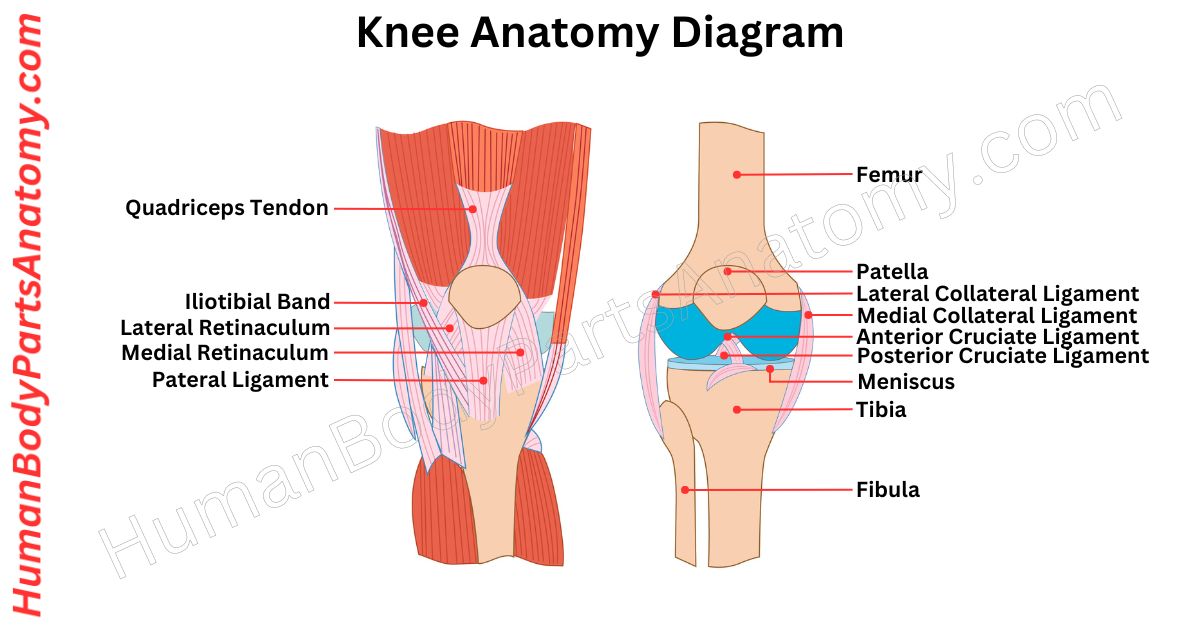
Knee Anatomy Diagram

Parts of the Knee
Bones
- Femur (Thigh bone)
- Tibia (Shin Bone)
- Patella (Kneecap)
Joints
- Patellofemoral joint
- Tibiofemoral joint
- Tibiofibular joint
- Superior Tibiofibular Joint
- Inferior Tibiofibular Joint
- Interosseous Membrane
Muscles
- Flexion Muscles (Hamstring)
- Biceps femoris
- Semitendinosus
- Semimembranosus
- Extension Muscles
- Rectus femoris
- Vastus lateralis
- Vastus medialis
- Vastus intermedius
Innervation
- Femoral nerve
- Tibial nerve
- Common fibular nerves
- Posterior division of the obturator nerve
Ligaments
- Anterior Cruciate Ligament (ACL)
- Posterior Cruciate Ligament (PCL)
- Medial Collateral Ligament (MCL)
- Lateral Collateral Ligament (LCL)
Cartilage
- Hyaline Cartilage
- Fibrocartilage
- Articular Cartilage
Knee Anatomy – Bones
Femur (Thigh bone)
The femur, or thigh bone, is your body’s longest and strongest bone. It helps you stand, walk, and move around by supporting your weight and working with muscles, tendons, and ligaments.[1]
The top part of the femur connects to a special socket in the pelvis called the hip joint. At the bottom of the femur, it joins with the shinbone (tibia) and the kneecap (patella) to create the knee joint.[1]
Surgery may be needed to fix it if it breaks, usually from a big impact like a fall or car crash. Afterward, physical therapy can help you regain strength and movement.[1]
Tibia (Shin Bone)
The tibia, or shinbone, is a key bone in the lower leg. It is located at the front and closer to the body’s midline. It works with the fibula and is linked by a tough membrane that allows limited movement.[1]
The main function of the tibia is to connect the knee joint to the ankle. It is the second-largest bone in the body after the femur. This strong bone is crucial for weight-bearing because of its sturdy structure.[1]
Patella (Kneecap)
The patella, also known as the kneecap, is like a shield for your knee joint. It is a flat, triangular bone in front of your knee. It is a protective cover for the part where your thigh and lower leg bones meet.[1][4]
Many animals, like mice, cats, birds, and dogs, also have a patella. But whales and most reptiles don’t have a patella.[4]
The patella is a special bone in humans because it starts as soft cartilage when we are born. But as we grow, it turns into a hard bone.[4]
The patella is a triangle with its point facing down. That pointy part is called the apex, and it connects to the patellar ligament, which helps hold everything together around your knee.[4]
- Patella Fractures: Approach to Treatment.
- Patella fractures treated with suture tension band fixation.
Knee Anatomy – Joint
Patellofemoral Joint
Your patellofemoral joint is where your kneecap and thigh bone meet at the front of your knee. The femur has a groove where the patella sits; both have smooth, cartilage-covered surfaces.[1][4]
The patella is a small, triangular bone attached to the thigh muscle via a tough tendon. This tendon continues below the patella, forming the patellar ligament, which connects to a groove on the shin bone.[1][4]
This joint is important for activities like climbing, walking on inclines, and moving your knees. Everyday movements like walking up or down stairs, kneeling, or standing up from sitting engage this joint.[5]
Tibiofemoral Joint
The tibiofemoral joint connects the lower end of the thigh bone to the top of the shinbone. It is like a hinge between the rounded ends of the thigh bone and the flat tops of the shinbone.[1]
On the bottom of the thigh bone are two condyles, one on the inside and one on the outside. They are separated by a groove at the back. The inside one is bigger and sticks out.[1]
The top of the shinbone has two slightly curved parts that fit the thigh bone’s condyles, and a vertical ridge separates them.[1]
Because the bone ends don’t fit perfectly, two cartilage wedges are in between to even out the pressure. They are called meniscus and act like cushions.[1][6]
Tibiofibular Joint
The tibia and fibula, the two bones in the leg, connect at three key points: the superior tibiofibular joint, the inferior tibiofibular joint, and the interosseous membrane. These junctions work together to maintain stability while allowing for limited movement to maintain ankle flexibility.[1]
Superior Tibiofibular Joint
This joint connects the upper ends of the tibia and fibula and is classified as a plane synovial joint. The tibial facet is on the lateral side of the top of the tibia, facing toward the back, down, and to the side.[1]
The fibular facet sits on the head of the fibula, facing forward, up, and inward. These flat surfaces are coated with a slick layer of hyaline cartilage, letting them glide against each other.[1]
Although this joint doesn’t facilitate active movements, it allows minor gliding motions to accommodate the ankle’s movement.[1]
Inferior Tibiofibular Joint
This joint connects the lower ends of the tibia and fibula. It is a syndesmosis, a type of fibrous joint. Here, the fibula’s convex surface aligns with the tibia’s concave fibular notch.[1]
This firm connection is critical for ankle stability but can stretch slightly to adapt to ankle movement.[1]
Interosseous Membrane
This membrane links the shafts of the tibia and fibula along the length of the leg. It plays a crucial role in keeping the two bones together and helps distribute force between them.[1]
Knee Muscle Anatomy – Flexion Muscles (Hamstring)
Biceps femoris
The Biceps Femoris is a muscle in the back of your thigh, part of the hamstring group. It helps bend your knees and extend your hips. It has two parts: the long and short heads, starting from the pelvis.[7]
The long head goes around the back of your leg, crossing both the hip and knee joints, ending at the head of the fibula. The short head starts from the femur and joins the fibula head.[7]
Its main jobs are bending the knee (bringing your heel toward your buttocks) and extending the hip (pushing your thigh backward).[7]
This muscle is crucial for running and jumping, and it helps stabilize your knee during various movements.[7]
Semitendinosus
The semitendinosus is a muscle in the back of your thigh. It works alongside the biceps femoris and semimembranosus.[7]
It helps with bending your knee, straightening your hip, and rotating your thigh and shin. Also, it helps prevent over-bending at the hips.[7]
This muscle starts from the bottom of your pelvis and connects to the inside of your shinbone. The tibial nerve and other muscles in the hamstring group control its actions.[7]
Athletes often injure this muscle, especially during fast running or sudden movements.[7]
Semimembranosus
The semimembranosus is a thigh muscle at the back of your leg, below another muscle called the semitendinosus. It helps you bend your knee, straighten your hip, and turn your thigh and knee inward.[7]
It starts at a bone in your pelvis called the ischial tuberosity and goes down to the inner part of your shinbone. The deep femoral artery supplies it with blood, and it is controlled by the sciatic nerve, which helps you move.[7]
This muscle works with other muscles in your thigh to keep your knee steady when you run or jump. Strengthening these muscles is important to avoid injuries and improve at sports.[7]
Knee Muscle Anatomy – Extension Muscles
Rectus femoris
The rectus femoris is a key thigh muscle in the quadriceps group. It is special because it connects to both the hip and knee joints. This muscle is also known as the “kicking muscle” because it powers forceful knee extension, like in soccer kicks.[1]
It starts at the front of the hip bone and goes down to the kneecap and shin bone. The femoral nerve controls it.[1]
Vastus Lateralis
The vastus lateralis (VL) is a big muscle that helps straighten your knee. It is on the outer side of your thigh and connects your thigh bone to your kneecap.[1]
The VL is extending your knee, helping you move, getting stronger, and keeping your hip and knee steady. It starts from your hip and thigh bones and goes down to your kneecap through the quadriceps tendon.[1]
Vastus Medialis
The vastus medialis (VM) helps straighten the knee and stabilize the kneecap. It is part of a group of four muscles, the quadriceps femoris.[1]
The VM has two parts: the upper part called the Vastus Medialis Longus (VML) and the lower part called the Vastus Medialis Obliquus (VMO), each playing a role in knee movement.[1]
The VM receives nourishment for the blood supply from the femoral artery, the deep femoral artery, and a branch of the popliteal artery.[1]
Nerve signals to the VM originate from lower femoral nerve roots and higher lumbar spinal segments, ensuring smooth coordination between the muscle and the central nervous system.[1]
Knee Anatomy – Ligaments
Anterior Cruciate Ligament (ACL)
The ACL is a ligament in your knee that starts from the bottom of your leg bone (tibia), between and in front of the intercondylar eminences.[8]
It connects to the back inner side of the outer leg bone (femur), just behind the middle of the knee. This connection involves a mix of collagen fibers and solid bone through in-between tissue called fibrocartilage and mineralized fibrocartilage.[8]
The ACL runs downward, inward, and a bit forward. It attaches to two spots on the outer side of the knee bone: one called the lateral intercondylar ridge and another called the lateral bifurcate ridge.[8]
On the tibia, it hooks onto two medial and lateral intercondylar tubercles. These spots act as anchors for the anteromedial and posterolateral bundles.[8]
Posterior Cruciate Ligament (PCL)
The posterior cruciate ligament in your knee runs along the back and connects your thigh to your lower leg bone. It helps keep your knee stable and allows for smooth movement.[1]
It has two parts: the bigger anterolateral bundle (ALB) and the smaller posteromedial bundle (PMB). The ALB’s attachment on the thigh bone is about twice as big as its tibial attachment. [1]
The PCL starts from the front and side of the medial femoral condyle and connects to the back of the tibial plateau, about a centimeter below the knee joint.[1]
The research found that the PCL’s attachment areas on the femoral and tibial footprints are about three times bigger than the middle part of the ligament.[1]
Medial Collateral Ligament (MCL)
The medial collateral ligament is a strong, flat band inside your knee that stretches from the medial epicondyle of the femur to the condyle of the tibia. It is a crucial part of the knee to prevent bending too much sideways.[1]
When you hear about knee injuries in sports, the MCL is often injured. It is the most common type of knee ligament injury in activities like skiing, where there is much side-to-side movement.[4]
The MCL has two main parts: the superficial ligament, also called the tibiofemoral ligament, and the deep ligament, known as the mid-third capsular ligament. These parts work together to keep your knee stable and safe during movement.[1]
Lateral Collateral Ligament (LCL)
The fibular or lateral collateral ligament (LCL) is a strong cord in your knee that helps to keep it stable. It is one of four important ligaments that hold your knee joint steady.[1]
The LCL mainly works on the outside of your knee, especially when pressure pushes your knee inward or your lower leg twists outward.[1]
When your knee is straight, the LCL gets stretched out. It joins with the iliotibial band, adding to its strength. The popliteus tendon lies beneath the LCL, keeping it apart from the lateral meniscus. Plus, it splits the biceps femoris muscle into two parts.[1]
As your knee bends, the LCL is less important for stability, especially past 30 degrees of bending. If the crucial ligaments in your knee are torn, the LCL steps in to help control your lower leg’s front and back movement.[1]
Cartilage
Cartilage is a smooth, rubbery connective tissue that acts like a cushion between bones and helps your body move with ease.[2][3]
There are three main types of cartilage found in your body: hyaline cartilage, fibrocartilage, and elastic cartilage. Each has a unique structure and function depending on where it’s located.[2][3]
Hyaline Cartilage
Hyaline cartilage is the most common type of cartilage in your body. It covers the ends of your bones where they meet to form joints, creating a smooth, slippery surface that reduces friction and allows bones to glide easily during movement.[1][2][3]
This same type of cartilage is also known as articular cartilage when it’s found in joints.[1][2][3]
It’s flexible yet durable, providing both support and shock absorption. Without hyaline cartilage, your joints would feel stiff and painful every time you move.[2][3]
Common locations of hyaline cartilage include:
- At the ends of bones in joints (like your knees, elbows, and shoulders).[1][2][3]
- Between your ribs, where it connects to your sternum (breastbone) and helps your chest expand while breathing.[2][3]
- Inside your nose, it gives it shape and structure.[2][3]
Fun fact: Hyaline cartilage has a glassy, bluish-white appearance when viewed under a microscope—hence the name “hyaline,” which means “glassy” in Greek.[2][3]
Fibrocartilage
Fibrocartilage is the toughest and strongest type of cartilage.[2][3] It’s made up of dense collagen fibers that can withstand heavy pressure and tension.[9] This type of cartilage acts like a shock absorber and helps hold bones and joints firmly in place, especially in areas that experience a lot of stress.[3][6][9]
You’ll find fibrocartilage in:
- The menisci of your knees which stabilize and cushion your joint.[1][3][6][9]
- The intervertebral discs between your spinal bones help your spine flex and twist without damage.[2][3]
- The places where tendons and ligaments attach to bones provide extra strength and stability.[2][3]
Did you know? Fibrocartilage is less flexible than other types, but its toughness makes it essential for protecting high-impact joints like the spine and knees.[2][3][9]
Elastic Cartilage
Elastic cartilage is the most flexible type of cartilage in your body. It contains a special network of elastic fibers that allows it to bend and spring back to its original shape — kind of like a rubber band. This flexibility is important for body parts that need to move freely without losing structure.[2][3]
Elastic cartilage is found in:
- Your outer ears (pinnae) can bend without breaking.[2][3]
- Your Eustachian tubes help equalize pressure between your middle ear and throat.[2][3]
- Your larynx (voice box) plays a key role in speech and breathing.[2][3]
Interesting note: Because of its elasticity, this cartilage helps your ears and throat maintain their shape even after repeated movement or force.[2][3]
FAQ’s
The knee connects three bones — the femur (thigh bone), tibia (shin bone), and patella (kneecap). These form the tibiofemoral and patellofemoral joints, allowing bending, straightening, and stability during movement.[1][2]
Four main ligaments (ACL, PCL, MCL, LCL) stabilize the knee,[1][2] while the menisci and articular cartilage cushion and protect the bones, absorbing shock and reducing friction during motion.[2][10]
The knee mainly acts as a hinge joint, allowing flexion and extension with slight rotation when bent.[1][2] These movements enable walking, running, squatting, and other leg activities.[2]
The knee bears much of your body weight and handles twisting forces.[2][12] Damage or wear to ligaments, cartilage, or menisci from injury or aging often causes pain, swelling, or limited motion.[11][12]
The patella improves the leverage of thigh muscles, helping you straighten your leg efficiently. It also shields the front of the knee and aids smooth joint movement.[2][10]
An ACL tear happens from sudden stops or pivots, causing instability.[1][11] A meniscus tear often results from twisting or heavy loads, leading to pain, swelling, or locking sensations.[2][11]
Over time, cartilage wears down and menisci weaken, causing bones to rub together.[1][2] This leads to osteoarthritis, marked by knee pain, stiffness, and reduced flexibility.[1][11]
Strengthen your quadriceps and hamstrings to support the joint.[2][10] Maintain proper posture, healthy weight, and avoid over-twisting to protect the cartilage and menisci from damage.[10][12]
References –
- Gupton M, et al. (2023). Anatomy, Bony Pelvis and Lower Limb, Knee. In: StatPearls [Internet]. Treasure Island (FL): StatPearls Publishing; Nov 5, 2023. NBK500017. https://www.ncbi.nlm.nih.gov/books/NBK500017/
- InformedHealth.org (IQWiG). (2024). In brief: How does the knee work? Cologne, Germany: Institute for Quality and Efficiency in Health Care (IQWiG); Updated Mar 8, 2024. NBK561512. https://www.ncbi.nlm.nih.gov/books/NBK561512/
- Halpern B, et al. (2022). Anatomy, Cartilage. StatPearls [Internet]. Treasure Island (FL): StatPearls Publishing; Oct 17, 2022. NBK532964. https://www.ncbi.nlm.nih.gov/books/NBK532964/
- Cox CF, et al. (2023). Anatomy, Bony Pelvis and Lower Limb, Knee, Patella. In: StatPearls [Internet]. Treasure Island (FL): StatPearls Publishing; Oct 27, 2023. NBK519534. https://www.ncbi.nlm.nih.gov/books/NBK519534/
- American Academy of Orthopaedic Surgeons (AAOS). (2024). Patellofemoral Pain Syndrome (Runner’s Knee). OrthoInfo; Reviewed Feb 2024. https://orthoinfo.aaos.org/en/diseases–conditions/patellofemoral-pain-syndrome/
- Raj MA, et al. (2023). Knee Meniscal Tears. In: StatPearls [Internet]. Treasure Island (FL): StatPearls Publishing; Jul 17, 2023. NBK431067. https://www.ncbi.nlm.nih.gov/books/NBK431067/
- Rodgers CD, et al. (2023). Anatomy, Bony Pelvis and Lower Limb, Hamstring Muscle. In: StatPearls [Internet]. Treasure Island (FL): StatPearls Publishing; Apr 1, 2023. NBK546688. https://www.ncbi.nlm.nih.gov/books/NBK546688/
- Yoo H, et al. (2023). Anatomy, Bony Pelvis and Lower Limb, Knee Anterior Cruciate Ligament. In: StatPearls [Internet]. Treasure Island (FL): StatPearls Publishing; 2023. NBK559233. https://www.ncbi.nlm.nih.gov/books/NBK559233/
- Mameri ES, et al. (2022). Review of Meniscus Anatomy and Biomechanics. J Clin Orthop Trauma. 32:101984. PMCID: PMC9463428. https://www.ncbi.nlm.nih.gov/pmc/articles/PMC9463428/ — PMID: 35947336
- Cleveland Clinic. (2024). Knee Joint: Function & Anatomy. https://my.clevelandclinic.org/health/body/24777-knee-joint
- American Academy of Orthopaedic Surgeons (AAOS). (2024). Common Knee Injuries. OrthoInfo. https://orthoinfo.aaos.org/en/diseases–conditions/common-knee-injuries/
- Johns Hopkins Medicine. (2024). Knee Pain and Problems. https://www.hopkinsmedicine.org/health/conditions-and-diseases/knee-pain-and-problems
Medical Disclaimer
All content on HumanBodyPartsAnatomy.com is educational and based on verified, peer-reviewed medical sources. Articles are authored or reviewed by qualified medical or biomedical professionals to ensure accuracy.
This website does not provide medical advice, diagnosis, or treatment. Always consult a licensed healthcare professional for personal medical guidance.
No commercial or promotional interests influence the medical content published on this site.