📅 Published on December 26, 2024 | 🕒 Last updated on May 20, 2026
Overview of Human Heart Anatomy
The heart is a vital muscular organ in most animals that powers the circulation of blood through the body.[1] In heart anatomy, blood vessels help form the circulatory system, which delivers oxygen and nutrients to tissues and removes waste like carbon dioxide.[1] This waste is carried to the lungs for expulsion. In human anatomy, the heart is roughly the size of a closed fist and lies between the lungs in the chest’s central area.[2] This area is known as the mediastinum.[2] It has four chambers: two upper atria (right and left) and two lower ventricles (right and left).[1][2] The right side (right atrium and ventricle) and the left side (left atrium and ventricle) function together to ensure that blood flows in the proper direction.[1]
Heart Function
The heart’s primary job is to pump blood, deliver oxygen and essential nutrients to cells, and carry away waste products for other organs to process.[1][4] Beyond pumping, the heart also:
- Regulates heart rate and rhythm.[4]
- Maintains blood pressure.[4]
This organ works closely with other systems to support these functions:
- Nervous System: The nervous system controls heart rate by signaling it to slow down during rest or speed up under stress.[4]
- Endocrine System: Hormones released by the endocrine system influence blood pressure by signaling blood vessels to constrict or relax. The thyroid gland, for example, releases hormones that can alter the heart’s speed.[4]
In this article, we will see the detailed anatomy of the heart with its different parts & systems that make every living thing breathe.
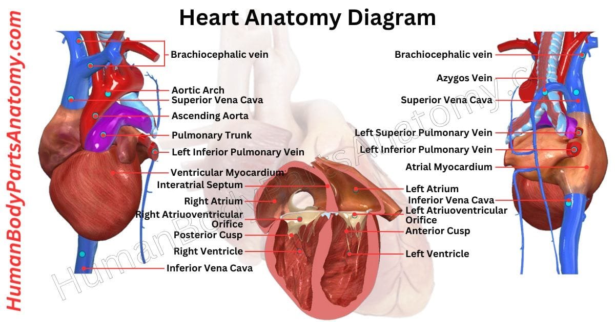
Heart Anatomy Diagram

Anatomy of the Heart
External Structure
- Apex
- Base
- Coronary Sulcus
- Anterior and Posterior Interventricular Sulcus
- Pericardium
Layers of the Heart Wall
- Epicardium
- Myocardium
- Endocardium
Heart Chambers
- Right Atrium
- Left Atrium
- Right Ventricle
- Left Ventricle
Valves of the Heart
- Atrioventricular (AV) Valves
- Tricuspid Valve
- Mitral (Bicuspid) Valve
- Semilunar Valves
- Pulmonary Valve
- Aortic Valve
Blood Vessels Connected to the Heart
- Pulmonary Circuit
- Pulmonary Artery
- Pulmonary Veins
- Systemic Circuit
- Aorta
- Superior Vena Cava
- Inferior Vena Cava
Supporting Structures
- Chordae Tendineae
- Papillary Muscles
Coronary Circulation
- Coronary Arteries
- Left Coronary Artery
- Right Coronary Artery
- Cardiac Veins
- Great Cardiac Vein
- Middle and Small Cardiac Veins
- Coronary Sinus
Conduction System of the Heart
- Sinoatrial (SA) Node
- Atrioventricular (AV) Node
- Atrioventricular Bundle
- Purkinje Fibers
External Structure of Heart Anatomy
Apex
The apex of the heart is located at the tip of the left and right ventricles, opposite the base of the heart. It is facing toward the left side of the chest and angled slightly forward.[2][3]
The apex plays a key role in blood circulation. With each heartbeat, it twists and makes contact with the front of the chest, creating the apex beat. You can feel the apex beat by placing a hand just below the left nipple line.[2][3]
The apex helps the ventricles efficiently pump blood: The left ventricular apex pushes oxygen-rich blood to the body, while the right ventricular apex channels blood to the lungs for oxygenation.[1][2]
This movement aids in wringing out the ventricles and helps them push blood upward and out of the heart, ensuring a steady flow through the body and lungs.[3]
Base
The base of the heart lies upward, slightly backward, and to the right, aligning with the fifth to eighth thoracic vertebrae. It closely neighbors the esophagus, aorta, and thoracic duct.[2][3]
The left atrium primarily forms the base, while the right atrium contributes a small portion. The base presents a quadrilateral shape.[2]
It lies just below the point where the pulmonary artery divides and meets the coronary sulcus at its lower border. The coronary sulcus forms a groove that contains the coronary sinus.[2][5]
- On the right side, the sulcus terminalis of the right atrium defines the base.[2]
- On the left, the ligament of the left vena cava and the oblique vein of the left atrium mark its boundary.[2]
The left atrium receives four pulmonary veins, with two on each side, and the right atrium takes in blood from the superior vena cava at the top and the inferior vena cava below.[1][2]
Coronary Sulcus
The coronary sulcus is a shallow groove on the heart’s surface that separates the upper chambers (atria) from the lower chambers (ventricles).[5]
It is located near the base of the right auricle and contains key blood vessels that supply the heart with oxygen. In the front, this groove is interrupted by the pulmonary trunk, the large vessel carrying blood to the lungs.[5]
On the back side of the heart, the coronary sulcus houses the coronary sinus. This large vein collects blood returning from the heart’s muscle tissue. This sinus extends from the area near the third left rib to the middle of the right sixth rib.[5]
There are two coronary sulci in the heart anatomy.
- The left portion of the coronary sulcus starts behind the pulmonary trunk. It runs down between the left atrium and left ventricle. The circumflex branch of the left coronary artery and the coronary sinus follow this path.[5][12]
- On the right side, the coronary sulcus is visible from the front. It marks the path of the right coronary artery and the small cardiac vein. This part of the sulcus separates the right atrium from the right ventricle. It continues down to wrap around the heart’s underside.[5]
Anterior Interventricular Sulcus
The anterior interventricular sulcus is a groove on the front side of the heart that helps separate the two lower chambers, called ventricles. It works alongside another groove at the back of the heart, known as the posterior interventricular sulcus.[5]
Sometimes, these grooves are called the paraconal and subsinosal interventricular grooves, respectively. The anterior interventricular sulcus runs from the coronary sulcus (a groove encircling the heart) down to the tip of the heart, known as the apex.[5]
When it reaches the underside of the heart, it ends at a small indentation called the cardiac apex notch. This groove holds the anterior interventricular artery (a branch of the left coronary artery) and the great cardiac vein.[5][12]
Posterior Interventricular Sulcus
The posterior interventricular sulcus is a distinct groove on the back of the heart. It marks the boundary between the two lower chambers or ventricles.[5]
The posterior interventricular sulcus is paired with the anterior interventricular sulcus on the heart’s front and provides a pathway for critical blood vessels. It is the subsinosal interventricular groove and extends from the heart’s base near the right side to the apex.[5]
This sulcus protects the posterior interventricular artery and the middle cardiac vein, which are essential in delivering oxygenated blood to the heart tissue and carrying deoxygenated blood away.[5]
Pericardium
The pericardium is a protective sac made of tough, flexible tissue that surrounds the heart. It has two layers: the outer fibrous pericardium, which is strong and helps keep the heart in place, and the inner serous pericardium, which is smooth and slippery to reduce friction as the heart beats.[3]
This design protects the heart, anchors it in the chest, and allows it to move easily while pumping blood. The pericardium doesn’t cover the heart entirely—it leaves openings where the large blood vessels connect and where it rests on the diaphragm.[3]
1. Fibrous Pericardium
The fibrous pericardium is a tough, non-flexible layer of connective tissue directly connected to the central tendon of the diaphragm. Its rigid nature limits the heart’s ability to overfill quickly, providing structural support.[3]
However, this inflexibility can lead to life-threatening complications, such as cardiac tamponade, when fluid builds up and compresses the heart.[3]
2. Serous Pericardium
It is housed within the fibrous pericardium, and the serous pericardium has two distinct layers. The outer parietal layer adheres to the inside of the fibrous pericardium. In contrast, the inner visceral layer, also called the epicardium, lies directly on the heart’s surface.[3]
Both layers consist of a single layer of specialized epithelial cells called mesothelium, which help reduce friction during heart movement.[3]
Layers of the Heart Wall
In the heart anatomy, the heart wall has three main layers: the epicardium, myocardium, and endocardium. These layers are similar to the three layers found in blood vessels, which are called the tunica adventitia, tunica media, and tunica intima.[1][2]
In both structures, each layer serves a comparable purpose: the outer layer protects, the middle layer is muscular and controls movement, and the inner layer provides a smooth lining for blood flow.
Epicardium
The epicardium is the outer protective layer of the heart. It is made up of mesothelial cells, connective tissue, and fat. It covers the heart and the beginnings of major blood vessels like the aorta, superior vena cava, and inferior vena cava.[3]
As part of the pericardium—the heart’s enclosing membrane—the epicardium shields the heart from external harm and friction. Beyond protection, it has several key roles in heart health and development.[3]
During embryonic growth, it sends important signals that guide the formation and maturation of the heart.
Additionally, it secretes factors crucial for the growth and survival of cardiomyocytes, the muscle cells of the heart.[1]
Epicardial cells can also act as progenitor cells, with the potential to develop into various cell types needed by the heart.[1]
Myocardium
The myocardium, located in the middle, is the thickest layer. It sits between the thin, inner endocardium layer and the outer epicardium, part of the pericardium that protects the heart.[1][2]
The myocardium is made up of special muscle cells called cardiomyocytes, which are designed for the heart’s pumping action. These cells have unique structures called intercalated discs that contain gap junctions.[1]
These junctions allow quick communication between cells, helping the heart muscle contract well-coordinately.[1]
The myocardium’s primary job is to help the heart contract and relax, allowing it to pump blood effectively through the body.[1][4]
Additionally, the myocardium supports the heart’s structure and helps transmit electrical signals needed for a steady heartbeat.[1][4]
Endocardium
The endocardium is the heart’s innermost tissue layer, lining its chambers and protecting the valves. Structurally, it resembles the endothelial cells that line blood vessels, reflecting shared biological origins.[1][2]
It is positioned beneath the thicker myocardium—the heart’s muscular layer responsible for pumping. The endocardium is a smooth interface, promoting efficient blood flow through the heart.[1]
The epicardium is the heart’s outermost layer. It has a small volume of lubricating fluid within the pericardium, a protective fibrous sac. This layered structure enables the heart’s smooth and powerful contractions.[3]
Heart Chambers Anatomy
Right Atrium
The right atrium receives deoxygenated blood from three main sources:[1][2]
- Superior vena cava
- Inferior vena cava
- Coronary veins.
The tricuspid valve then funnels this blood into the right ventricle, controlling its flow.[10]
The right side of the heart houses the right atrium, which connects to a small, expandable pouch called the right auricle, or right atrial appendage.[1][2]
This pouch helps increase the atrium’s blood-holding capacity. Inside the right atrium, there are two main areas separated by a ridge called the crista terminalis. Each area has a specific role and developmental origin:[2]
- Sinus Venarum: This smooth-walled part, located behind the crista terminalis, receives blood from the superior and inferior vena cavae. It originates from a fetal structure known as the sinus venosus.[2]
- Atrium Proper: It is found in front of the crista terminalis. This area includes the right auricle and has rough, muscular walls due to pectinate muscles.[2]
Each part of the right atrium is specialized to support blood movement through the heart.
Left Atrium
The left atrium is a chamber in the heart that receives oxygenated blood from the lungs through four pulmonary veins.[1][2]
It pumps this blood into the left ventricle via the left atrioventricular orifice. This orifice is an opening regulated by the mitral valve to ensure one-way blood flow.[10][11]
The left atrium forms the heart’s base and sits at the back of the heart. Its upper part bears the left auricle, a small, ear-shaped pouch that partially covers the base of the pulmonary trunk. The main artery carries blood from the heart to the lungs.[2]
The inner surface of the left atrium has two distinct regions with different origins:
- Inflow area: This smooth-surfaced section receives blood from the pulmonary veins, forming directly from them during development.[2]
- Outflow area: Located toward the front and containing the left auricle, this region has muscle ridges known as pectinate muscles, developed from the embryonic atrium.[2]
Each part of the left atrium plays a role in efficiently moving oxygen-rich blood through the heart and into the body’s circulation.
Right Ventricle
The right ventricle receives deoxygenated blood from the right atrium. It pumps it into the pulmonary artery via the pulmonary valve.[1][2] It is triangular in shape & forms most of the heart’s front border.[2]
This ventricle has two main sections: the inflow section, where blood enters, and the outflow section, where blood exits to the lungs. These sections are divided by a muscular ridge called the supraventricular crest.[2]
- Inflow Section: The inner surface of the inflow section is lined with muscular ridges called trabeculae carneae, which give it a sponge-like appearance. These trabeculae carneae have three main structures:
- Ridges – muscles attached along their entire length, forming raised lines along the ventricle walls.
- Bridges – muscles connected at both ends but free in the middle. Among these is the moderator band, which contains parts of the heart’s right bundle branch, helping to transmit electrical signals efficiently.[4][8]
- Papillary Muscles – These muscles anchor to the ventricle walls and connect to thin, fibrous strings called chordae tendineae. The chordae tendineae attach to the flaps of the tricuspid valve. It prevents them from flipping backward during contraction by tightening when the papillary muscles contract.[10][11]
- Outflow Section (Conus Arteriosus): The outflow section, located at the top of the right ventricle, leads to the pulmonary artery. This part is known as the conus arteriosus and originates from the fetal heart structure called the bulbus cordis. It has smooth walls free of trabeculae carneae, which gives it a distinctly different appearance from the rest of the right ventricle.[2]
Left Ventricle
The left ventricle receives oxygen-rich blood from the left atrium. It pumps it through the aortic valve into the aorta.[1][2] It is positioned at the apex of the heart in anatomical terms and contributes to the heart’s left and diaphragmatic borders.[2]
Like the right ventricle, it has two main sections:
- Inflow Portion: The muscular ridges called trabeculae carneae line the inner walls of the inflow portion, similar to those in the right ventricle. It connects to the mitral valve, and the two papillary muscles actively stabilize the valve, preventing blood from flowing backward.[10][11]
- Outflow Portion: The outflow portion, called the aortic vestibule, is a smooth-walled region free of trabeculae carneae. This part originates from the embryonic bulbus cordis and is specially structured to ensure efficient blood flow into the aorta.[2][13]
Valves of the Heart Anatomy
The heart is a powerful muscle that pumps blood through the entire body, supplying each organ with oxygen and nutrients. Inside the heart, some valves act like gates, opening and closing with each heartbeat to control blood flow between its chambers and maintain a steady rhythm.[10]
These valves ensure that blood moves in the right direction and at the right moment, preventing any backward flow. As they open and shut, they produce two distinct sounds—the familiar “lub-dub” of a heartbeat.[10]
Atrioventricular (AV) Valves
The atrioventricular valves, positioned between the atria and ventricles, play a crucial role in heart function. At the onset of ventricular contraction (systole), these valves close tightly to prevent blood from flowing backward, creating the distinct first heart sound.[10]
1. Tricuspid Valve
The tricuspid valve is positioned between the right atrium and right ventricle, regulating blood flow through the right atrioventricular opening.[10]
It features three distinct cusps—anterior, septal, and posterior—each securely attached at its base to a fibrous ring encircling the orifice. This structure ensures efficient, one-way blood movement within the heart.[10]
2. Mitral (Bicuspid) Valve
The mitral valve is found between the left atrium and the left ventricle. It helps to control blood flow through the left atrioventricular opening.[10][11]
Often called the bicuspid valve, it has two flaps or cusps—one at the front (anterior) and one at the back (posterior). Each cusp is anchored to a strong fibrous ring that encircles the opening, ensuring it stays securely in place.[10][11]
Semilunar Valves
The semilunar valves sit between the heart’s ventricles and the major blood vessels that carry blood out of the heart. They close at the start of the heart’s relaxation phase, known as diastole, and create the second “dub” sound in the heartbeat. There are two of these valves.[10]
1. Pulmonary Valve
The pulmonary valve sits at the junction of the right ventricle and the pulmonary trunk. It comprises three cusps—left, right, and anterior. These are named for their original orientation during fetal heart development before rotation occurs.[14]
2. Aortic Valve
The aortic valve is positioned between the left ventricle and the ascending aorta. It also consists of three cusps: right, left, and posterior.[13]
The left and right aortic sinuses are small pockets in the aortic wall that give rise to the left and right coronary arteries. During diastole, as the heart relaxes, blood briefly flows backward, filling these sinuses.[13]
It ensures that blood enters the coronary arteries to supply oxygen and nutrients to the heart muscle, supporting its function.[5][13]
Blood Vessels Connected to the Heart
The great vessels of the heart are important blood vessels that connect directly to the heart. These include arteries and veins that carry blood between the heart and the lungs, as well as to the rest of the body.[1][2]
Pulmonary Circuit
The pulmonary circuit connects the heart and lungs, ensuring blood is refreshed with oxygen.[1][9]
- Main Pulmonary Artery – The main pulmonary artery carries oxygen-poor blood from the right ventricle. This artery splits into two branches, one for each lung. In the lungs, the blood releases carbon dioxide and absorbs oxygen.[1][9]
- Pulmonary Veins – Once oxygenated, the blood travels back to the heart through the pulmonary veins. Typically, there are four pulmonary veins—two from each lung—which deliver the oxygen-rich blood into the left atrium. This continuous cycle keeps the body supplied with oxygen for its essential functions.[1][2]
Systemic Circuit
- The ascending aorta, the initial section of your aorta, transports oxygen-rich blood from the left ventricle of your heart. This blood then moves through various aorta branches, supplying oxygen and nutrients to the entire body.[1][9]
- The superior vena cava is a large vein responsible for carrying oxygen-depleted blood from the upper part of your body to the right atrium of your heart.[1][2]
- Similarly, the inferior vena cava, another major vein, brings oxygen-depleted blood from the lower parts of your body into the right atrium.[1][2]
Supporting Structures of Heart Anatomy
The chordae tendineae and papillary muscles are important parts of the heart that help the valves work correctly. They work together to keep the valves in place and ensure that blood flows in the right direction.[10][11]
Chordae Tendineae
The chordae tendineae are thin, string-like structures inside the heart’s ventricles. They connect the small muscles on the inner walls of the heart, called papillary muscles, to the flaps of the heart valves. Some of these strings branch into multiple strands, while others stay as single cords.[10][11]
In the right ventricle, they attach to the three flaps of the tricuspid valve, and in the left ventricle, they connect to the two flaps of the mitral valve.[10][11]
Their main job is to hold the valve flaps in place during heartbeats, stopping them from flipping backward into the atria and making sure blood flows in the right direction.[10][11]
Papillary Muscles
Papillary muscles are small but vital parts inside the heart’s ventricles. They play a key role in helping the heart’s valves work correctly by connecting to them with thin, string-like fibers called chordae tendineae. These muscles ensure that blood flows in the right direction.[10][11]
In the heart anatomy, there are a total of five papillary muscles.
- In the right ventricle, there are three—named anterior, posterior, and septal papillary muscles—that support the tricuspid valve.[10]
- In the left ventricle, there are two—anterolateral and posteromedial papillary muscles—that assist the mitral valve.[10][11]
When the heart beats and pumps blood (a phase called ventricular systole), the papillary muscles contract. This action pulls on the chordae tendineae, keeping the valves tightly shut so blood doesn’t leak backward into the atria. This coordinated effort is essential for proper blood circulation and helps prevent issues like valve leakage.[10][11]
Coronary Circulation – Parts of the Heart
Coronary circulation refers to the flow of blood through a network of arteries and veins that deliver oxygen and nutrients to the heart muscle (myocardium) while removing waste products. This system ensures that the heart functions efficiently by meeting its constant energy demands.[5][9]
Coronary Arteries
- Left Main Coronary Artery (LMCA): This plays a crucial role in heart function by delivering oxygen-rich blood to the left atrium and left ventricle. These chambers receive oxygenated blood from the lungs and pump it throughout the body. Additionally, the LMCA branches supply most of the interventricular septum, supporting the heart’s ability to circulate blood efficiently.[5][12]
- Right Coronary Artery (RCA): This artery supplies blood to the right atrium and right ventricle, which are essential for pumping deoxygenated blood to the lungs. It also supplies the sinoatrial (SA) and atrioventricular (AV) nodes, which control the heart’s electrical signals and ensure coordinated muscle contractions. Additionally, branches of the RCA nourish about one-third of the interventricular septum, the wall separating the heart’s lower chambers and supporting its structural and functional integrity.[5][6][7]
Cardiac Veins
The heart’s veins play an essential role in carrying deoxygenated blood back to the heart chambers.[5]
- Anterior Cardiac Veins: Usually numbering 2 to 5, these veins carry blood from the front of the right ventricle directly into the right atrium.[5]
- Great Cardiac Vein: This vein starts at the heart’s apex (the tip) and moves upward along the front groove between the ventricles. It joins with a small vein from the left atrium to form the coronary sinus, which is located on the back of the heart.[5]
- Middle Cardiac Vein: It is at the beginning of the apex. This vein travels along the lower groove between the ventricles and empties into the coronary sinus near its end.[5]
- Small Cardiac Vein: Also called the right coronary vein, it collects blood from parts of the right atrium and right ventricle.[5]
- Thebesian Veins: These are very small, valve-free veins found in the walls of all four chambers of the heart. They directly drain blood from the heart muscle (myocardium) into the corresponding chamber.[5]
Coronary Sinus
The coronary sinus is the heart’s largest vein, carrying most of the heart muscle’s deoxygenated blood back to the right atrium.[5]
It begins at the back of the heart, where the great cardiac and the oblique vein of the left atrium meet. It is positioned in the groove between the left atrium and left ventricle and gathers blood from several smaller veins.[5]
Finally, it delivers blood to the right atrium through an opening, often guarded by a tiny flap called the valve of the coronary sinus.[5]
Conduction System of the Heart Anatomy
The heart’s conduction system is an internal electrical network that ensures a steady heartbeat by coordinating the contraction and relaxation of heart muscles.[4]
It generates and transmits electrical impulses along precise pathways and synchronizes the movements of the heart’s chambers. This system ensures that blood is pumped efficiently and delivers oxygen and nutrients to the body.[4]
Sinoatrial (SA) Node
The sinoatrial (SA) node is a cluster of specialized pacemaker cells located in the upper wall of the right atrium, near the entry point of the superior vena cava. These cells have the unique ability to spontaneously produce electrical signals to initiate the heart’s rhythmic contractions.[6]
The electrical impulses generated by the SA node propagate through gap junctions across both atria and trigger their contraction (atrial systole). This action pumps blood from the atria into the ventricles to ensure continuous blood flow.[6]
The autonomic nervous system modulates the activity of the SA node to regulate heart rate:[4][6]
- The sympathetic nervous system accelerates the SA node’s firing, increasing heart rate.[6]
- The parasympathetic nervous system slows its activity, decreasing heart rate.[6]
This dynamic regulation ensures that the heart adapts to the body’s changing needs.
Atrioventricular (AV) Node & Bundle
The atrioventricular (AV) node and the bundle of His are essential parts of the heart’s electrical system. They control how electrical signals move from the atria (upper chambers) to the ventricles (lower chambers), helping the heart beat in a steady and coordinated way.[7][8]
The AV node works like a checkpoint. It briefly slows down the electrical signal before sending it to the ventricles. This delay allows the ventricles enough time to fill with blood before they contract. The AV node is composed of specialized cells connected by small channels known as gap junctions.[7]
These channels are formed by proteins known as connexins, including Cx43, Cx40, Cx45, and Cx30.2/31.9. These proteins help regulate the spread of electrical signals through the heart.[7]
During procedures like cardiac catheterization, doctors carefully locate the AV node and the bundle of His. This helps them understand the heart’s electrical activity and safely perform treatments such as cardiac ablation. Ablation is often used to treat abnormal heart rhythms, including certain types of fast heartbeats and atrial fibrillation.[7][8]
- The bundle of His is located just below the AV node, near the tricuspid valve.[8]
- It begins where the AV node enters a firm area called the central fibrous body.[8]
- The bundle of His has three parts: the penetrating bundle, the nonbranching bundle, and the branching bundle.[8]
- The final portion divides into the right and left bundle branches, which carry electrical signals to the ventricles so they can contract properly.[8]
Although the bundle of His receives some nerve supply, it has fewer nerve connections than the AV node and does not have large dedicated blood vessels.[8]
Purkinje Fibers
Purkinje fibers, called the sub-endocardial conduction network, are special heart cells that play a key role in keeping the heart working smoothly. These fibers store glycogen and connect tightly through gap junctions, enabling quick and efficient communication between cells.[4]
Purkinje fibers lie just beneath the inner surface of the ventricles and transmit electrical signals from the atrioventricular (AV) bundle to the ventricular walls of the heart. This fast signal ensures that the ventricles contract together during a heartbeat (ventricular systole).[4]
As a result, the heart pumps blood efficiently—sending oxygen-poor blood to the lungs through the pulmonary artery and oxygen-rich blood to the rest of the body through the aorta.[1][4]
FAQ’s
The human heart is a four-chambered muscular organ with valves, major blood vessels, and an electrical system that pumps blood throughout the body.[1][2]
Heart anatomy includes two atria and two ventricles, along with four valves (tricuspid, pulmonary, mitral, and aortic). It also contains coronary arteries that supply oxygen to the heart muscle. This structure allows continuous circulation, delivering oxygen and nutrients to organs across the body.[1][2][5][10]
The heart is located slightly left of the center of the chest, behind the breastbone and between the lungs.[2]
It sits in the mediastinum, above the diaphragm, with about two-thirds of its mass on the left side. The lower pointed tip, called the apex, angles toward the left hip. This location is important for understanding chest pain and CPR hand placement.[2]
The heart has four chambers: two atria and two ventricles.[1][2]
The right atrium and right ventricle handle oxygen-poor blood going to the lungs. The left atrium and left ventricle manage oxygen-rich blood pumped to the body. The left ventricle has thicker walls because it generates higher pressure for systemic circulation.[1][2]
The heart has four valves that keep blood flowing in one direction.[10]
The tricuspid and mitral valves control blood flow between atria and ventricles. The pulmonary and aortic valves regulate blood leaving the heart. Proper valve function prevents backflow and maintains healthy circulation. Valve disorders can lead to murmurs or heart disease.[10][11][13][14]
The right side pumps blood to the lungs, and the left side pumps blood to the rest of the body.[1]
The right heart supports pulmonary circulation by sending oxygen-poor blood to the lungs. The left heart supports systemic circulation by distributing oxygen-rich blood throughout the body. The left ventricle is thicker because it works against higher pressure.[1][9]
An adult human heart is about the size of a clenched fist and weighs 7–15 ounces (200–425 grams).[2]
Heart size varies by age, sex, and fitness level. Athletes may have slightly enlarged hearts due to training. However, abnormal enlargement can signal conditions like cardiomyopathy or high blood pressure.[2][4]
The heart’s electrical conduction system controls heartbeat and rhythm.[4]
Electrical signals start in the sinoatrial (SA) node, the natural pacemaker. Signals pass through the atrioventricular (AV) node and into the ventricles, causing coordinated contractions. Disruptions in this system can cause arrhythmias or irregular heartbeats.[4][6][7][8]
Changes or damage in heart anatomy can lead to heart disease.[1]
Blocked coronary arteries reduce oxygen supply to heart muscle, causing heart attacks. Damaged valves can impair blood flow. Thickened or weakened heart walls may lead to heart failure. Understanding heart structure helps with early detection and prevention of cardiovascular disease in the United States.[1][5][9]
References-
- National Heart, Lung, and Blood Institute (NHLBI). (2022). How the Heart Works. U.S. Department of Health & Human Services, National Institutes of Health (NIH). Reviewed 2022. Available from: https://www.nhlbi.nih.gov/health/heart
- Volpe JK, Makaryus AN. (2023). Anatomy, Thorax, Heart and Pericardial Cavity. In: StatPearls. Treasure Island (FL): StatPearls Publishing; Updated July 25, 2023. PMID: 29494059. Available from: https://www.ncbi.nlm.nih.gov/books/NBK482452/
- Rehman I, Nassereddin A, Rehman A. (2023). Anatomy, Thorax, Pericardium. In: StatPearls. Treasure Island (FL): StatPearls Publishing; Updated July 24, 2023. PMID: 29489245. Available from: https://www.ncbi.nlm.nih.gov/books/NBK482256/
- Oberman R, Shumway KR, Bhardwaj A. (2023). Physiology, Cardiac. In: StatPearls. Treasure Island (FL): StatPearls Publishing; Updated July 30, 2023. Available from: https://www.ncbi.nlm.nih.gov/books/NBK526089/
- Ogobuiro I, Wehrle CJ, Tuma F. (2023). Anatomy, Thorax, Heart Coronary Arteries. In: StatPearls. Treasure Island (FL): StatPearls Publishing; Updated July 24, 2023. PMID: 30521211. Available from: https://www.ncbi.nlm.nih.gov/books/NBK534790/
- Hafeez Y, Grossman SA. (2025). Physiology, Sinoatrial Node. In: StatPearls. Treasure Island (FL): StatPearls Publishing; January 2025 edition. Available from: https://www.ncbi.nlm.nih.gov/books/NBK459238/
- Heaton J, Goyal A. (2023). Atrioventricular Node. In: StatPearls. Treasure Island (FL): StatPearls Publishing; Updated July 25, 2023. Available from: https://www.ncbi.nlm.nih.gov/books/NBK557664/
- Patra C, Zhang X, Brady MF. (2023). Physiology, Bundle of His. In: StatPearls. Treasure Island (FL): StatPearls Publishing; Updated May 1, 2023. Available from: https://www.ncbi.nlm.nih.gov/books/NBK531498/
- Rehman S, Khan A, Rehman A. (2023). Physiology, Coronary Circulation. In: StatPearls. Treasure Island (FL): StatPearls Publishing; Updated May 1, 2023. Available from: https://www.ncbi.nlm.nih.gov/books/NBK482413/
- Mancini MC, et al. (2023). Physiology, Heart Sounds. In: StatPearls. Treasure Island (FL): StatPearls Publishing; Updated July 17, 2023. PMID: 30725927. Available from: https://www.ncbi.nlm.nih.gov/books/NBK541010/
- Sharma A, Dey A, Gupta N, et al. (2024). Anatomy, Thorax, Mitral Valve. In: StatPearls. Treasure Island (FL): StatPearls Publishing; January 2024 edition. Available from: https://www.ncbi.nlm.nih.gov/books/NBK549884/
- Rehman I, Rehman A. (2023). Anatomy, Thorax, Heart Left Anterior Descending (LAD) Artery. In: StatPearls. Treasure Island (FL): StatPearls Publishing; Updated January 27, 2023. Available from: https://www.ncbi.nlm.nih.gov/books/NBK482375/
- Moradi M, Dalen H, Hou Y. (2023). Anatomy, Thorax, Aortic Valve. In: StatPearls. Treasure Island (FL): StatPearls Publishing; Updated September 4, 2023. Available from: https://www.ncbi.nlm.nih.gov/books/NBK559384/
- Bhimji SS. (2023). Anatomy, Thorax, Heart Pulmonic Valve. In: StatPearls. Treasure Island (FL): StatPearls Publishing; Updated July 24, 2023. Available from: https://www.ncbi.nlm.nih.gov/books/NBK547706/
Read More-
Human Head
- Skull Anatomy: Complete Guide with Parts, Names, Functions & Diagram
- Ultimate Guide to Eye Anatomy: Parts, Structure, Functions & Diagram
- Tongue Anatomy: Complete Guide with Parts, Names, Functions & Diagram
- Mouth Anatomy: Complete Guide with Parts, Names, Functions & Diagram
- Complete Guide to Tooth Anatomy: Learn Parts, Names & Diagram
- Ultimate Guide to Ear Anatomy: Parts, Structure, Functions & Diagram
- Nose Anatomy: Complete Guide with Parts, Names, Functions & Diagram
Brain
- Basal Ganglia Anatomy: Complete Guide with Names, Functions & Diagram
- Lobes of the Brain: Complete Guide with Names, Functions & Diagram
Organs
- Kidney Anatomy: Complete Guide with Parts, Names, Functions & Diagram
- Liver Anatomy: Complete Guide with Parts, Names, Functions & Diagram
- Heart Anatomy: Complete Guide with Parts, Names, Functions & Diagram
Medical Disclaimer
All content on HumanBodyPartsAnatomy.com is educational and based on verified, peer-reviewed medical sources. Articles are authored or reviewed by qualified medical or biomedical professionals to ensure accuracy.
This website does not provide medical advice, diagnosis, or treatment. Always consult a licensed healthcare professional for personal medical guidance.
No commercial or promotional interests influence the medical content published on this site.