
📅 Published on September 19, 2025 | 🕒 Last updated on May 20, 2026
Overview of Pancreas Anatomy
The pancreas is a specialized organ that plays a crucial role in both digestion and hormone regulation.[1] It functions as an exocrine gland by releasing digestive juices and as an endocrine gland by producing essential hormones, such as insulin.[1][2] Pancreas anatomy is divided into four parts, with the uncinate process often described as a distinct extension of the head.[4][1] This organ sits behind the stomach in a space called the retroperitoneum.[3][1] Inside, it has a system of small ducts that carry its digestive fluids.[1] The pancreas gets its blood supply from nearby arteries, known as pancreatic arteries.[1] It receives nerve signals from the vagus nerve (cranial nerve X), the celiac plexus, and the superior mesenteric plexus.[2]
This organ is very powerful. If it becomes overactive and releases its digestive enzymes uncontrollably, it can begin to digest itself—a dangerous condition known as autodigestion.[7] On the other hand, if it doesn’t work well enough, especially in people with diabetes, it can lead to serious issues like high blood sugar and diabetic ketoacidosis (with fruity-smelling breath) or, if over-treated with insulin, low blood sugar coma, where a person may become unconscious.[8]
In this article, we will take a closer look at the pancreas anatomy—its structure, location, blood and nerve supply, lymph drainage, and how it works.
Anatomy of Pancreas Diagram
Anatomy of the Pancreas
Anatomical Parts of the Pancreas
- Head
- Neck
- Body
- Tail
Ductal System
- Main Pancreatic Duct (Duct of Wirsung)
- Accessory Pancreatic Duct (Duct of Santorini)
Histological (Microscopic) Anatomy
1. Exocrine Portion (approximately 99%)
- Acini
- Ducts
2. Endocrine Portion (approximately 1%)
- Islets of Langerhans
- Alpha cells
- Beta cells
- Delta cells
- PP cells
- Epsilon cells
Blood Supply
- Arterial Supply:
- Head
- Body & Tail
- Venous Drainage:
Innervation
- Sympathetic
- Parasympathetic
Lymphatic Drainage
Pancreas Anatomy
Anatomical Parts
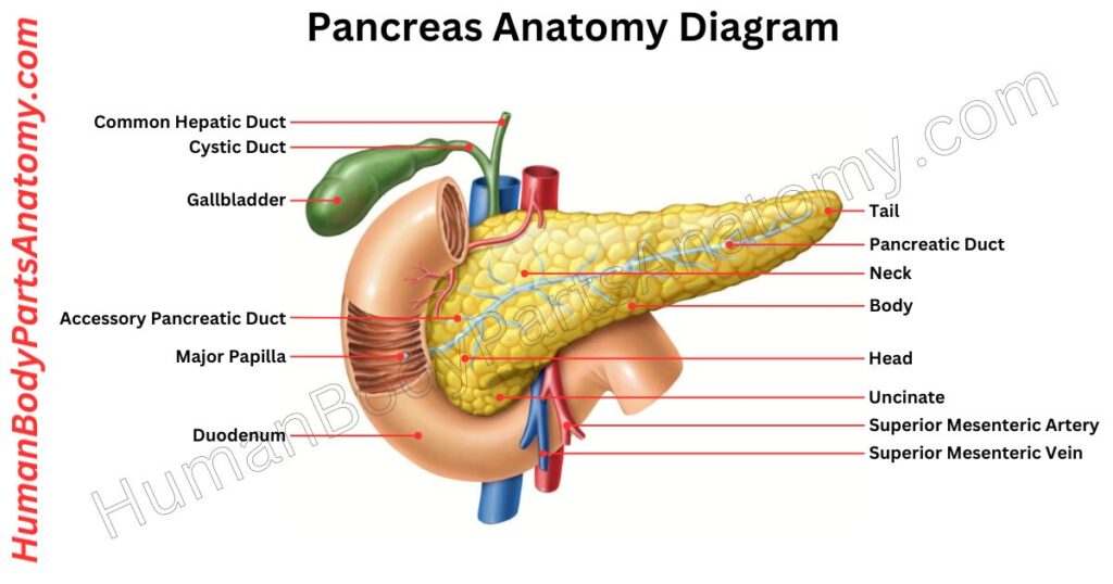
The pancreas is a vital gland of the digestive system located deep in the abdomen, behind the stomach.[1][3][6] It lies in a retroperitoneal position, crossing the lower back at the level of the L1 and L2 vertebrae along the posterior abdominal wall. Structurally, the pancreas is divided into four main parts: the head, neck, body, and tail.[1][4]
1. Head
The head of the pancreas is its broad inner portion that fits snugly into the curve of the C-shaped duodenum.[1][4]
This part of the small intestine bends around the head, with both the descending and horizontal sections closely touching it.[1]
It extends downward from the head and is a hook-like projection called the uncinate process, which curves behind and reaches toward the superior mesenteric artery.[1][4]
2. Neck
To the left of the head lies the neck of the pancreas — a short, bridge-like segment about 2 cm long that links the head to the body.[1] Behind the neck run the superior mesenteric artery and vein.[1][4]
It is also where the hepatic portal vein begins, formed by joining the superior mesenteric and splenic veins.[1]
3. Body
After the neck, the pancreas continues into the body, which has two main surfaces — front (anterior) and back (posterior) — along with upper and lower borders.[1]
It lies in front of the second lumbar (L2) vertebra and forms part of the floor of the lesser sac (also called the omental bursa).[1]
Behind the body of the pancreas are several important structures, including the aorta, superior mesenteric artery, left kidney, left adrenal (suprarenal) gland, and left renal blood vessels.[1]
4. Tail
The final portion of the pancreas is the tail, which is located within the peritoneal cavity (intraperitoneal).[1] It stretches toward the spleen, lying close to its hilum, and travels along with the splenic blood vessels inside a fold of tissue called the splenorenal ligament.[1][4]
Ductal System of Pancreas
1. Main Pancreatic Duct (Duct of Wirsung)
The main pancreatic duct (MPD), also known as the duct of Wirsung, runs through the middle of the pancreas, starting from the tail and ending at the head.[1][5][10]
It carries digestive enzymes that the pancreas makes and drains them into the small intestine.[1][2] This duct collects fluid from nearly the entire pancreas, including the uncinate process and the lower portion of the head.[1]
During early development, the MPD begins in the dorsal pancreatic bud. As the pancreas grows, this duct joins with a smaller duct from the ventral bud in the head region.[1] Near its end, the MPD usually joins the common bile duct at an angle of about 45–60°.[1]
Together, they release digestive juices into the duodenum through a small opening called the ampulla of Vater. This flow is controlled by the sphincter of Oddi, a ring of muscle that acts like a valve.[1][5][10]
The diameter of the MPD becomes narrower as it moves from head to tail—about 3 mm wide in the head, 2 mm in the body, and 1 mm in the tail.[1]
2. Accessory Pancreatic Duct (Duct of Santorini)
Most people have a single pancreatic duct that carries digestive juices from the pancreas to the small intestine.[1]
However, some individuals also have a second duct called the accessory pancreatic duct, or Duct of Santorini. This extra duct may or may not function.[1][5][10]
When it does work, it usually drains directly into the second part of the duodenum, known as the dorsal side, through a small opening called the minor duodenal papilla.[1][5][10] This thing happens in about 70% of cases.[5][10]
In the remaining 30%, the accessory duct connects to the main pancreatic duct, and together, they empty into the duodenum through the major duodenal papilla.[5][10]
No matter the route, both ducts eventually deliver enzymes into the vertical part of the duodenum, helping with digestion.[1][10]
Histological (Microscopic) Anatomy of Pancreas
1. Exocrine Portion (approximately 99%)
The exocrine part of the pancreas makes up about ~98-99% of the entire organ.[2]
Acini
The exocrine part of the pancreas contains many tightly packed groups of cells called pancreatic acini, which are the main sites for producing digestive enzymes.[2]
Each acinus is made up of a single layer of pyramidal-shaped acinar cells. These cells are wider at the base and taper toward the top, surrounding a small central space called the lumen.[2]
These acinar cells are specialized for secreting enzymes that help break down food in the small intestine. They have lots of rough endoplasmic reticulum and a Golgi apparatus, which help make and package these enzymes.[2]
Under the microscope, the lower part of the cell (the base) stains blue due to its high protein-making activity, while the top part (the apex) contains pink-stained granules known as zymogen granules.[2]
Zymogen granules store inactive forms of digestive enzymes called zymogens or proenzymes.[2]
When the pancreas is stimulated, these granules move to the cell surface and release their contents into the acinus lumen through a process called exocytosis. Once released, the enzymes become active and help with digestion.[2]
Ducts
After they are produced, pancreatic secretions leave the small clusters of cells called acini through tiny passageways known as intercalated ducts.[2]
These ducts begin right inside the acini and are lined by special flat cells called centroacinar cells, which mark the start of the pancreatic duct system.[2]
These cells have flat, centrally placed nuclei and appear pale when viewed under a microscope with H&E staining.[2]
- As the duct continues outside the acinus, it is lined by low cuboidal cells. These intercalated ducts then connect to slightly larger channels called intralobular ducts, which are found within the small sections (lobules) of the pancreas and are lined with low columnar epithelial cells.[2]
- From there, the secretions flow into the interlobular ducts, which lie in the connective tissue between lobules. These ducts have taller columnar epithelium, and as they get larger, their lining can become more layered or stratified.[2]
Eventually, these ducts empty into the main pancreatic duct, also called the duct of Wirsung, or sometimes into the accessory pancreatic duct, known as the duct of Santorini.[1][2] Both ducts are lined with tall, often stratified columnar cells.[2]
2. Endocrine Portion (approximately 1-2%)
The endocrine part of the pancreas makes up only about 1-2% of the entire organ. This portion is made up of around 1 to 2 million clusters of cells called pancreatic islets (or islets of Langerhans), which are scattered throughout the pancreas, mostly in the tail region.[2]
These islets are separated from the surrounding tissue by a thin layer of reticular fibers.[2]
The main tissue of the pancreas, especially in its duct system, includes different types of epithelial cells.
Each pancreatic islet is a small, round cluster of hormone-producing cells with a polygonal shape.[2]
On a histology slide stained with H&E (hematoxylin and eosin), these islets appear as large, lightly colored areas surrounded by darker-staining pancreatic acini.[2]
The islet cells are linked together by structures called desmosomes and gap junctions, forming rows or groups. These islets are rich in fenestrated capillaries, which help hormones quickly enter the bloodstream.[2]
Islets of Langerhans
The pancreatic islets (also called islets of Langerhans) are clusters of hormone-producing cells that help regulate blood sugar and digestive functions. These islets contain four main types of cells, each with a specific role:[2]
1. Beta (B) cells
Beta cells make up about 70% of the islet and are mainly found in the center. They release insulin, a hormone that lowers blood glucose levels. Inside these cells are secretory granules with a dense core of crystallized insulin, surrounded by a clear area, giving them a distinct appearance under the microscope.[2]
2. Alpha (A) cells
These cells account for 15–20% of the islet and are typically located around the edges. Alpha cells produce glucagon, which raises blood sugar levels. Their granules are evenly sized, with a dark center and a thinner, clear rim than those in beta cells.[2]
3. Delta (D) cells
It makes up 5–10% of the islet delta cells are scattered throughout, often near the periphery. They secrete somatostatin, a hormone that helps control the release of both insulin and glucagon. Delta cell granules are generally larger than those in alpha or beta cells.[2]
4. PP (pancreatic polypeptide) cells
These are the least common, forming less than 5% of the islet cells. PP cells are mostly found in the head of the pancreas and release pancreatic polypeptide, which influences digestive functions and appetite regulation.[2]
Each cell type plays a key role in maintaining balance in blood sugar and digestive health. Their structure, location, and hormone content help distinguish them from one another.[2]
Blood Supply
The pancreas is supplied by a network of arteries and veins that deliver oxygen-rich blood and carry away used blood.[1]
Arterial Supply
- Head and Uncinate Process: The superior and inferior pancreaticoduodenal arteries wrap around this part of the gland. The superior branch comes from the gastroduodenal artery, and the inferior branch comes from the superior mesenteric artery. Each of these splits into a front and a back branch, creating arcade-like loops around the pancreatic neck.[1]
- Body and Tail: The splenic artery runs along the top edge of the pancreas and sends multiple branches directly into the body and tail. A few small extra branches from the gastroduodenal and superior mesenteric arteries also help. However, the splenic artery is the main supplier of these parts.[1]
Venous Drainage
- Head Region: Blood from the front of the head drains into the superior mesenteric vein. Blood from the back of the head flows straight into the portal vein.[1]
- Lower Head: Veins from the lower head region follow the inferior pancreaticoduodenal arteries and empty into the superior mesenteric vein.[1]
- Body and Tail: Veins running alongside the splenic artery collect blood from the body and tail and drain it into the splenic vein.[1]
Innervation
The pancreas is regulated by the autonomic nervous system, which functions automatically without conscious control. It receives two types of nerve signals: parasympathetic and sympathetic.[11]
Parasympathetic signals travel through the vagus nerve (cranial nerve X), while sympathetic signals come from the spinal cord segments T5 to T12.[11]
These sympathetic nerves pass through the greater and lesser splanchnic nerves, relay at the celiac ganglion, and continue through the superior mesenteric plexus to reach the pancreas.[11]
- Parasympathetic – Its stimulation activates the pancreas, encouraging acinar cells to release digestive enzymes. It also boosts the secretion of insulin and glucagon, helping in both digestion and blood sugar regulation.[11]
- Sympathetic – Its stimulation prepares the body for stress or high energy needs. It reduces blood flow to the pancreas by causing vasoconstriction, limits enzyme secretion, slows insulin release, and increases glucagon output. This response helps raise blood sugar levels during emergencies or intense physical activity.[11]
Inside the pancreas, these nerves affect two important structures:
- Acinar cells, which produce enzymes for digestion
- Pancreatic islets, which secrete hormones like insulin and glucagon
Lymphatic Drainage
Lymph from the pancreas flows along its arteries, draining into two main groups of lymph nodes: the pancreaticosplenic nodes near the spleen and the pyloric nodes near the stomach.[9]
These then pass lymph into larger nodes—specifically, the superior mesenteric and coeliac lymph nodes—helping to filter and manage immune responses in the abdomen.[9]
FAQ’s
The pancreas is a long, flat gland located deep in your upper abdomen, behind the stomach and extending toward the left side near the spleen. It has two main parts: the “head” on the right side (near the duodenum) and the “tail” on the left side (near the spleen).
The pancreas is commonly divided into several parts: the head, neck, body and tail (and some sources also list the uncinate process). The head lies in the curve of the duodenum, the body crosses behind the stomach, and the tail reaches toward the spleen.
The pancreas has a dual role:
Exocrine function: It produces digestive enzymes (like lipase, amylase, proteases) and secretes these into the duct system to the small intestine to help digest fats, proteins and carbohydrates.
Endocrine function: It contains clusters of cells (islets of Langerhans) that release hormones like insulin and glucagon into the bloodstream to regulate blood sugar.
Knowing the anatomy helps you understand how the pancreas works, how it relates to other organs (like stomach, duodenum, spleen and major blood vessels) and why diseases of the pancreas (such as Pancreatitis or Pancreatic cancer) can cause specific symptoms. It also helps medical professionals plan surgeries or interpret imaging of this organ.
The pancreas lies mostly in the mid-upper abdomen. The head of the pancreas is on the right side (in the curve of the duodenum) and the tail extends to the left side toward the spleen. Because it’s behind the stomach and near major blood vessels, its exact location can feel “deep” in the belly.
The pancreas has a network of ducts that collect the digestive enzymes produced by its exocrine tissue. The main pancreatic duct (also called the duct of Wirsung) runs through it and joins with the common bile duct before entering the duodenum (via the ampulla of Vater). Some people also have an accessory duct (duct of Santorini) that drains part of the pancreas.
Yes — there are anatomical variants such as:
Pancreas divisum, where the dorsal and ventral pancreatic ducts don’t fully fuse, which may predispose to pancreatitis.
Annular pancreas, a rare congenital condition where pancreatic tissue wraps around the duodenum and may cause obstruction. Understanding these variants is helpful in imaging, surgery and understanding certain disease presentations.
References-
- StatPearls Publishing. (2023). Anatomy, Abdomen and Pelvis, Pancreas. PMCID: — https://www.ncbi.nlm.nih.gov/books/NBK532912/ — PMID: 30422507 — DOI: 10.4103/0974-2077.110136
- StatPearls Publishing. (2023). Physiology, Pancreas. PMCID: — https://www.ncbi.nlm.nih.gov/books/NBK459261/ — PMID: 27929636
- Mayo Clinic. (2024). Pancreatic Cancer – Symptoms and Causes. PMCID: — https://www.mayoclinic.org/diseases-conditions/pancreatic-cancer/symptoms-causes/syc-20355421
- Johns Hopkins University School of Medicine. (2023). Pancreas Basics. PMCID: — https://pathology.jhu.edu/pancreas/basics
- Al-Haddad M, et al. (2018). The Main Anatomical Variations of the Pancreatic Duct System: Review of the Literature and Its Importance in Surgical Practice. Gastroenterology Research and Practice. PMCID: PMC5862083. https://pmc.ncbi.nlm.nih.gov/articles/PMC5862083/ — PMID: 29581798 — DOI: 10.1155/2018/1614529
- MedlinePlus, National Library of Medicine (NLM). (2024). Pancreatitis – Series: Normal Anatomy. PMCID: — https://medlineplus.gov/ency/presentations/100149_1.htm
- StatPearls Publishing. (2023). Pancreatitis. PMCID: — https://www.ncbi.nlm.nih.gov/books/NBK538337/ — PMID: 30726023
- MedlinePlus, National Library of Medicine (NLM). (2025). Diabetic Ketoacidosis. PMCID: — https://medlineplus.gov/ency/article/000320.htm
- Munoz-Bongrand N, et al. (2014). The Surgical Anatomy of the Lymphatic System of the Pancreas. HPB Journal. PMCID: — https://pubmed.ncbi.nlm.nih.gov/25220721/ — PMID: 25220721 — DOI: 10.1016/j.hpb.2014.07.004
- Kamiya M, et al. (2015). Accessory Pancreatic Duct Patterns. Dig Surg. PMC4413057. PMID: 25824501. DOI: 10.1159/000371591. https://pmc.ncbi.nlm.nih.gov/articles/PMC4413057/
- Rodriguez-Diaz R, et al. (2021). Optical Imaging of Pancreatic Innervation. Diabetes. PMC8112238. PMID: 33707228. DOI: 10.2337/db20-0988. https://pmc.ncbi.nlm.nih.gov/articles/PMC8112238/
Read More-
Human Head
- Skull Anatomy: Complete Guide with Parts, Names, Functions & Diagram
- Ultimate Guide to Eye Anatomy: Parts, Structure, Functions & Diagram
- Tongue Anatomy: Complete Guide with Parts, Names, Functions & Diagram
- Mouth Anatomy: Complete Guide with Parts, Names, Functions & Diagram
- Complete Guide to Tooth Anatomy: Learn Parts, Names & Diagram
- Ultimate Guide to Ear Anatomy: Parts, Structure, Functions & Diagram
- Nose Anatomy: Complete Guide with Parts, Names, Functions & Diagram
Brain
- Basal Ganglia Anatomy: Complete Guide with Names, Functions & Diagram
- Lobes of the Brain: Complete Guide with Names, Functions & Diagram
Organs
- Kidney Anatomy: Complete Guide with Parts, Names, Functions & Diagram
- Liver Anatomy: Complete Guide with Parts, Names, Functions & Diagram
- Heart Anatomy: Complete Guide with Parts, Names, Functions & Diagram
Medical Disclaimer
All content on HumanBodyPartsAnatomy.com is educational and based on verified, peer-reviewed medical sources. Articles are authored or reviewed by qualified medical or biomedical professionals to ensure accuracy.
This website does not provide medical advice, diagnosis, or treatment. Always consult a licensed healthcare professional for personal medical guidance.
No commercial or promotional interests influence the medical content published on this site.