📅 Published on July 1, 2026 | 🕒 Last Reviewed & Updated on October 14, 2026
Overview of Human Anatomy and Physiology
The human body has 206 bones,[1] more than 600 muscles,[2] and a wide network of organs and blood vessels. All of these parts work together, with each cell playing its own role to keep us alive. Two main fields help us understand how our bodies function: physiology, which looks at how the body works inside, and anatomy, which studies its structure. Anatomy examines everything from the smallest cells to tissues, organs, and entire systems. By learning about human anatomy, we better understand how our bodies are built and how all the parts work together to keep us alive.
On organ counts. Popular articles often claim the body has “78 organs.” No U.S. federal health agency publishes an official organ count, because the total depends on how an organ is defined and whether paired structures, glands, and accessory organs are counted separately. Authoritative anatomical resources describe the body by organ system rather than by organ tally, and this article follows that convention.
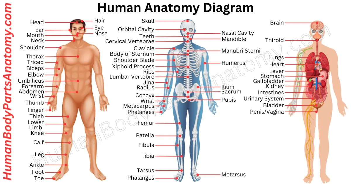
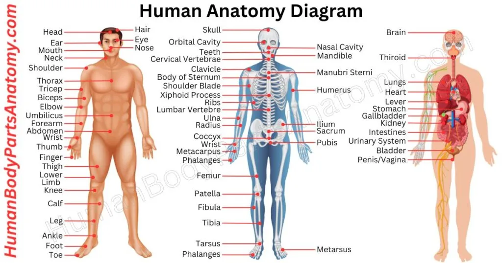
Human Anatomy Diagram
Detailed Human Anatomy
Skeletal System
Lymphatic & Immune System
- Lymph Nodes
- Lymphatic Vessels
- Spleen
- Thymus
- Tonsils
- Bone Marrow
Integumentary System
- Skin
- Hair
- Nails
- Sweat Glands
- Sebaceous Glands
Urinary System
- Kidneys
- Ureters
- Bladder
- Urethra
Nervous System
- Brain
- Spinal Cord
- Cranial Nerves
- Peripheral Nerves
- Sensory Organs
Cardiovascular System
- Heart
- Arteries
- Veins
- Capillaries
- Blood
Digestive System
- Mouth
- Teeth
- Tongue
- Salivary Glands
- Esophagus
- Stomach
- Small Intestine
- Large Intestine
- Liver
- Gallbladder
- Pancreas
- Anus
Reproductive System
- Male
- Female
Muscular System
- Head Muscles
- Neck Muscles
- Chest Muscles
- Back Muscles
- Shoulder Muscles
- Arm Muscles
- Hand Muscles
- Abdominal Muscles
- Hip Muscles
- Leg Muscles
- Tendons
Respiratory System
- Nose
- Sinuses
- Pharynx
- Larynx
- Trachea
- Bronchi
- Lungs
- Diaphragm
Endocrine System
- Hypothalamus
- Pituitary
- Pineal
- Thyroid
- Parathyroids
- Adrenal Glands
- Pancreatic Islets
- Ovaries
- Testes
Human Bone Anatomy
In human anatomy, the skeleton is the internal framework of the body. It is responsible for both structure and function. At birth, it is composed of approximately 270 bones. However, by adulthood, this number reduces to roughly 206 due to bone fusions.[1] Bone is living tissue that is constantly renewed: older bone is broken down and new bone formed in its place, a process called remodelling, and almost all bone in adults is replaced about every 10 years.[3] Bone mass is gained during growth and typically peaks during a person’s twenties.[3]
Skull
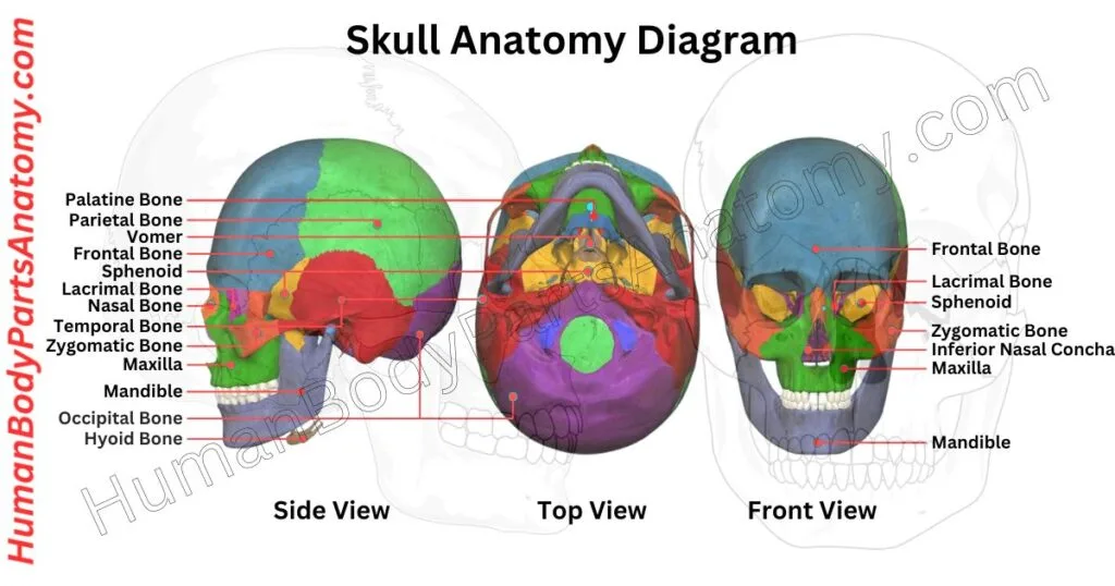
The skull is a bony structure that covers and protects the brain. It comprises three main types of bones: cranial bones, facial bones, and ear ossicles.[4]
In humans, the skull is divided into the neurocranium (the braincase) and the viscerocranium (the facial skeleton), which includes the mandible.[4] This structure is an example of cephalization, where the brain and sensory organs are concentrated at the head.
The skull is located at the uppermost part of the skeleton, a result of cephalization.[4] It houses the brain along with key sensory organs such as the eyes, ears, nose, and mouth.
The human skull is made up of 22 bones,[4] or 29 if you include the six middle ear ossicles and the hyoid bone. These bones are mainly connected by ossified joints known as sutures.[4]
The skull has several crucial functions: it protects the brain, provides attachment for muscles, and allows blood vessels and nerves to pass between the inside and outside of the cranium.[4] Its structure also maintains the proper distance between the eyes for stereoscopic vision and positions the ears to help with sound localization.
Read More – Skull Anatomy: Parts of the Skull, Structure, Cranial, Facial Bones & Functions
Vertebral Column or Spine
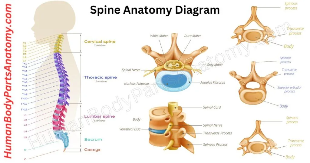
The vertebral column, or the spine, is an essential human body part of the axial skeleton. It safeguards the spinal cord and nerves while maintaining an upright posture.[5]
This complex skeletal framework bears most of the body’s weight to maintain a vertical pose.[5] Its distinctive feature is the transformation of the flexible rod found in all chordates into a segmented array of bones referred to as vertebrae. In humans there are 33 vertebrae: 7 cervical, 12 thoracic, 5 lumbar, 5 sacral, and 4 coccygeal.[5]
These vertebrae are interposed with intervertebral discs, which enhance the spine’s durability and flexibility.[5] Each vertebra is named according to its position within the spinal column.[5]
The spinal canal is enclosed within the vertebral column, a protective cavity that envelops and shields the spinal cord.[5]
Read More – Spine Anatomy: Parts of the Spine, Vertebrae, Curves, Spinal Cord & Diagram
Hip Bone
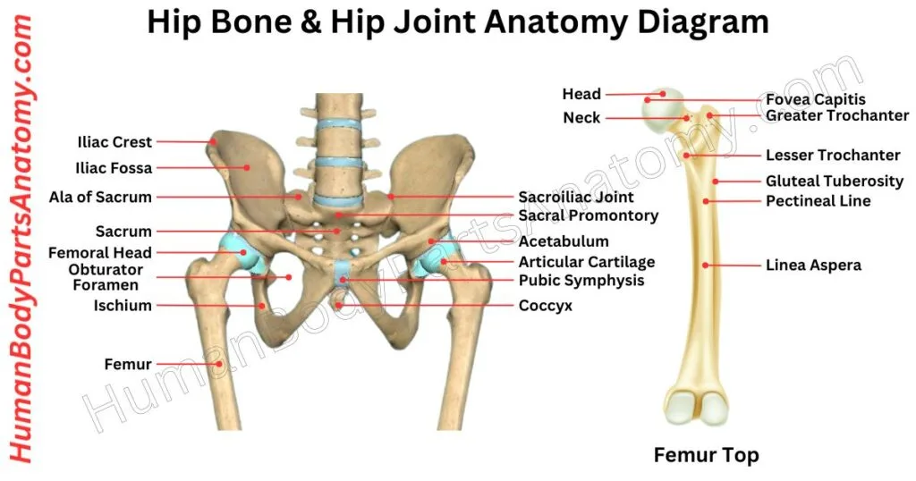
The hip is also known as the coxa in medical terms. It is a key area in vertebrate anatomy found on the outer side of the pelvis. It is located to the side and front of the buttocks, below the bony ridge of the iliac crest, and beside the obturator foramen.[6]
This area includes muscles, tendons, and soft tissues that cover the prominent greater trochanter of the femur.[7]
In adults, the hip bones forms from the fusion of three pelvic bones (the ilium, ischium, and pubis).[6] It creates the sturdy inner and upper walls of the hip region.
Read More – Hip Bone Anatomy: Complete Guide with Parts, Names, Functions & Diagram
Femur
The femur, commonly called the thigh bone, is essential within the human skeletal system. It is the bone in the lower limb between the hip joint and knee joints, and it is the longest, heaviest, and strongest bone in the body.[7] This bone shapes the hip joint at its proximal end and forms an articulation point with the pelvic socket.[7]
Moreover, the femur’s distal end engages with the tibia and patella to form a knee joint structure.[7] Beyond this, the femur bears the human body’s weight during stationary and dynamic activities.[7]
Additionally, the femur is an essential anchor point for muscles, tendons, and ligaments that help move the hip joint and knee joints.[7]

Read More – Femur Anatomy: Complete Guide with Parts, Names, Functions & Diagram
Rib Cage
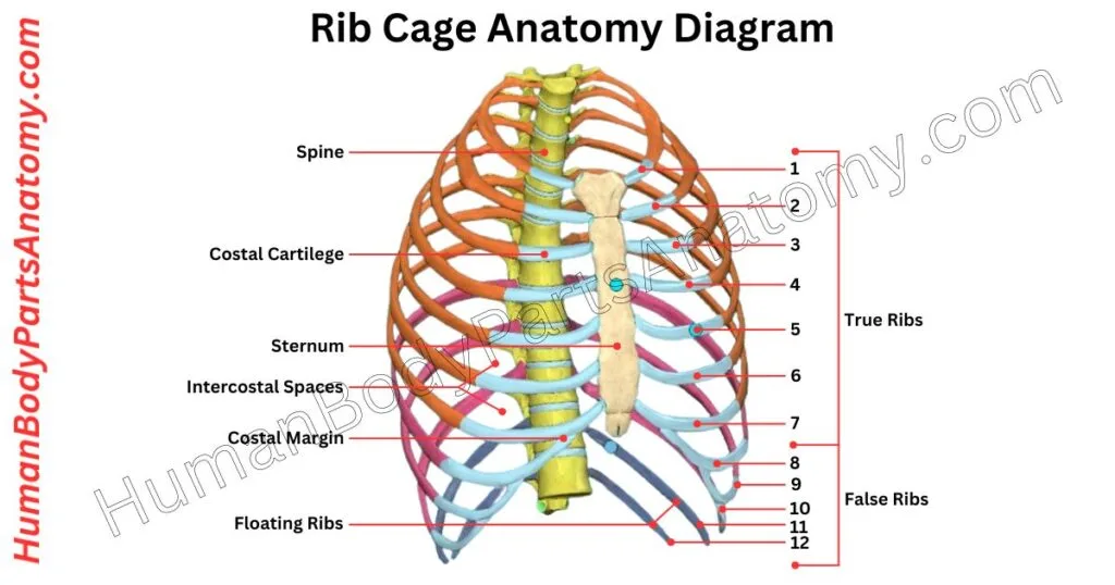
The rib cage, or thoracic cage, forms a vital part of the vertebrate skeleton. It consists of the ribs, sternum, and thoracic vertebrae.[8] This structure protects the heart, lungs, and major blood vessels while supporting the shoulder girdle and axial skeleton.
In humans, the thoracic cage has 12 pairs of ribs, but they do not all connect to the sternum in the same way. Ribs 1–7 (the true ribs) connect directly to the sternum through their own costal cartilage; ribs 8–10 (the false ribs) attach indirectly, their costal cartilages joining the seventh costal cartilage; and ribs 11–12 (the floating ribs) do not attach to the sternum at all.[8] The sternum includes three parts: the manubrium, body, and xiphoid process.[8]
Together with the 12 thoracic vertebrae, the rib cage provides muscle attachment sites for the neck, upper limbs, abdomen, and back.[8] It also forms the chest wall with the skin and surrounding tissues.
Sternum
The sternum, or breastbone, is a flat, vertical bone situated in the center of your chest. It forms the front of the rib cage and has three parts:
- Manubrium: The upper section with the suprasternal notch and clavicular notches that form the sternoclavicular joints.
- Body (Gladiolus): The longest section, where the costal cartilages of ribs 2–7 attach. It joins the manubrium at the sternal angle (Angle of Louis).[8]
- Xiphoid Process: The small, triangular lower section that varies in shape and size.[8]
The sternum protects vital organs, including the heart and lungs.[8]

Human Muscle Anatomy
In human anatomy, muscle tissues are made up of specialized cells that can contract and allow movement.[2] This movement includes not just the motion of body parts and limbs but also the flow of blood, food, and other substances within the body.
Muscles are essential for moving the skeleton and making the heart beat. They are found in the walls of organs like the intestines, uterus, and stomach.[2]
Biceps
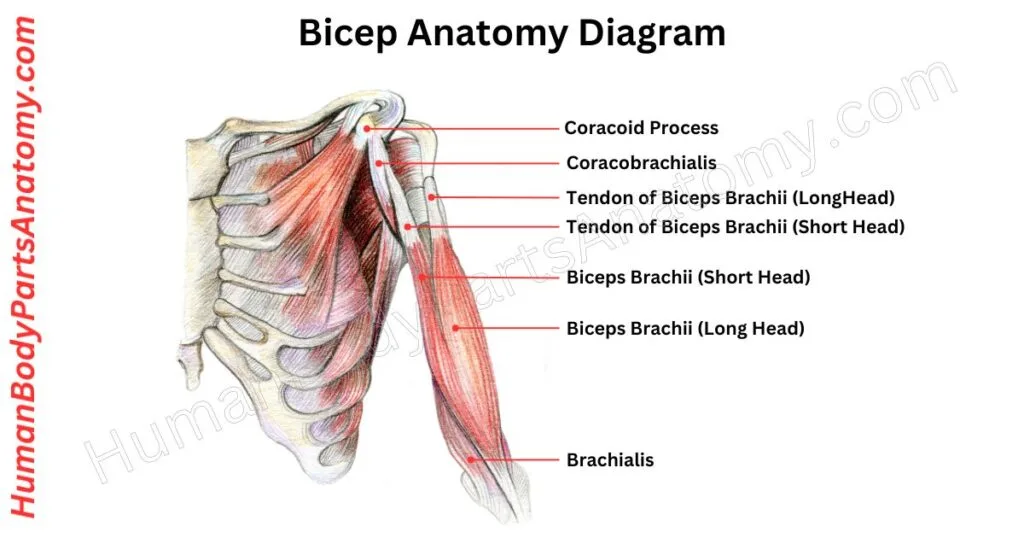
The biceps brachii is a large muscle in the anterior upper arm that extends from the shoulder to the elbow. It has two unique heads, the long and short heads, which emerge from the scapula.[9] These heads join together to produce a muscular system that joins to the upper section of the forearm.[9]
Function—The biceps brachii is a strong supinator of the forearm and also flexes the elbow, although the brachialis is the primary elbow flexor.[10] It helps with various activities and daily tasks. Curling the forearm at the elbow joint is referred to as forearm flexion.
Read More – Ultimate Guide to Bicep Anatomy: Parts, Names, Functions & Diagram
Triceps
The triceps brachii is an extensor muscle in various vertebrates at the back of the upper limb. These muscles originate from the humerus and scapula, and comprise three distinct parts: the medial, lateral, and long heads.[9]
Function—The triceps brachii muscle extends the forearm at the elbow joint [9]. Its long head helps extend and adduct the arm at the shoulder joint.[9]
Forearm
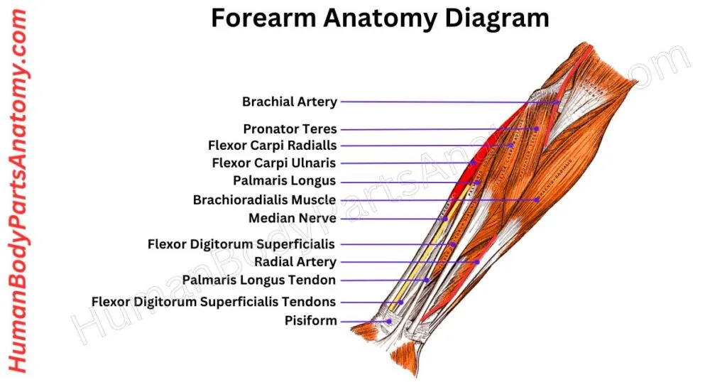
The forearm is the part of your arm between the elbow and wrist. It is made up of two bones: the outer radius and the inner ulna.[11]
It has 20 muscles grouped into front (flexor) and back (extensor) compartments, which control elbow, wrist, and hand movements.[11]
There are two types of muscles: front flexors and back extensors.[11] Fascia organizes and supports these muscles around the ulna and radius.
Two structures, the intermuscular septum and interosseous membrane, create compartments and offer extra support.[12]
Read More – Complete Guide to Forearm Anatomy: Parts, Names, Functions & Diagram
Hip Muscles
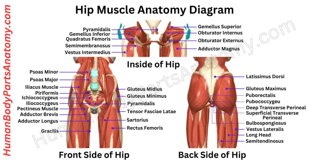
The muscles around the hip joint are essential for movement. Anatomists group them into four categories based on their location: the gluteal, lateral rotator, adductor, and iliopsoas groups.[13] The exact number counted as a “hip muscle” varies between classification schemes.
Hip movements result from the coordinated action of multiple muscles, and most muscles assist in more than one movement.[13]
- Flexion — chiefly iliopsoas, assisted by rectus femoris and sartorius.
- Lateral rotation — obturators, quadratus femoris, gemelli; assisted by gluteus maximus, sartorius, piriformis.
- Medial rotation — tensor fasciae latae, fibres of gluteus medius and minimus.
- Abduction — gluteus medius and minimus.
- Adduction — adductor longus, brevis, magnus; assisted by gracilis and pectineus.
Read More – Hip Muscle Anatomy: Complete Guide with Parts, Names, Functions & Diagram
Thigh
The thigh is a significant part of human anatomy in the lower limb. It lies between the hip joint and the knee joint. The femur is the prominent bone within the thigh and has exceptional strength, density, and robustness.[7]
Functionally, the femur forms a ball and socket joint at the hip[13] and a modified hinge joint at the knee[16]. Remarkably, the thigh region houses various main muscles in the human body, divided into anterior, medial, and posterior compartments.[14]
These muscles enable various body movements, including flexion, extension, adduction, and rotation.[14]
Additionally, they bear most of the body’s total weight. Furthermore, these muscles help maintain the structural integrity of the hips and legs.[14]

Read More – Complete Guide to Thigh Muscle Anatomy: Learn Parts, Names & Diagram
Human Body Parts – Joints
Wrist Joint
In human anatomy, the wrist is scientifically termed the carpus or carpal bones. It is a crucial part of the hand’s structure, consisting of eight distinct bones that create the foundational framework for the upper part of the hand.[15]
The wrist joint is scientifically known as the radiocarpal joint. It acts as the vital connection between the radius and the carpal bones.[15] It includes both the carpus and the lower portions of the forearm bones.
The metacarpus is formed by the five metacarpal bones. A network of interconnected joints exists among these anatomical components, making hand movement possible.[15]

Read More – Wrist Anatomy: Ultimate Guide to Parts, Names, Functions & Diagram
Hip Joint
The hip joint connects your thigh bone (femur) to your hip bone (pelvis). It is a crucial body part, second in size only to your knee joint.[13][16]
This ball-and-socket joint consists of the rounded head of the femur fitting snugly into a cup-like cavity in the pelvis, known as the acetabulum.[13] This structure allows for extensive movement and helps your legs support your body weight.
This universal joint is essential for everyday activities, enabling a wide range of motions and providing stability and support.[13]
Read More – Hip Bone Anatomy: Complete Guide with Parts, Names, Functions & Diagram
Knee Joint
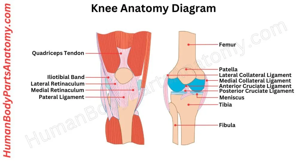
The knee joint, or a synovial joint, is an essential link between the femur, tibia, and patella bones. It is the body’s largest joint, mainly allowing leg bending and straightening.[16] It contains two primary components: the tibiofemoral and patellofemoral articulations.[16]
The tibiofemoral joint forms a connection between the tibia and the femur, while the patellofemoral joint forms between the patella and the femur.[16]
Your knees are vital in supporting your body weight and allowing leg movement. This joint helps in activities like walking, running, and jumping.[16]
Read More – Knee Anatomy: Complete Guide to Parts, Names, Functions & Diagram
Ankle Joint
Your ankle is a hinge joint connecting your lower leg and foot. It is a hinge-like joint formed by the talus, tibia, and fibula bones.[17]
The bony bump on the lower fibula (lateral malleolus) forms the outer boundary on one side, and the bony bump on the lower tibia (medial malleolus) creates the inner boundary. Together, they make up the ankle mortise.[17]
The talus bone acts like a connector, linking with the calcaneus below and the navicular in front.[17] The top part of the talus has a smooth surface, allowing comfortable up-and-down movement of your foot.
It snugly fits between the bony bumps, making the ankle most stable when you lift your toes towards your shin (dorsiflexion).[17]
Shoulder Joint
The human shoulder anatomy has three bones: the collarbone, shoulder blade, and upper arm bone. These bones are connected by joints, with the main one being the shoulder joint or glenohumeral joint.[18]
Other joints, like the acromioclavicular joint, are also part of the shoulder.[18] The shoulder joint allows circular rotation and lifting of the arm away from the body.
It is like a ball in a socket formed by the shoulder blade. A soft tissue envelope called the joint capsule surrounds the shoulder joint, lined with a smooth synovial membrane.[18]
A group of four muscles maintains the shoulder’s stability, called the rotator cuff. These muscles attach to the shoulder blade and the upper arm bone. They are the supraspinatus, subscapularis, infraspinatus, and teres minor.[19]

Human Anatomy – Alimentary System
Mouth
The mouth is necessary for digestion.[20] It is a complex structure with different parts that work together to make the digestion system more efficient.
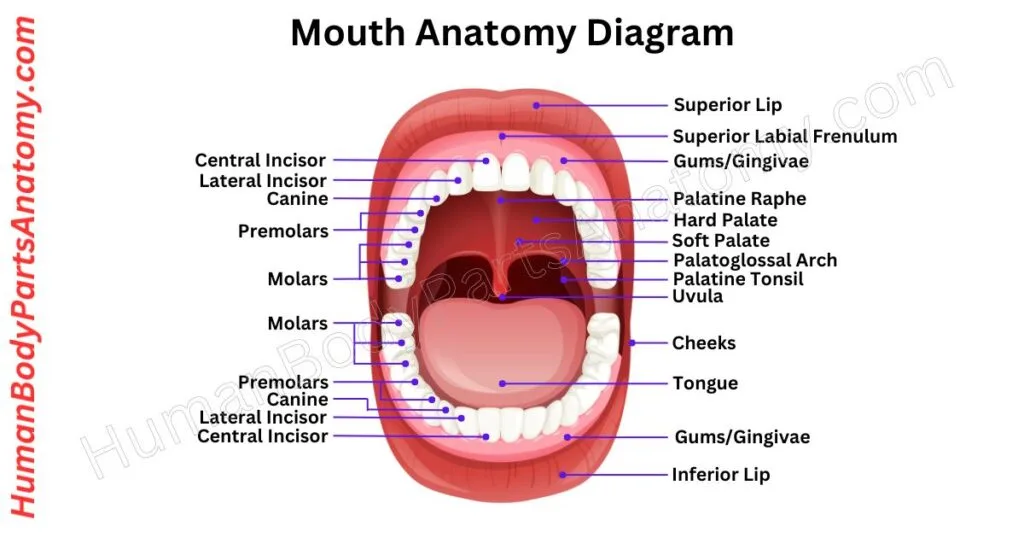
The lips create two regions: the vestibule and the oral cavity. The tongue occupies the central cavity and is surrounded by teeth, cheeks, and the isthmus of the fauces at the back.
The hard palate forms the front roof, and the soft palate makes up the rear, with the uvula hanging down. The inner lining is called the oral mucosa. It is made of stratified squamous epithelium.
Salivary glands provide fluid to keep the mouth moist and begin the chemical breakdown of food.[20] Nerves and blood vessels form a network essential for the mouth’s diverse functions in human life.
Read More – Mouth Anatomy: Complete Guide with Parts, Names, Functions & Diagram
Teeth
Teeth are essential for chewing food and helping with digestion. Although they may look like bones, they develop from ectoderm and ectomesenchyme, with the enamel arising specifically from the ectoderm.[21]
In adults, the 32 permanent teeth work together to cut, tear, mix, and grind food into smaller pieces.[21] The tongue and oropharynx shape the food into a ball for easy swallowing.
Teeth have four main layers. The outer layer, called enamel, is the hardest and most highly mineralized tissue in the body and protects against cavity-causing bacteria.[22]
Below the enamel is dentin, which is softer than enamel but harder than bone.[22] If enamel wears away, it exposes dentin, increasing the risk of cavities.
The tooth root is covered by cementum, which, along with periodontal tissues, anchors the tooth in the jaw.[21] The innermost layer, tooth pulp, houses nerves, blood vessels, and connective tissues, contributing to overall tooth health.[21]

Read More – Complete Guide to Tooth Anatomy: Learn Parts, Names & Diagram
Lips
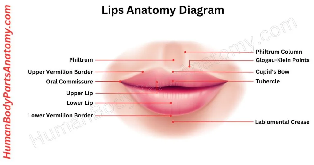
The lips are an essential part of the human face, pivotal in expressing emotions, talking, feeling, chewing, and romantic moments. Soft structures connected to the jaws are visible in many animals, including humans.
The upper and lower lips are scientifically called labium superius oris and labium inferius oris. Both lips have inner mucosal membranes, a colored vermilion layer, and outer skin.
In animals, including humans, lips are soft and flexible, helping with tasks like eating (such as sucking and swallowing) and forming sounds for speech.[20]
Stomach
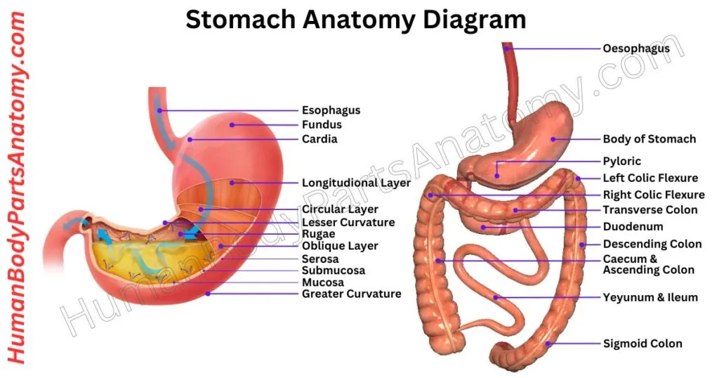
The stomach is a J-shaped crucial component of the digestive system, made up of four regions: the cardia, fundus, body, and pylorus.[23] It makes enzymes and acids that chemically decompose food.[23]
This process helps digestion before the food passes into the small intestine via the gastrointestinal (GI) tract.[20] This tube extends from the mouth to the anus, through which food travels and waste exits.
The primary function of the stomach is to temporarily store food, mixing and breaking it down through muscular contractions and producing specialized cells and enzymes necessary for digestion.[23]
Read More – Stomach Anatomy: Complete Guide with Parts, Names, Functions & Diagram
Intestine
The intestine, or bowel, is a long muscular tube that runs from the stomach to the anus. It digests food, produces hormones, and supports the immune system.[20]
The small intestine connects to the stomach and measures about 10–16 feet (3 to 5 metres) long. It has three parts: the duodenum, jejunum, and ileum.[24] Its folded lining increases the surface area for nutrient absorption.
The small intestine uses enzymes to break food into sugars, amino acids, and fatty acids. It then absorbs these nutrients into the bloodstream for distribution throughout the body.[20]
The large intestine is shorter than the small intestine but has a much larger lumen. It includes the cecum, appendix, entire colon, rectum, and anal canal.[25]
The large intestine absorbs water and salts from digested food to form stool.[25] Muscular contractions move the stool to the anus for elimination.[20]

Liver
The liver is a critical organ found only in vertebrate animals that helps maintain the body healthy. It performs multiple critical functions, like removing toxins from the blood and producing proteins and other compounds required for digestion and development.[26]
In humans, the liver is positioned in the upper right abdomen, just below the diaphragm, and protected by the lower ribs.[26]
One of the liver’s primary functions is to assist in controlling the body’s carbohydrate utilization, which includes storing glycogen and releasing glucose.[27] It also promotes the breakdown of old red blood cells and the production of proteins such as albumin and clotting factors.[27]

Read More – Liver Anatomy: Complete Guide with Parts, Names, Functions & Diagram
Pancreas
The pancreas is a big gland found deep within the belly. It works in both your digestive and endocrine systems.[20] This dual-role organ functions as a factory with two independent manufacturing lines:
- Enzymes for Digestion: It creates enzymes that help break down the food you ingest.[20]
- Hormones for Blood Sugar Regulation: It secretes hormones that control blood sugar levels in your body.[20]
Each day, it secretes about 1 to 2 liters of enzyme-rich juice[28][29], with the exact amount depending on your food intake — flow rises from roughly 0.2 to 0.3 mL per minute at rest to about 4.0 mL per minute during a meal.[28]

Human Anatomy – Respiratory System
Nose
The nose is an essential facial organ that allows air to enter the body. It filters, warms, and humidifies the air before it reaches the lungs.[30] Bones and cartilage give the nose its unique shape.[30]
Inside the nose, shell-like nasal conchae help condition the air [30], while tiny nasal hairs trap dust and other large particles.[30] When irritants such as dust or allergens enter the nose, the body triggers a sneeze to remove them.
The nose also enables the sense of smell[31] and plays a key role in facial appearance. Common conditions, such as nasal congestion and nosebleeds, can affect breathing, smell, and overall comfort.

Read More –Nose Anatomy: Complete Guide with Parts, Names, Functions & Diagram
Human Anatomy – Sense Organs
Eye
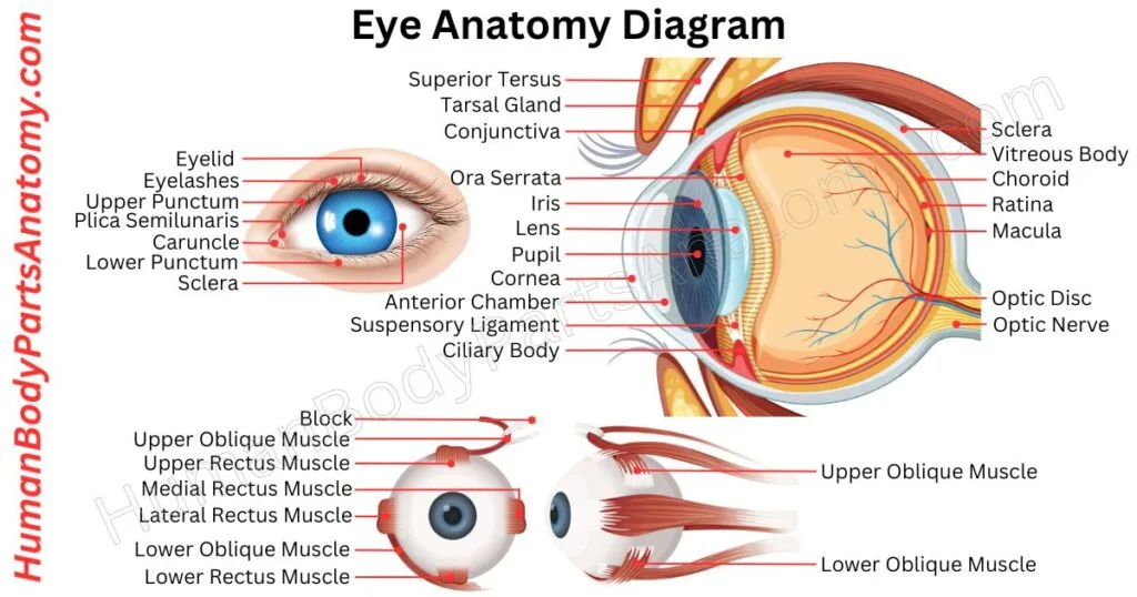
Our eyes are incredible organs that respond to light and allow us to see and understand the world around us. The human brain cannot sense the environment directly.
Our eyes collect crucial information about what’s happening and help us to see things and keep our body balanced.
For each eye, the normal field of vision extends about 60 degrees upward, 70 to 75 degrees downward, 60 degrees toward the nose, and 100 to 110 degrees outward.[32] Because the outward field of each eye reaches well past the midline, the two fields overlap in the centre — and it is that overlap which lets us perceive depth and see in 3D. Each eye also has a small physiological blind spot, corresponding to the optic disc, about 15 degrees to the outer side of the point of focus.[32]
Read More – Ultimate Guide to Eye Anatomy: Parts, Structure, Functions & Diagram
Ear
Your ears help us hear and stay balanced. When sound enters your ear, it makes your eardrum vibrate. This vibration passes through tiny bones in your middle ear, making the sound louder.[33] Then, in your inner ear, small hair cells turn the vibrations into electrical signals and send them to your brain.[33]
Your inner ear also has fluid-filled canals that help you stay balanced. These canals have hair-like sensors. When you move, the fluid shifts and sends signals to your brain.
Your brain uses these signals to help your muscles keep you steady. So, your ears do much more than hear—they help you stay on your feet!

Read More – Ultimate Guide to Ear Anatomy: Parts, Structure, Functions & Diagram
Tongue
The tongue is a muscular organ that helps you eat, speak, and taste food. Tiny bumps on its surface contain taste buds, which detect sweet, sour, salty, bitter, and umami (savoury) tastes.[34] At birth there are about 10,000 taste buds, and after age 50 they may begin to be lost.[34] Saliva keeps the tongue moist and helps you taste and chew food.[20]
The tongue moves food around your mouth for chewing and pushes it into your throat for swallowing.[20]
In humans, the tongue forms words and speech sounds. In other animals, it helps produce different vocalizations.
The tongue has two main parts: the front part inside the mouth and the back part near the throat. A groove down the middle divides it into left and right halves.

Read More – Tongue Anatomy: Complete Guide with Parts, Names, Functions & Diagram
Human Body Parts – Integument
Nails
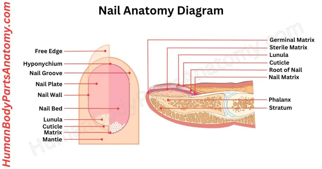
Nails, found on our fingers and toes, are rigid plates made of keratin.[35] This protein is also in other animals’ claws, hooves, and horns.
Nails are attached to the nail bed and can be used for scratching. The visible part is the “nail plate,” made of compact, hardened cells called onychocytes.[35]
They have lateral folds on each side and a proximal nail fold at the base.[35] The cuticle, a thin layer of skin, protects and enhances sensory experiences.[35]
Read More – Complete Guide to Nail Anatomy with all Parts, Names & Diagrams
Hair
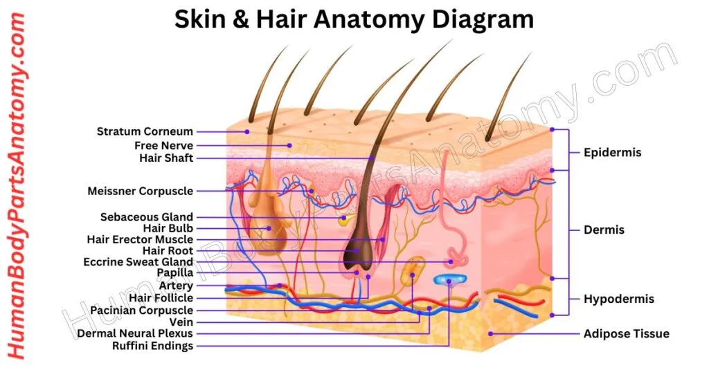
Hair is a protein-based filament that emerges from follicles embedded in the dermis layer of the skin.[36] It is a distinctive feature of mammals.
Except for areas of smooth, hairless skin — the palms, soles, lips, and parts of the external genitalia — the human body is largely covered with follicles that produce two types of hair: thick terminal hair and fine vellus hair.[36][37]
While much attention is given to hair growth, types, and care, hair also serves a protective role, enhancing sensation and helping regulate temperature.[37]
Skin
Skin is the soft outer layer that covers and protects the bodies of humans and many animals, and it is the largest organ of the body.[38] It has three main jobs: protecting, controlling, and sensing[39]
First, the skin acts as a shield, keeping out harmful things like germs and preventing the body from losing too much water.[39] It also helps keep us warm or cool by controlling our body temperature.[38]
Additionally, the skin lets us feel sensations like touch.[39] When exposed to sunlight, skin helps make vitamin D, which is important for our health.[39]
The thickness of the skin changes depending on where it is on the body.[38] For example, the skin of the eyelids has no underlying fat layer and is the thinnest skin on the body, making it more prone to wrinkles.[40]
Human Anatomy – Nervous System
Brain
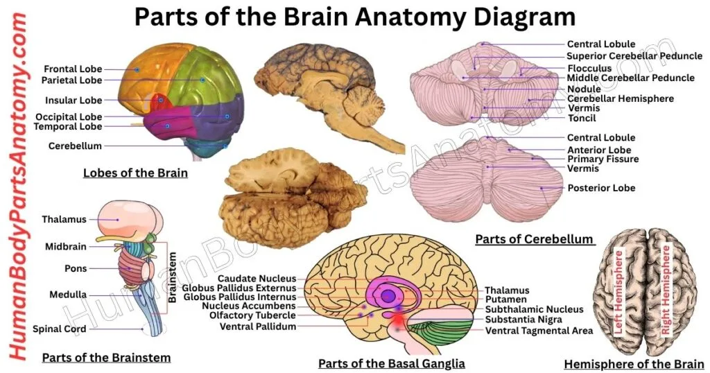
The brain is the body’s control center and a vital part of the nervous system. It lies inside the skull, where cerebrospinal fluid cushions and protects it.[41]
The brain and spinal cord together form the central nervous system, which processes sensory information and controls almost every body function.[41]
An adult brain weighs roughly 1.4 to 1.5 kg and contains an estimated 86 billion nerve cells, along with a similar number of non-neuronal cells.[42] At birth, the brain is only about one-quarter to one-third of its adult volume.[43]
The brain grows rapidly during the first years of life, reaching about 80% of adult volume by age three[43] and approximately 95% by age six[44] Growth after that involves refinement rather than bulk — unused connections are pruned away and nerve fibres become progressively insulated.[44]
Read More – Parts of the Brain Anatomy: Complete Guide with Names, Functions & Diagram
Lobes of the Brain
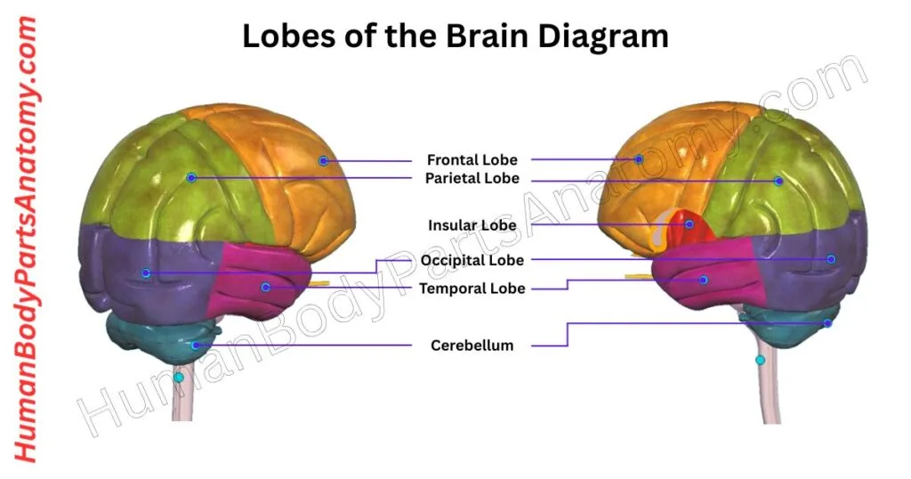
The cerebrum is the brain’s largest part and sits at the top of the skull. Its folded surface, made of sulci (grooves) and gyri (ridges), increases surface area without enlarging the skull.
A thin layer of gray matter processes information, while the white matter beneath carries signals throughout the brain.
The cerebrum has two hemispheres connected by the corpus callosum, which allows communication between them. Each hemisphere controls the opposite side of the body.[41]
Each hemisphere contains four lobes: the frontal lobe controls movement, planning, and problem-solving; the parietal lobe processes touch, temperature, and taste; the temporal lobe handles hearing, language, and memory; and the occipital lobe processes vision.[41]
Read More – Lobes of the Brain: Complete Guide with Names, Functions & Diagram
Human Anatomy – Cardiovascular System
Heart
The heart is a vital organ of muscles that pumps blood throughout the body and delivers oxygen and nutrients to every human body part.[46] While doing this, it removes waste like carbon dioxide from the body.
In humans, the heart is a muscular organ situated in the center of the chest behind the sternum, between the lungs and leaning to the left.[45] It is roughly the size of a closed fist, though its size varies with body size, age, and sex.
Humans, birds, and mammals have four heart chambers: the two upper chambers are the right and left atria, and the two lower chambers are the right and left ventricles.[45] The right atrium and right ventricle together are often called the right heart, and the left atrium and left ventricle form the left heart.[45]
The heart is separated by the muscular wall called the septum.[46] Blood is pumped from the right side of the heart through the pulmonary artery for oxygen, and this blood goes to the lungs.[47]
Four valves — the tricuspid, pulmonary, mitral, and aortic — open and close in time with the pumping chambers, preventing blood from flowing backward as it moves through the heart.[47] After the lungs add oxygen, the left side receives the blood through the pulmonary veins.[47]

Read More – Heart Anatomy: Complete Guide with Parts, Names, Functions & Diagram
Arteries
In the human body, arteries carry blood away from the heart to all our organs, while veins carry blood back toward the heart.[47] They work closely with the heart, like tubes that transport blood from the heart to all parts of the body.
This blood, with oxygen and nutrients, is essential for the proper functioning of the different organs. Arteries can change based on signals from the nervous system and outside factors like pressure and temperature.
Nerves in the arteries help them respond to these signals. Hormones like catecholamines can narrow or widen arteries, influencing blood pressure and flow. So, arteries are dynamic vessels that ensure our body gets the oxygen and nutrients it needs.
Human Anatomy – Urinary System
Ureter
The ureters are two muscular tubes that carry urine from the kidneys to the bladder.[48] The kidneys filter blood to produce urine, which flows through the collecting ducts, calyces, and renal pelvis before entering the ureters.[48]
Blood reaches the ureters from several vessels, including the renal arteries, the abdominal aorta, and the common iliac and gonadal arteries.[48] Sympathetic fibres from T10 to L2 and parasympathetic fibres from S2 to S4 supply them, although the peristaltic wave itself is generated by pacemaker cells in the minor calyces rather than by these nerves.[48]
In adults, the ureters average about 26 cm long, with a range of 22 to 30 cm, and are 3 to 4 mm wide.[48] Their urothelial lining and smooth muscle walls use peristaltic contractions to move urine to the bladder.[48]

Kidney
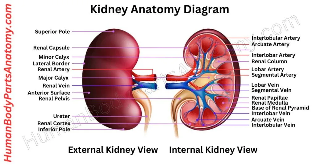
The kidneys are two bean-shaped organs in your urinary system that filter your blood, each about the size of a fist.[50] Each day, they filter about 120 to 150 quarts of blood.[50]
They remove waste by producing about 1 to 2 quarts of urine, while your body reabsorbs and reuses the rest.[49][50]
The kidneys also maintain fluid balance, regulate electrolytes such as sodium, calcium, phosphorus, and potassium, and remove acid and waste products.[49] Together, they filter about half a cup of blood every minute.[49]
Read More – Kidney Anatomy: Complete Guide with Parts, Names, Functions & Diagram
Human Body Parts
Leg
The leg is part of your body between your knee and foot. It is made up of two bones: the tibia and the fibula.[51] These bones give support and balance to your body, and they work with muscles to help you move around.[51]
The tibia connects with the femur at your knee, and at the bottom, it joins with the fibula to form the ankle joint with the talus bone.[52] The tibia is the weight-bearing bone of the leg and the second largest bone in the body.[52]
When your ankle joint works properly, it lets your foot move smoothly while staying stable, which makes walking and moving around comfortable.[17]

Read More – Complete Guide on Leg Anatomy with Parts, Functions & Diagram
Arm
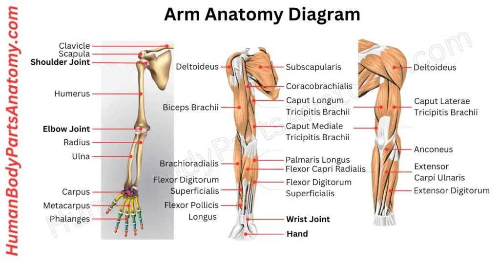
The upper extremity, or arm, is a crucial part of the human anatomy. It has three main sections: the upper arm, forearm, and hand. It starts from the shoulder and reaches to the fingers, and its skeleton is made up of the humerus, the radius and ulna,[12] and the 27 bones of the hand[15] — 30 bones in all, together with nerves, blood vessels, and muscles.
Starting at the shoulder joint, a ball-and-socket joint.[18] It allows for a wide range of movement, though it is less stable than the hip joint because the socket is shallow and holds less than a third of the head of the upper arm bone.[18]
Next is the elbow joint, a hinge joint that facilitates arm bending and straightening. Because the radius can pivot around the ulna, the forearm also has the abilities of pronation and supination.[12]
Read More – Comprehensive Guide to Arm Anatomy: Parts, Names & Diagram
Hand
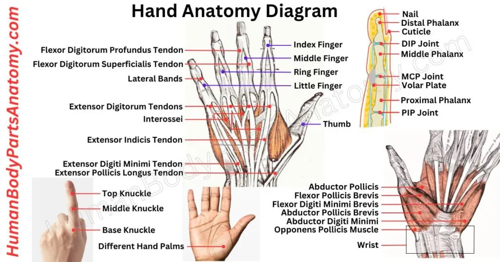
A hand is a helpful part at the end of our arm. Humans and some animals like monkeys and koalas have hands. Even raccoons are said to have hands but don’t have thumbs like we do.
A human hand usually has five parts called fingers. We count the thumb as one of them. There are 27 bones in a hand, not depending on a particular bone. There are 14 finger bones connecting to the wrist bones.[15]
Each hand has five long metacarpal bones and eight small carpal bones.[15] Thus, a hand comprises fingers, thumbs, and bones that help it move and work.
Read More – Comprehensive Guide to Hand Anatomy: Parts, Functions & Diagram
Finger
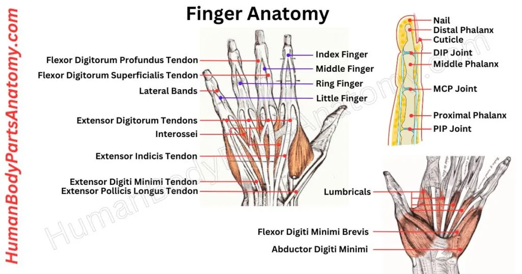
Fingers are essential parts of our hands and similar limbs in many animals. Most animals with limbs, like humans and primates, have five fingers, while shorter ones are called toes.
Fingers are flexible and opposable in humans. They help us feel things and make precise movements, and they are vital for skills like grabbing and moving objects.
The thumb is the first digit, followed by the index finger, the middle finger, the ring finger, and the little finger, also known as the pinkie.
Read More – Complete Guide to Finger Anatomy with Parts, Names, Functions & Diagram
FAQ’s
The human body has more than 600 muscles, which together make up about 40% of a person’s weight.[2] These muscles help with movement, posture, breathing, and essential internal functions like digestion and blood circulation. Muscles are divided into three types: skeletal (movement), smooth (internal organs), and cardiac (heart).
The human body has about 86 billion neurons (nerve cells).[42] These neurons form the central nervous system (brain and spinal cord) and the peripheral nervous system, which includes 12 pairs of cranial nerves and 31 pairs of spinal nerves.[53] The 31 spinal nerve pairs consist of 8 cervical, 12 thoracic, 5 lumbar, 5 sacral, and 1 coccygeal.[54]
There is no official count. No health authority publishes a definitive number, because the total depends on how an organ is defined and whether paired structures, glands, and accessory organs are counted separately. Figures such as “78 organs” are widely repeated conventions rather than measured values, which is why anatomical references describe the body by organ system instead.
The human body has over 78 organs, but the most vital ones include the brain, heart, lungs, liver, and kidneys.[26][41][45][49] Together with bones, muscles, and joints, they maintain life functions such as movement, circulation, and digestion.
An adult human has 206 bones, while a newborn has about 270 bones that gradually fuse as the body grows.[1] Bones provide structure, protect organs, and store essential minerals like calcium and house the marrow where blood cells are made.[1]
Muscle pain, or myalgia, is most often related to tension, overuse, or muscle injury from exercise or hard physical work.[55] It can also be a sign of conditions affecting the whole body, such as some infections including influenza, connective tissue disorders such as lupus, or fibromyalgia.[55]
References-
- Kamel-ElSayed S, et al.; StatPearls, NCBI Bookshelf/NLM. Physiology, Bone. StatPearls 2026 edition. PMID 28722997. https://pubmed.ncbi.nlm.nih.gov/28722997/
- National Cancer Institute, NIH. SEER Training Modules: Muscle Groups. Cited 2026. https://training.seer.cancer.gov/anatomy/muscular/groups/
- NIAMS, NIH. Osteoporosis in Men. Updated June 2025. https://www.niams.nih.gov/health-topics/osteoporosis-men
- Anderson BW, Kortz MW, Black AC, Al Kharazi KA; StatPearls. Anatomy, Head and Neck, Skull. Updated 2023. NBK499834; PMID 29763009. https://www.ncbi.nlm.nih.gov/books/NBK499834/
- DeSai C, Reddy V, Agarwal A; StatPearls. Anatomy, Back, Vertebral Column. Updated 8 August 2023. NBK525969; PMID 30247844. https://www.ncbi.nlm.nih.gov/books/NBK525969/
- Burgess MD, Lui F; StatPearls. Anatomy, Bony Pelvis and Lower Limb: Pelvic Bones. Updated 24 July 2023. NBK551580. https://www.ncbi.nlm.nih.gov/books/NBK551580/
- Chang A, Breeland G, Black AC, Hubbard JB; StatPearls. Anatomy, Bony Pelvis and Lower Limb: Femur. Updated 17 November 2023. NBK532982; PMID 30422577. https://www.ncbi.nlm.nih.gov/books/NBK532982/
- Safarini OA, Bordoni B; StatPearls. Anatomy, Thorax, Ribs. Updated 10 July 2023. NBK538328; PMID 30855912. https://www.ncbi.nlm.nih.gov/books/NBK538328/
- StatPearls, NCBI Bookshelf. Anatomy, Shoulder and Upper Limb, Arm Muscles. Updated 24 July 2023. NBK554420. https://www.ncbi.nlm.nih.gov/books/NBK554420/
- StatPearls, NCBI Bookshelf. Anatomy, Shoulder and Upper Limb, Biceps Muscle. Updated 2024. NBK519538. https://www.ncbi.nlm.nih.gov/books/NBK519538/
- Mitchell B, Whited L; StatPearls. Anatomy, Shoulder and Upper Limb, Forearm Muscles. Updated 5 June 2023. NBK536975. https://www.ncbi.nlm.nih.gov/books/NBK536975/
- StatPearls, NCBI Bookshelf. Anatomy, Shoulder and Upper Limb, Forearm Bones. Updated 20 June 2023. NBK545260. https://www.ncbi.nlm.nih.gov/books/NBK545260/
- Gold M, Munjal A, Varacallo MA; StatPearls. Anatomy, Bony Pelvis and Lower Limb, Hip Joint. Updated 25 July 2023. NBK470555; PMID 29262200. https://www.ncbi.nlm.nih.gov/books/NBK470555/
- Launico MV, Sinkler MA, Nallamothu SV; StatPearls. Anatomy, Bony Pelvis and Lower Limb: Femoral Muscles. Updated 24 October 2023. NBK500008. https://www.ncbi.nlm.nih.gov/books/NBK500008/
- StatPearls, NCBI Bookshelf. Anatomy, Shoulder and Upper Limb, Hand Carpal Bones. Updated 2022. NBK535382. https://www.ncbi.nlm.nih.gov/books/NBK535382/
- Gupton M, Imonugo O, Black AC, Launico MV, Terreberry RR; StatPearls. Anatomy, Bony Pelvis and Lower Limb, Knee. Updated 2023. NBK500017; PMID 29763193. https://www.ncbi.nlm.nih.gov/books/NBK500017/
- Manganaro D, Alsayouri K; StatPearls. Anatomy, Bony Pelvis and Lower Limb: Ankle Joint. Updated 23 May 2023. NBK545158; PMID 31424742. https://www.ncbi.nlm.nih.gov/books/NBK545158/
- Miniato MA, Anand P, Varacallo MA; StatPearls. Anatomy, Shoulder and Upper Limb, Shoulder. Updated 24 July 2023. NBK536933. https://www.ncbi.nlm.nih.gov/books/NBK536933/
- La Rose J, Madrazo-Ibarra A, Varacallo MA; StatPearls. Anatomy, Rotator Cuff. Updated 18 June 2026. NBK441844. https://www.ncbi.nlm.nih.gov/books/NBK441844/
- NIDDK, NIH. Your Digestive System & How It Works. Reviewed 2024. https://www.niddk.nih.gov/health-information/digestive-diseases/digestive-system-how-it-works
- StatPearls, NCBI Bookshelf. Anatomy, Head and Neck, Teeth. Updated 24 July 2023. NBK557543. https://www.ncbi.nlm.nih.gov/books/NBK557543/
- StatPearls, NCBI Bookshelf. Histology, Tooth. Updated 26 June 2023. NBK572055. https://www.ncbi.nlm.nih.gov/books/NBK572055/
- Chaudhry SR, Liman MNP, Omole AE, Peterson DC; StatPearls. Anatomy, Abdomen and Pelvis: Stomach. Updated 17 July 2024. NBK482334; PMID 29493959. https://www.ncbi.nlm.nih.gov/books/NBK482334/
- Collins JT, Nguyen A, Omole AE, Badireddy M; StatPearls. Anatomy, Abdomen and Pelvis, Small Intestine. Updated 18 February 2025. NBK459366; PMID 29083773. https://www.ncbi.nlm.nih.gov/books/NBK459366/
- Omole AE, Mandiga P, Kahai P, Lobo S; StatPearls. Anatomy, Abdomen and Pelvis: Large Intestine. Updated 6 April 2025. NBK470577; PMID 29261962. https://www.ncbi.nlm.nih.gov/books/NBK470577/
- StatPearls, NCBI Bookshelf. Anatomy, Abdomen and Pelvis: Liver. Updated 2022. NBK500014. https://www.ncbi.nlm.nih.gov/books/NBK500014/
- Kalra A, Yetiskul E, Wehrle CJ, Tuma F; StatPearls. Physiology, Liver. Updated 1 May 2023. NBK535438. https://www.ncbi.nlm.nih.gov/books/NBK535438/
- NCBI Bookshelf / NLM. The Exocrine Pancreas — Water and Ion Secretion from the Pancreatic Ductal System. NBK54130. https://www.ncbi.nlm.nih.gov/books/NBK54130/
- Lee MG, Ohana E, Park HW, Yang D, Muallem S. Molecular mechanism of pancreatic and salivary gland fluid and HCO₃⁻ secretion. Physiological Reviews. 2012;92(1):39–74. PMID 22298651; DOI 10.1152/physrev.00011.2011. https://journals.physiology.org/doi/full/10.1152/physrev.00011.2011
- Galarza-Paez L, Marston G, Downs BW; StatPearls. Anatomy, Head and Neck, Nose. Updated 24 July 2023. NBK532870. https://www.ncbi.nlm.nih.gov/books/NBK532870/
- Sobiesk JL, Munakomi S; StatPearls. Anatomy, Head and Neck, Nasal Cavity. StatPearls 2026 edition. PMID 31334952. https://pubmed.ncbi.nlm.nih.gov/31334952/
- Stanford Medicine, Department of Ophthalmology. Neuro-ophthalmology Illustrated — Chapter 3, Visual Fields. 2018. https://neuro-ophthalmology.stanford.edu/2018/12/neuro-ophthalmology-illustrated-chapter-3-visual-fields/
- NIDCD, NIH. How Do We Hear? Reviewed by NIDCD. https://www.nidcd.nih.gov/health/how-do-we-hear
- NIDCD, NIH. Taste Disorders. Reviewed by NIDCD. https://www.nidcd.nih.gov/health/taste-disorders
- Brahs AB, Bolla SR; StatPearls. Histology, Nail. Updated 29 May 2023. NBK539733. https://www.ncbi.nlm.nih.gov/books/NBK539733/
- Martel JL, Miao JH, Badri T, Fakoya AO; StatPearls. Anatomy, Hair Follicle. Updated 22 June 2024. NBK470321. https://www.ncbi.nlm.nih.gov/books/NBK470321/
- StatPearls, NCBI Bookshelf. Physiology, Integument. Updated 1 May 2023. NBK554386. https://www.ncbi.nlm.nih.gov/books/NBK554386/
- Yousef H, Alhajj M, Fakoya AO, Sharma S; StatPearls. Anatomy, Skin (Integument), Epidermis. Updated 8 June 2024. NBK470464. https://www.ncbi.nlm.nih.gov/books/NBK470464/
- StatPearls, NCBI Bookshelf. Anatomy, Skin (Integument). Updated 2022. NBK441980. https://www.ncbi.nlm.nih.gov/books/NBK441980/
- Cochran ML, Lopez MJ, Czyz CN; StatPearls. Anatomy, Head and Neck: Eyelid. Updated 14 August 2023. NBK482304. https://www.ncbi.nlm.nih.gov/books/NBK482304/
- NINDS, NIH. Brain Basics: Know Your Brain. Reviewed by NINDS. https://www.ninds.nih.gov/health-information/public-education/brain-basics/brain-basics-know-your-brain
- Azevedo FAC, Carvalho LRB, Grinberg LT, et al. Equal numbers of neuronal and nonneuronal cells make the human brain an isometrically scaled-up primate brain. Journal of Comparative Neurology. 2009;513(5):532–541. PMID 19226510; DOI 10.1002/cne.21974. https://onlinelibrary.wiley.com/doi/10.1002/cne.21974
- Choe MS, Ortiz-Mantilla S, Makris N, et al. Regional Infant Brain Development: An MRI-Based Morphometric Analysis in 3 to 13 Month Olds. Cerebral Cortex. PMC3729199. https://pmc.ncbi.nlm.nih.gov/articles/PMC3729199/
- NCBI Bookshelf / Disease Control Priorities — Child and Adolescent Health and Development, 3rd edition. Brain Development: The Effect of Interventions on Children and Adolescents. 2017. NBK525261. https://www.ncbi.nlm.nih.gov/books/NBK525261/
- Rehman I, Rehman A; StatPearls. Anatomy, Thorax, Heart. Updated 28 August 2023. NBK470256. https://www.ncbi.nlm.nih.gov/books/NBK470256/
- NHLBI, NIH. How the Heart Works — Anatomy of the Heart. Reviewed by NHLBI. https://www.nhlbi.nih.gov/health/heart/anatomy
- NHLBI, NIH. How the Heart Works — How Blood Flows Through the Heart. Reviewed by NHLBI. https://www.nhlbi.nih.gov/health/heart/blood-flow
- Lescay HA, Jiang J, Leslie SW, Tuma F; StatPearls. Anatomy, Abdomen and Pelvis Ureter. Updated 5 May 2024. NBK532980; PMID 30422575. https://www.ncbi.nlm.nih.gov/books/NBK532980/
- NIDDK, NIH. Your Kidneys & How They Work. Reviewed 2025. https://www.niddk.nih.gov/health-information/kidney-disease/kidneys-how-they-work
- NIDDK, NIH. The Urinary Tract & How It Works. Reviewed 2025. https://www.niddk.nih.gov/health-information/urologic-diseases/urinary-tract-how-it-works
- Cantrell AJ, Imonugo O, Varacallo MA; StatPearls. Anatomy, Bony Pelvis and Lower Limb: Leg Bones. Updated 14 August 2023. NBK537024. https://www.ncbi.nlm.nih.gov/books/NBK537024/
- Bourne M, Sinkler MA, Murphy PB; StatPearls. Anatomy, Bony Pelvis and Lower Limb: Tibia. Updated 8 August 2023. NBK526053. https://www.ncbi.nlm.nih.gov/books/NBK526053/
- StatPearls, NCBI Bookshelf. Neuroanatomy, Somatic Nervous System. Updated 2022. NBK556027. https://www.ncbi.nlm.nih.gov/books/NBK556027/
- Kaiser JT, Lugo-Pico JG; StatPearls. Neuroanatomy, Spinal Nerves. Updated 14 August 2023. NBK542218. https://www.ncbi.nlm.nih.gov/books/NBK542218/
- MedlinePlus Medical Encyclopedia, NLM/NIH. Muscle aches. Reviewed 2025. https://medlineplus.gov/ency/article/003178.htm
Read More-
Human Body-
- Human Skeleton Anatomy: All 206 Bones Explained with Functions & Diagrams
- Human Muscle Anatomy: Validated Guide to Every Major Muscles & Functions
Head, Face & Senses-
- Nose Anatomy: Parts of the Nose, Structure, Nasal Cavity & Sinuses Explained
- Skull Anatomy: Parts of the Skull, Structure, Cranial, Facial Bones & Functions
- Mouth Anatomy: Guide on Parts of Mouth, Lips, Palate, Gums & Oral Cavity
- Eye Anatomy: Parts of the Eye, Cornea, Lens, Retina, Optic Nerve & Diagram
- Ear Anatomy: Parts of the Ear, Outer, Middle & Inner Ear & Structures
Brain & Nervous System-
- Brain Anatomy: Parts of the Brain, Structure, Functions & Regions Explained
- The 4 Lobes of the Brain: Complete Guide with Locations & Functions
Spine & Back-
- Cervical Spine Anatomy: C1–C7 Vertebrae, Muscles & Nerves Explained
- Spine Anatomy: Parts of the Spine, Vertebrae, Curves, Spinal Cord & Diagram
- Neck Muscle Anatomy: Guide with Key Muscles, Groups, Functions & Diagrams
- Rib Cage Anatomy: Ribs, Sternum, Thoracic, Vertebrae & Functions Explained
Organs-
- Pancreas Anatomy: Parts of Pancreas, Structure, Location, Functions & Role
- Thyroid Anatomy: Guide on Key Parts, Location, Structure & Functions
- Stomach Anatomy: Parts of Stomach, Regions, Layers & Digestive Function
- Heart Anatomy: Guide on Parts of Heart, Chambers, Valves & Blood Flow
- Liver Anatomy: Key Parts of Liver, Functions, Lobes, Segments & Diagram
- Kidney Anatomy: Guide on Parts of Kidney, Structure, Functions & Diagram
Upper Limb-
- Shoulder Anatomy: Parts of the Shoulder, Bones, Joint Structure & Diagram
- Hand Anatomy: Parts of the Hand, Bones, Muscles with Functions & Diagram
- Arm Anatomy: Parts of Arm, Bones, Muscles & Joints with Functions & Diagram
Lower Limb-
- Hip Muscle Anatomy: Guide on Key Muscle Groups, Names, Functions & Diagram
- Hip Bone Anatomy: Parts of Hip Bone, Ilium, Pubis, Functions & Diagram
- Leg Anatomy: Parts of the Leg, Bones, Muscles & Lower Leg with Functions
- Knee Anatomy: Parts of Knee, Bones, Ligaments, Cartilage & Joint Structure
- Thigh Muscle Anatomy: Key Muscle Groups, Names, Functions & Diagram
Medical Disclaimer
All content on HumanBodyPartsAnatomy.com is educational and based on verified, peer-reviewed medical sources. Articles are authored or reviewed by qualified medical or biomedical professionals to ensure accuracy.
This website does not provide medical advice, diagnosis, or treatment. Always consult a licensed healthcare professional for personal medical guidance.
No commercial or promotional interests influence the medical content published on this site.