
📅 Published on August 21, 2026 | 🕒 Last Reviewed & Updated on October 1, 2026
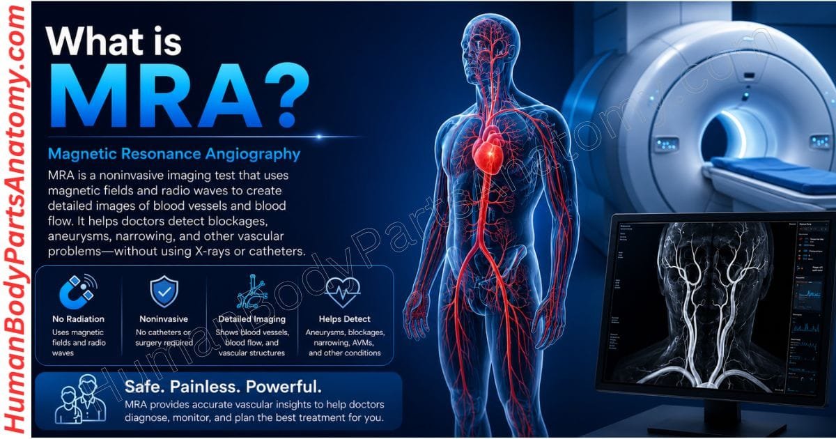
Magnetic resonance angiography, commonly abbreviated as MRA, is a noninvasive imaging test that uses magnetic fields and radio waves to produce detailed images of blood vessels and the blood flowing through them.[1] Doctors use it mainly to look for problems in the arteries and veins — narrowing, blockages, aneurysms, or unusual vessel structures.[2][1] Depending on which part of the body is being studied, the scan may be done with or without a contrast agent.[1] Unlike conventional angiography, MRA usually does not require threading a catheter into a blood vessel.[1]
What Is MRA?
MRA is a specialized form of MRI that images the body’s blood vessels.[1] While standard MRI focuses on organs, bones, and soft tissues, MRA uses specialized sequences to show the size, shape, and course of arteries and veins, as well as the blood flowing through them.[2][14]
The test can reveal narrowed arteries, blocked vessels, enlarged segments, and abnormal connections between vessels.[1][14][15] Doctors order it to investigate symptoms, confirm a suspected vascular disorder, monitor a condition already diagnosed, or plan treatment.[2][14][15]
The underlying physics is straightforward enough to summarize: a strong magnetic field aligns protons in the body’s tissues, radiofrequency pulses briefly knock them out of alignment, and sensors detect the energy they release as they realign and reconstruct it into an image.[3] Because none of this involves ionizing radiation, an MRA carries no radiation exposure.[4]
Which Blood Vessels Can an MRA Examine?
MRA can be used almost anywhere in the body, including:[1][14][15]
- The aorta
- Blood vessels in the brain
- Carotid arteries in the neck
- The heart and the large vessels around it
- Peripheral arteries in the arms and legs
- Pulmonary arteries in the lungs
- Renal arteries supplying the kidneys
- Blood vessels in the abdomen and pelvis
What Conditions Can an MRA Help Detect?
An MRA may be used to evaluate several blood vessel conditions, including:
- Other disorders that affect normal blood flow
- Aneurysms
- Aortic dissection
- Atherosclerosis
- Arterial stenosis or narrowing
- Blood vessel blockages
- Arteriovenous malformations (AVMs)
- Certain congenital vascular abnormalities
One caveat about the coronary arteries. MRA can assess the heart and the large vessels around it, and coronary MRA is an established technique.[2] But imaging the coronary arteries themselves in detail is harder with MRA than with other tests, so cardiac CT or a catheter procedure is usually chosen for that purpose.[14] MRA may still be used to help plan treatments such as coronary bypass or stenting.[14]
What Conditions Can an MRA Help Detect?
An MRA may be used to evaluate a range of blood vessel conditions:
- Aneurysms — abnormal widening or ballooning of part of an artery. In the aorta these appear as balloon-like bulges where the vessel wall has been damaged or weakened.[1][5]
- Aortic dissection — a tear in the inner wall of the aorta that lets blood flow between the layers of the vessel wall, which can lead to rupture or reduced blood flow to organs.[1][6]
- Atherosclerosis — a waxy plaque of fat, cholesterol, calcium, and other substances building up on the inner lining of arteries, narrowing and hardening them.[1][7]
- Arterial stenosis, other narrowing, and outright blockages.[1][14]
- Arteriovenous malformations (AVMs) — tangles of vessels in which blood passes straight from arteries to veins, bypassing the capillary network that normally slows it down and delivers oxygen to tissue.[8][14]
- Congenital vascular abnormalities, including malformations associated with congenital heart disease.[1][2][14]
- Other conditions affecting blood flow, such as mesenteric artery ischemia, renal artery stenosis, and pulmonary embolism.[1][2][14]
For a suspected blood clot in the lungs, doctors typically use CT angiography as the first test. They usually reserve MRA for patients who cannot undergo CT and perform it at centers with appropriate expertise.[14]
What Can a Brain MRA Show?
A brain MRA focuses on the arteries supplying the brain.[14][15] It helps doctors look for brain aneurysms, vascular malformations, narrowed or blocked arteries, and vessel abnormalities linked to stroke.[1][8][9][15]
It produces detailed images that show an aneurysm’s size, location, and shape, specifically helping doctors identify unruptured aneurysms.[9] Most brain aneurysms form in the major arteries along the base of the skull.[9]
When symptoms suggest a rupture, MRA is not the test
If a ruptured aneurysm is suspected—for example, after a sudden, severe headache that people often describe as the worst headache of their life—doctors usually order a CT scan first because it can detect bleeding around the brain quickly.[9] A headache like that is a medical emergency and warrants immediate care, not a scheduled outpatient scan.[9]
A brain MRA may also be ordered when a provider is investigating secondary headaches that could have a vascular cause.[15]
Does an MRA Use Contrast Dye?
Some MRA studies use no contrast, while others use a gadolinium-based agent to make certain vessels stand out more clearly.[1][14] The choice depends on the area being studied, the suspected condition, and what the provider needs to see.[2][14] Gadolinium contrast is less likely to cause an allergic reaction than the iodine-based contrast used for CT.[2][14]
How Is MRA Different From Traditional Angiography?
MRA images blood vessels from outside the body and generally avoids putting a catheter into an artery or vein.[1][14] Catheter angiography, by contrast, is invasive: a thin tube is guided through a blood vessel to obtain the images. That makes MRA the less invasive and less painful option for many indications.[1]
MRA also takes less time than catheter angiography, costs less, and needs no recovery period unless sedation was used.[2][14] Even so, catheter angiography remains the reference standard for some indications because of the image detail it provides.[14]
Types of Magnetic Resonance Angiography
MRA is really an umbrella term covering several techniques built on different physical principles. Which one is used depends on the vessels being examined, the clinical question, and the patient’s circumstances.[2][15]
1. Contrast-enhanced MRA
A gadolinium-based agent is injected into a vein before or during the scan. Gadolinium shortens the T1 relaxation time of blood, so vessels appear bright on rapid gradient-echo images — useful for assessing narrowed arteries, blocked vessels, and aneurysms.[15][2] Because the technique relies on the contrast agent rather than on flow physics, it largely avoids the flow-related artifacts that affect other MRA methods.[2]
2. Non-contrast MRA
This approach produces images without any injection, exploiting inflow effects, the T1 and T2 relaxation properties of blood, or the phase of the MRI signal to separate flowing blood from stationary tissue.[2][16]
It is particularly valuable for pediatric and pregnant patients and for people with impaired kidney function, and it can supply functional and hemodynamic information that neither CT angiography nor contrast-enhanced MRA provides.[16]
3. 2D MRA
Two-dimensional acquisition suits situations where a provider needs to follow a long stretch of a single vessel — peripheral or cervical carotid imaging, for instance, where blood flows largely along the vessel’s length.[2][15]
4. 3D MRA
Three-dimensional acquisition captures a volume of data that can be rotated and viewed from any angle. For time-of-flight imaging in particular, moving from 2D to 3D produced a marked gain in diagnostic accuracy.[2] It is especially useful for a compact cluster of arteries, such as the vessels at the base of the brain known as the circle of Willis.[2][15]
Why Might I Need an MRA?
Your provider may recommend an MRA when they suspect that an artery or other blood vessel has narrowed, become blocked, sustained damage, or enlarged abnormally and need detailed images without performing an invasive angiogram.[1][14]
Depending on the area examined, an MRA may be used to look for:
- Aneurysms: places where an artery wall has weakened and bulged outward.[5][9]
- Aortic coarctation: a narrowing of part of the aorta. This is a congenital heart defect, present from birth and caused by abnormal development of the aorta before birth. MRI or MR angiography of the chest may be used in the workup, particularly in older children.[10]
- Aortic dissection: a tear in the inner layer of the aorta that allows blood to separate the layers of the artery wall.[6]
- Vessel problems linked to stroke: narrowed, blocked, or abnormal arteries in the brain and neck, including carotid plaque that can restrict blood flow to the brain.[1][14]
- Cardiovascular conditions: the state of major arteries and vessels around the heart, including in congenital heart disease.[1][2][14]
- Peripheral artery disease (PAD): reduced blood flow or narrowing in the arteries supplying the arms or legs. PAD is caused by plaque buildup and most often affects the legs and feet.[1][7]
- Renal artery stenosis: narrowing of one or both arteries carrying blood to the kidneys. Roughly 90% of cases are caused by atherosclerosis. The condition can raise blood pressure — renovascular hypertension — and, when severe, contribute to declining kidney function.[11]
What Are the Risks of Magnetic Resonance Angiography?
MRA is generally safe and poses almost no risk to the average patient when standard safety procedures are followed.[14] It involves no ionizing radiation.[4] The MR environment does, however, combine a powerful static magnetic field, rapidly switching gradient fields, and radiofrequency energy — and each of those brings its own considerations, covered below.[4]
When contrast is needed, a small IV line goes into a vein.[1][14] You may feel a brief pinch, some pressure, or mild soreness at the site, and some people notice nausea, headache, a warm sensation, or a metallic taste.[15]
Serious reactions to gadolinium are uncommon. A systematic review and meta-analysis pooling 716,978 gadolinium administrations across nine studies found allergic-like reactions at roughly 9 per 10,000 doses, with severe reactions at about 0.5 per 10,000.[17] Even so, tell your provider about any previous reaction to contrast.[14]
The scanner is an enclosed space, and some people find that uncomfortable — particularly anyone with claustrophobia.[3][4] Say so before the appointment. Your team may suggest relaxation or distraction techniques, an open-configuration scanner where one is available and suitable, or medication to help you stay settled.[3][4][14]
Potential Risks and Safety Concerns
Metal objects and implants
The magnetic field extends beyond the machine and pulls hard on iron, some steels, and other magnetizable materials.[3] Loose metal can become a projectile; implanted devices may shift or malfunction; and radiofrequency energy can heat an implant and the tissue around it, potentially causing burns.[4]
For this reason, patients with implanted devices should not undergo an MRI unless they can positively identify the device as MR Safe or MR Conditional. Treat any device with an unknown status as MR Unsafe.[4]
Tell the MRI team about pacemakers, defibrillators, neurostimulators, implanted pumps, cochlear implants, aneurysm clips, surgical hardware, and any retained metal fragments.[1][2][3][4]
Noise, nerve stimulation, and heating
The switching gradient fields generate loud knocking that can damage hearing without ear protection, and can produce a harmless twitching sensation from peripheral nerve stimulation.[3][4] Radiofrequency energy heats tissue, and the potential for heating rises with longer examinations.[4]
Adverse events are rare overall. When patients report them to the FDA, most involve heating or burns, with second-degree burns being the most common patient problem. Other reported events include projectile injuries, pinched or crushed fingers from the patient table, falls, and hearing effects.[4]
Kidney disease and contrast
Gadolinium agents are cleared mainly by the kidneys.[12] People with severe kidney impairment were historically at risk of nephrogenic systemic fibrosis (NSF), which remains the only adverse health effect definitively tied to gadolinium retention.[12]
That risk has fallen dramatically with modern agents: consensus statements from the American College of Radiology and the National kidney Foundation report zero NSF events across 4,931 administrations of group II agents to patients with an eGFR below 30 mL/min/1.73 m².[18]
As a result, clinicians can now consider kidney-function screening before contrast-enhanced MRI optional when they use a Group II agent, but they should still perform it when using a Group III agent.[18] Your provider decides what is appropriate in your case and may recommend a non-contrast MRA or a different test entirely.[11][14]
Gadolinium retention
The FDA requires a class warning on all gadolinium-based contrast agents noting that trace amounts can remain in the body — including the brain, bone, and skin — for months to years after a dose.[12]
The FDA has not directly linked retention to adverse health effects in people with normal kidney function and has concluded that the benefits of approved agents continue to outweigh the potential risks.[12]
Every patient receiving one of these agents should receive an FDA-required Medication Guide, and patients expecting repeated doses over their lifetime should discuss retention with their doctor.[12][14]
Pregnancy and breastfeeding
Tell your provider if you are or might be pregnant.[14] MRI uses no X-rays, and current data show no convincing evidence that non-contrast MRI harms a fetus — but if the scan is not time-sensitive, your doctor may prefer to wait.[14]
Gadolinium crosses the placenta, so clinicians generally avoid it during pregnancy unless medically necessary. This makes non-contrast MRA techniques particularly useful in this setting.[2][16]
For breastfeeding, professional organizations agree that mothers can continue nursing after receiving a gadolinium contrast medium because only a very small amount of the maternal dose enters breast milk, and infants absorb very little of it orally.[13]
Sedation
If a sedative is used there is a small risk of oversedation, so your vital signs are monitored.[14] Sedation and anesthesia carry risks that are not specific to MRI, including slowed or difficult breathing and low blood pressure.[4]
For most patients, MRA is well tolerated and carries few risks. Your provider can explain what applies to you based on your medical history and whether they use contrast.[14]
How Should You Prepare for an MRA?
Preparation varies by facility and by the type of MRA. Follow the instructions your provider or imaging center gives you.[14][15]
Food, drinks, and medications
Rules about eating and drinking differ between facilities.[14] Many patients can eat and drink normally and, unless told otherwise, should take their usual medications.[14][15] Some studies — particularly those involving sedation or certain contrast protocols — do require several hours without food or drink.[1] Go by the instructions for your specific appointment.[14]
What to wear
You will probably be asked to change into a gown.[1][14] Remove jewelry, watches, piercings, hair accessories, credit cards, and hearing aids; certain metals also blur the images.[1] Wear loose, comfortable clothing and leave valuables at home.[14]
Tell the team about implants and metal
Before the scan, give a complete account of any pacemakers, implanted pumps, aneurysm clips, cochlear implants, surgical hardware, metal fragments, or other devices in your body.[1][4][14] Some are MRI-compatible, others need special handling, and a few make the scan unsafe.[4]
If you were given a card with your implant’s details, bring it and hand it to the technologist.[14] Anyone who has worked with sheet metal may need imaging to check for tiny fragments in the eyes.[1] And mention implants or past surgeries again even if you have had an MRI before, so the team can re-verify.[4][14]
If contrast is used
Contrast goes in through an IV in your arm or hand, and the injection itself takes very little time.[1][14] Some people briefly notice:
- A warm or cool sensation
- A strange or metallic taste
- Mild nausea
- Headache
- Tingling near the injection site or in the arm
These usually pass quickly.[15] Tell the staff right away if you develop anything more serious — trouble breathing, swelling of the face or throat, severe dizziness, or widespread hives. A physician is on hand to respond immediately if an allergic reaction occurs.[14]
If you have reacted to contrast before
Tell your provider.[14] A previous allergic or anaphylactic reaction to gadolinium is a contraindication to further gadolinium, and your doctor may recommend an alternative agent, extra precautions, or premedication.[2] Do not take premedication unless it has been prescribed specifically for you.
If you have claustrophobia
Raise it before the appointment.[1][14] The team may offer relaxation or distraction techniques, an open MRI scanner, or a mild sedative.[3][14][15] If you take a sedative, arrange for someone to drive you home.[15]
What Happens During an MRA?
An MRA may be done at an outpatient imaging center or in hospital, and it generally follows the same sequence.[11][15]
You start by removing clothing and personal items that could interfere with the scan and changing into a gown.[1] If contrast is needed, an IV is placed.[14][15]
You then lie on a narrow table that slides into the scanner. MRI scanners are tunnel-shaped; depending on the model, the bore may be open at both ends or closed at one end, and some newer systems are built to feel more open.[1][11] The technologist works from an adjoining room and can see and speak with you throughout.[1][15]
Staying still matters a great deal — even small movements degrade the images and may mean repeating a sequence.[1][14] For some MRA studies you will be asked to hold your breath for 15 to 25 seconds at a time, or the scan may be synchronized to your heartbeat.[14][15]
The machine produces loud tapping, humming, and knocking while it works.[1] Earplugs or headphones are provided to protect your hearing and make the noise easier to tolerate.[1][4][15]
You will normally be able to reach the technologist during the scan, usually through an intercom or a squeeze ball you press for attention.[1][15]
How Long Does an MRA Take?
It depends on which part of the body is being studied and how many images are needed. Many MRA examinations run about 20 to 60 minutes.[15] Studies covering several vascular territories can take an hour or longer.[1]
The scan itself is painless.[1] Any discomfort usually comes from holding one position for a while, or from the IV.[1]
What Happens After the MRA?
Once the imaging is finished, the technologist checks that the images are good enough to interpret. If you had no sedation, there is no recovery period and you can go back to your usual activities straight away.[1][14]
If contrast was used, follow whatever instructions your team gives you. If you had a sedative, follow the safety advice and do not drive until your provider says it is fine.[1][15]
A radiologist — a physician trained to supervise and interpret imaging — reviews the images and sends a signed report to the doctor who ordered the scan, who then goes through the findings with you.[14] Sometimes a follow-up study is recommended, either to look at something more closely or to see whether it has changed over time.[14]
Understanding MRA Results
A normal MRA result means the vessels examined show no sign of narrowing or blockage.[1]
An abnormal result points to a problem in one or more vessels and may indicate atherosclerosis, a narrowed or blocked artery, an aneurysm, trauma, a congenital vascular condition, or another abnormality.[1] Depending on what is found, your doctor may recommend further imaging or tests before deciding on treatment.[14]
What Are the Limitations of MRA?
MRA is not the right tool for every question. Unlike CT angiography, it cannot show calcium deposits in vessel walls.[14] Images of some arteries are less crisp than catheter angiography images, small vessels can be difficult to assess, and separating arteries from veins is not always straightforward.[14]
Image quality also depends heavily on you. Anxiety, confusion, severe pain, involuntary movement, or a very irregular heartbeat can all reduce diagnostic quality.[14] Implants and other metal can degrade the images, and body size or weight may exceed a particular scanner’s limits.[4][14]
Key Points to Remember
Preparing for an MRA is usually simple. Follow your imaging center’s instructions, remove metal objects, and tell the team about implants, metal in your body, kidney problems, pregnancy or breastfeeding, previous contrast reactions, and claustrophobia.[1][4][14] Staying relaxed and still during the scan produces clearer images and a more accurate result.[1][14]
FAQ’s –
An MRA is a type of MRI. A standard MRI is optimized for organs and soft tissue; an MRA uses sequences built specifically to show blood vessels and blood flow.[1][15]
No. It uses magnetic fields and radio waves, not ionizing radiation.[4][14]
Gadolinium contrast is less likely to cause an allergic reaction than the iodine-based contrast used in CT, and severe reactions occur in roughly 0.5 per 10,000 administrations.[14][17] Anyone with significant kidney impairment should discuss the options with their provider.[18]
Only if the device has been positively identified as MR Safe or MR Conditional. Devices of unknown status are treated as unsafe, so disclose everything to the MRI team.[4]
Yes. Professional guidelines agree that breastfeeding does not need to be interrupted after a gadolinium contrast medium, because very little reaches breast milk and infant absorption is limited.[13]
A radiologist reviews the images and sends a signed report to your referring doctor, who discusses the findings with you.[14] Turnaround varies by facility and commonly takes a few days.[15]
References –
- National Library of Medicine, National Institutes of Health. Magnetic resonance angiography. MedlinePlus Medical Encyclopedia. Reviewed 10 May 2024. https://medlineplus.gov/ency/article/007269.htm
- De Leucio A, De Jesus O. MR Angiogram. In: StatPearls [Internet]. NCBI Bookshelf, National Library of Medicine; updated 23 August 2023. Bookshelf ID NBK558984. PMID: 32644410. https://www.ncbi.nlm.nih.gov/books/NBK558984/
- National Institute of Biomedical Imaging and Bioengineering (NIBIB), National Institutes of Health. Magnetic Resonance Imaging (MRI). https://www.nibib.nih.gov/science-education/science-topics/magnetic-resonance-imaging-mri
- U.S. Food and Drug Administration, Center for Devices and Radiological Health. MRI (Magnetic Resonance Imaging): Benefits and Risks. Content current as of 9 December 2017. https://www.fda.gov/radiation-emitting-products/mri-magnetic-resonance-imaging/benefits-and-risks
- National Heart, Lung, and Blood Institute (NHLBI), National Institutes of Health. Aortic Aneurysm. https://www.nhlbi.nih.gov/health/aortic-aneurysm
- National Library of Medicine, National Institutes of Health. Aortic dissection. MedlinePlus Medical Encyclopedia. Reviewed 28 April 2026. https://medlineplus.gov/ency/article/000181.htm
- National Heart, Lung, and Blood Institute (NHLBI), National Institutes of Health. Peripheral Artery Disease — Causes and Risk Factors. https://www.nhlbi.nih.gov/health/peripheral-artery-disease/causes
- National Institute of Neurological Disorders and Stroke (NINDS), National Institutes of Health. Arteriovenous Malformations (AVMs). Last reviewed 13 March 2026. https://www.ninds.nih.gov/health-information/disorders/arteriovenous-malformations-avms
- National Institute of Neurological Disorders and Stroke (NINDS), National Institutes of Health. Cerebral Aneurysms. Last reviewed 13 March 2026. https://www.ninds.nih.gov/health-information/disorders/cerebral-aneurysms
- National Library of Medicine, National Institutes of Health. Coarctation of the aorta. MedlinePlus Medical Encyclopedia. Reviewed 1 October 2025; editorial update 17 June 2026. https://medlineplus.gov/ency/article/000191.htm
- National Institute of Diabetes and Digestive and Kidney Diseases (NIDDK), National Institutes of Health. Renal Artery Stenosis. Last reviewed July 2014. https://www.niddk.nih.gov/health-information/kidney-disease/renal-artery-stenosis
- U.S. Food and Drug Administration. FDA Drug Safety Communication: FDA warns that gadolinium-based contrast agents (GBCAs) are retained in the body; requires new class warnings. Issued 19 December 2017; updated 16 May 2018. https://www.fda.gov/drugs/drug-safety-and-availability/fda-drug-safety-communication-fda-warns-gadolinium-based-contrast-agents-gbcas-are-retained-body
- Gadoterate. In: Drugs and Lactation Database (LactMed®) [Internet]. Bethesda (MD): National Institute of Child Health and Human Development; NCBI Bookshelf. Last revision 15 December 2025. Bookshelf ID NBK500619. https://www.ncbi.nlm.nih.gov/books/NBK500619/
- Radiological Society of North America (RSNA), American College of Radiology (ACR), and American Society of Radiologic Technologists (ASRT). MR Angiography (MRA). RadiologyInfo.org. Last reviewed 15 June 2026. https://www.radiologyinfo.org/en/info/angiomr
- Cleveland Clinic (non-profit academic medical center). MRA (Magnetic Resonance Angiography). Medically reviewed; last updated 24 April 2025. https://my.clevelandclinic.org/health/diagnostics/24024-mra
- Navot B, Hecht EM, Lim RP, Edelman RR, Koktzoglou I. MR Angiography Series: Fundamentals of Non–Contrast-enhanced MR Angiography. RadioGraphics. 2021 Sep–Oct;41(5):E157–E158. PMID: 34469213. DOI: 10.1148/rg.2021210141.
- Behzadi AH, Zhao Y, Farooq Z, Prince MR. Immediate Allergic Reactions to Gadolinium-based Contrast Agents: A Systematic Review and Meta-Analysis. Radiology. 2018 Feb;286(2):471–482. PMID: 28846495. DOI: 10.1148/radiol.2017162740.
- Weinreb JC, Rodby RA, Yee J, Wang CL, Fine D, McDonald RJ, Perazella MA, Dillman JR, Davenport MS. Use of Intravenous Gadolinium-based Contrast Media in Patients with Kidney Disease: Consensus Statements from the American College of Radiology and the National Kidney Foundation. Radiology. 2021 Jan;298(1):28–35. PMID: 33170103. DOI: 10.1148/radiol.2020202903.
Read More-
Pain Management –
- Lower Back Pain: An Overview of Types, Risk Factors, and When to Seek Medical Care
- Lower Back Pain in Women: Why It Affects Women Differently — Causes, Symptoms, Treatment & Prevention
- Lower Back Pain Treatments: Effective Relief Options, Tests & Expert Tips
- Lower Back Pain Causes & Symptoms: Common Reasons & Risk Factors
- RICE Method For Injuries – Rest, Ice, Compression & Elevation
- 13 Best Exercises for Knee Pain: Step-by-Step Guide with Images
- 15 Effective Home Remedies for Knee Pain with Pictures That Actually Work
Diagnostic Testing –
- PET Scan vs CT Scan: What’s the Difference and Which One Do You Need?
- CT Scan vs. MRI Scan: Key Differences, Uses, and Which One You Need
Medical Disclaimer
All content on HumanBodyPartsAnatomy.com is educational and based on verified, peer-reviewed medical sources. Articles are authored or reviewed by qualified medical or biomedical professionals to ensure accuracy.
This website does not provide medical advice, diagnosis, or treatment. Always consult a licensed healthcare professional for personal medical guidance.
No commercial or promotional interests influence the medical content published on this site.