📅 Published on July 29, 2026 | 🕒 Last Reviewed & Updated on September 15, 2026
Choosing the right imaging test is an important step in finding the cause of a health problem. If your doctor has recommended a CT scan or an MRI, you may be wondering what makes these tests different and why one is better suited for your condition. While both create detailed images of the inside of your body, they use different technology and are designed to answer different medical questions. Understanding the differences between CT scan vs MRI can help you feel more confident about your diagnosis and treatment plan.
Overview of CT Scan vs MRI
A CT scan uses X-rays and computer processing to quickly capture detailed cross-sectional images, making it especially useful in emergencies and for detecting injuries.[4][5] An MRI, on the other hand, uses powerful magnets and radio waves to produce highly detailed images of soft tissues, organs, and nerves without exposing you to ionizing radiation.[1] Choosing between imaging methods often starts with a simple CT scan vs MRI comparison, and understanding the basics makes the rest of this guide much easier to follow.
In this guide, we explain the key differences between CT scans and MRIs, how each imaging test works, the conditions each helps diagnose, and why your healthcare provider may recommend one over the other. By the end, you’ll have a better understanding of which imaging test is most appropriate for different medical situations and what you can expect during the procedure.
What Is an MRI?
Magnetic Resonance Imaging (MRI) is a safe, non-invasive imaging test that uses powerful magnets, radio waves, and a computer to create highly detailed images of the body’s internal organs and tissues.[1] When used to examine the brain, an MRI produces clear images of the brain’s structure, helping doctors identify a wide range of conditions such as tumors, strokes, multiple sclerosis, infections, inflammation, and abnormalities in the brain’s blood vessels.[3][13]
Unlike X-rays and CT scans, MRI does not use ionizing radiation, making it a preferred option when detailed imaging is needed or when repeated scans may be required.[1] During the procedure, you will lie on a padded table that slowly moves into a large, tube-shaped MRI machine.[3] It is important to remain as still as possible because movement can make the images blurry.[3]
Key points:
- The scan is painless[3] and most often takes 30 to 60 minutes, though it may take longer depending on the examination.[20]
- Uses strong magnetic fields and radio waves to create detailed images.[1]
- Does not expose the patient to ionizing radiation.[1]
- Provides excellent images of the brain and other soft tissues.[3]
- Helps detect tumors, stroke, infections, multiple sclerosis, and other brain disorders.[3][13]
This is one of the main reasons MRI often comes up first in any MRI vs CT scan brain imaging discussion.
What Is a CT Scan?
A Computed Tomography (CT) scan is a medical imaging test that combines X-rays and advanced computer technology to create detailed cross-sectional images of the body’s internal structures.[4] For brain imaging, a CT scan is commonly used to quickly detect conditions such as head injuries, brain bleeding, skull fractures, strokes, swelling, and certain brain tumors.[5][6][14]
During the scan, you lie on a motorized table that passes through a large, doughnut-shaped CT scanner. The scanner rotates around your head, taking multiple X-ray images from different angles.[4] A computer then combines these images to produce detailed cross-sectional views of the brain.[4]
One of the biggest advantages of a CT scan is its speed. Most brain CT scans are completed in a matter of minutes, making them an excellent choice during medical emergencies when rapid diagnosis is essential.[5]
Key points:
- Uses ionizing radiation, so scans are performed only when medically necessary.[7][8]
- Uses X-rays to create detailed cross-sectional images.[4]
- Ideal for quickly diagnosing emergency conditions, particularly stroke and traumatic brain injury.[5][14]
- Commonly used to detect brain hemorrhage, skull fractures, stroke, and traumatic injuries.[5][6][14]
- Most CT scans are completed within a few minutes.[5]
Speed is one of the clearest advantages CT holds in the CT vs MRI comparison.
MRI vs. CT Scan: What’s the Difference?
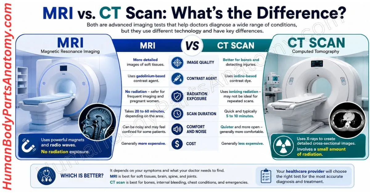
MRI (Magnetic Resonance Imaging) and CT (Computed Tomography) scans are two of the most common medical imaging tests used to diagnose a wide range of health conditions. While both create detailed images of the inside of the body, they use different technologies and are designed for different purposes.[1][4]
Understanding the difference between an MRI and a CT scan can help you know why your healthcare provider recommends one test over the other. The sections below break down the CT vs MRI scan comparison point by point.
1. Image Quality and Detail
Both MRI and CT scans can capture images of bones, organs, blood vessels, muscles, and other internal structures.
- However, an MRI produces more detailed images of soft tissues, making it especially useful for examining the brain, spinal cord, joints, ligaments, muscles, and internal organs.[1][2]
- CT scans provide excellent images of bones and are often the preferred choice for detecting fractures, internal bleeding, lung diseases, and traumatic injuries.[4][10] They also produce images much faster than MRI scans, making them ideal in emergency situations.[5]
In terms of image detail, the CT scan vs MRI comparison usually favors MRI for soft tissue and CT for bone.
2. Contrast Agents
Doctors may use contrast material during both MRI and CT scans to improve image clarity.
- CT scans typically use an iodine-based contrast dye.[10][11]
- MRI scans use a gadolinium-based contrast agent.[3][12]
The type of contrast agent used depends on the examination and the patient’s medical history. People with kidney disease, certain allergies, or other health conditions should discuss the safest option with their healthcare provider before the scan.[12] Contrast agents are just one small piece of the larger MRI vs CT scan puzzle.
3. Radiation Exposure
One of the biggest differences between MRI and CT scans is radiation exposure.
- CT scans use a small amount of ionizing radiation to create cross-sectional images of the body.[7][8]
- MRI scans use powerful magnets and radio waves instead of radiation.[1]
Because MRI does not expose patients to radiation, healthcare providers may recommend it for people who need repeated imaging or when avoiding radiation is preferable.[1] However, the medical condition being evaluated ultimately determines the most appropriate imaging test.
Patients often consider radiation the single biggest factor in the CT vs MRI debate. Healthcare providers keep the radiation dose from each CT scan as low as possible while still producing a diagnostic-quality image, and a single CT scan carries only a small risk.[7][8]
4. Scan Duration
A CT scan captures images in seconds to a few minutes, making it a fast imaging test for emergency care and rapid diagnosis.[5][6]
An MRI generally takes 30 to 60 minutes, depending on the body part being examined and whether contrast material is used.[20] During the scan, patients need to remain as still as possible to produce clear images.[3] Duration is a major consideration in any CT vs MRI scan comparison, especially for anxious or claustrophobic patients.
5. Comfort and Noise
CT scans are typically more comfortable because they are shorter and quieter. The scanner is also more open, which many patients find less intimidating.
MRI machines are loud, producing knocking or tapping sounds during the procedure, and the technician can provide earplugs to reduce the noise.[2]
Some people with claustrophobia may feel uncomfortable inside the MRI machine; in that case, a provider may offer medicine to help you feel sleepy and less anxious, or suggest an “open” MRI in which the machine is not as close to the body — though the images from an open scanner may not be as detailed.[3][20]
Comfort is a smaller but still meaningful factor in the MRI vs CT discussion for nervous patients.
6. Cost
In many healthcare settings, CT scans are generally less expensive than MRI scans because they are faster and require less scanning time.
The exact cost depends on factors such as the body part being examined, whether contrast material is used, the healthcare facility, and insurance coverage.
Patients should check with their imaging center or insurance provider for the most accurate pricing information. Cost differences are one more practical detail in the MRI scan vs CT scan comparison.
What Injuries Require an MRI?
A Magnetic Resonance Imaging (MRI) scan is one of the most effective imaging tests for diagnosing injuries that affect the body’s soft tissues, joints, muscles, ligaments, tendons, nerves, and spine.[1][2]
Unlike X-rays, which mainly show bones, an MRI provides detailed images of both soft tissues and bones, making it easier to identify injuries that may not be visible with other imaging tests.[1]
MRI uses a powerful magnetic field and radio waves to create high-resolution images of the inside of the body. It does not use ionizing radiation, making it a safe imaging option for most people when recommended by a healthcare provider.[1] For soft tissue injuries, the MRI vs CT decision usually leans toward MRI.
When Is an MRI Recommended?
Doctors often recommend an MRI when:
- X-rays do not clearly explain the source of pain or symptoms.
- A soft tissue injury is suspected.
- Joint pain, swelling, or limited movement continues after an injury.[20]
- Numbness, tingling, or weakness may be caused by nerve damage.
- A detailed evaluation is needed before surgery or to monitor recovery.[20]
Modern MRI technology can produce highly detailed images in a reasonable amount of time. Most MRI, including joint studies such as a knee MRI, are completed in about 30 to 60 minutes, though the exact time depends on the body part being examined and whether contrast is used.[20]
Common Injuries That May Require an MRI
MRI is commonly used to diagnose sports injuries, workplace injuries, and other musculoskeletal conditions, including:
- Cartilage damage or cartilage loss[16][20]
- Joint inflammation and swelling[20]
- Pinched or compressed nerves
- Spinal injuries, including damaged discs and soft tissue injuries
- Torn or stretched ligaments[17][20]
- Tendon injuries or ruptures[18]
- Muscle tears and strains
- Detached or damaged cartilage[16][20]
Doctors also rely on MRI to accurately diagnose several common orthopedic injuries, such as:
- Meniscus tears in the knee[16][20]
- Anterior cruciate ligament (ACL) injuries, where an MRI can confirm the diagnosis and reveal associated knee damage[17]
- Achilles tendon tears or ruptures — most are diagnosed on physical exam, with an MRI or ultrasound used to pinpoint the location of the tear[18]
- Moderate to severe sprains and muscle strains
- Rotator cuff tears in the shoulder[1][19]
What Injuries Require a CT Scan?
A CT (Computed Tomography) scan is an advanced imaging test that uses X-rays and computer technology to create detailed cross-sectional and 3D images of the inside of the body.[4]
While it uses more radiation than a standard X-ray, it provides much clearer images of bones, organs, blood vessels, and soft tissues, helping doctors quickly identify serious injuries and medical conditions.[4][7]
CT scans are commonly used in emergency rooms because they are fast, accurate, and can often be completed in just a few minutes.[5]
They are especially helpful after accidents, falls, or other traumatic injuries when doctors need to quickly determine the extent of internal damage. In emergency trauma situations, the CT vs MRI choice almost always favors CT because of its speed.
Injuries That May Require a CT Scan
Your healthcare provider may recommend a CT scan if you have experienced:
- Head injuries, including suspected skull fractures or brain bleeding[5][6]
- Facial fractures or jaw injuries
- Neck or spinal injuries after trauma
- Broken or complex bone fractures
- Chest injuries, such as rib fractures or lung damage[10]
- Abdominal or pelvic injuries that may involve internal bleeding or organ damage[11]
- Multiple injuries caused by car accidents, sports injuries, or significant falls
A CT scan can also help doctors evaluate injuries before surgery by providing detailed images of the affected area.
What Else Can a CT Scan Detect?
In addition to injuries, CT scans are widely used to diagnose and monitor many medical conditions, including:
- Bone fractures and joint problems
- CT scans can detect kidney stones, bladder stones, and some gallstones. However, CT often misses the most common type of gallstone (cholesterol stones), which abdominal ultrasound usually detects more effectively.[11][25]
- Internal bleeding and blood vessel disorders[11]
- Diseases affecting organs such as the liver, pancreas, kidneys, and spleen[11]
- Lung conditions, including infections and blood clots[10]
- Certain types of cancer, including tumor detection, staging, and treatment monitoring[9]
- Emergency abdominal conditions such as appendicitis, bowel obstruction, or diverticulitis[11]
This broad range of uses is a key part of the MRI scan vs CT scan comparison for cancer care.
When Is a CT Scan Recommended?
Doctors typically recommend a CT scan when they need more detailed images than a standard X-ray can provide. The scan helps identify injuries or medical conditions quickly, allowing healthcare providers to make accurate diagnoses and begin the most appropriate treatment as soon as possible.
Advantages of CT Scans
- Fast imaging: CT scans are completed within a few minutes, making them ideal for emergency situations.[5]
- Quick diagnosis: They help doctors rapidly identify the cause of symptoms and begin treatment sooner.
- Detailed body images: CT scans produce clear cross-sectional images of bones, organs, blood vessels, and soft tissues.[4]
- Accurate cancer staging: They help determine whether cancer has spread to the lungs, liver, bones, or other parts of the body.[9]
- Guides treatment planning: CT images help doctors plan surgeries, biopsies, and other medical treatments.[9]
Advantages of MRI
- Excellent soft tissue detail: MRI provides highly detailed images of organs, muscles, ligaments, nerves, and the brain.[1][2]
- Better detection of certain cancers: It can identify cancers of the prostate, breast, liver, brain, and other soft tissues that may be difficult to detect with CT.[15]
- Highly sensitive imaging: MRI can detect subtle changes in tissues, helping diagnose diseases at an earlier stage.[1]
- No ionizing radiation: MRI uses powerful magnets and radio waves instead of X-rays, making it a safer imaging option for many patients.[1]
- Supports long-term monitoring: It is well suited for people who need repeated imaging over time because it does not expose them to radiation.[1]
These benefits explain why MRI often wins the MRI vs CT scan for cancer comparison due to no ionizing radiation, superior soft-tissue contrast, and detailed local tracking.
What Are the Common Concerns About CT Scans and MRIs?
CT scans and MRI scans are both safe, noninvasive imaging tests that help doctors diagnose and monitor many health conditions. While each test has a few limitations, they are widely used because the benefits of an accurate diagnosis usually outweigh the potential risks.
Concerns About CT Scans
A CT (computed tomography) scan uses a small amount of ionizing radiation to create detailed images of the inside of the body.[7] Although many people worry about radiation exposure, the dose used during a medical CT scan is carefully controlled and kept as low as possible.[7][8] Your healthcare provider will only recommend a CT scan when the information it provides is important for making the right diagnosis or treatment plan.
Many CT scans also use a contrast dye to improve image quality.[10] Most people have no problems with the contrast, but some may experience mild side effects such as a warm sensation or a metallic taste.[10] In rare cases, an allergic reaction can occur, so it is important to tell your doctor if you have had a previous reaction to contrast material or have kidney disease.[11][10]
One advantage of CT is that it is very fast, making it especially useful during emergencies.[5] It is also suitable for most people, including many patients with medical implants that may not be compatible with MRI.[21] Weighing these concerns is an important part of any CT vs MRI scan decision.
Concerns About MRI Scans
MRI (magnetic resonance imaging) does not use radiation. Instead, it uses powerful magnets and radio waves to produce highly detailed images of organs, muscles, nerves, and other soft tissues.[1] Serious adverse events during MRI are very rare.[21]
Because an MRI scan takes longer than a CT scan, you will need to remain still throughout the exam to avoid blurry images.[3] The scanner is also enclosed, which may feel uncomfortable for people with claustrophobia.
If you are concerned about enclosed spaces, let your healthcare provider know before the exam, as options such as medicine to help you relax or an open MRI scanner may be available.[3][20]
MRI machines produce loud knocking or buzzing sounds during the scan, but earplugs or headphones are usually provided for comfort.[2][3]
Some people cannot undergo an MRI if they have certain medical devices or metal implants.[2] Many modern devices, including some pacemakers and joint replacements, are labeled “MR Safe” or “MR Conditional,” meaning they can be scanned safely — an MR Conditional device only within the specific conditions it was tested for.
Importantly, any device whose MRI safety status is unknown should be treated as unsafe, so your care team will identify your implant before scheduling a scan.[21] Depending on the reason for the scan, a contrast agent may also be used to improve image quality.
Your healthcare team will review your medical history to determine whether contrast is appropriate for you, particularly if you have kidney disease.[12]
How AI Is Improving MRI and CT Scan Imaging
Artificial intelligence (AI) is changing the way MRI and CT scans are analyzed by helping radiologists detect important details more accurately and efficiently.[22][23]
AI does not replace the expertise of a radiologist. Instead, it works as a supportive tool that highlights areas needing closer attention and assists in identifying patterns that may be difficult to notice during a routine review; in current radiology workflows, the radiologist reviews and, when needed, corrects the AI output before it becomes part of the official report.[23]
AI can also assist with image reconstruction and enhancement tasks such as noise reduction.[22] Clearer images help radiologists compare current and previous scans more effectively, making it easier to monitor changes in tumors, injuries, or other medical conditions over time. This can support earlier detection, better treatment planning, and more accurate follow-up care.
Despite these advancements, every MRI and CT scan is carefully interpreted by a board-certified radiologist. The final diagnosis is always based on the radiologist’s medical expertise, combined with the patient’s medical history, symptoms, and imaging results.[23]
AI simply provides additional insights that can improve speed, consistency, and diagnostic confidence, and any AI-enabled imaging device used in U.S. clinical practice must go through FDA review before it reaches the market.[22]
Is an MRI Better Than a CT Scan?
There is no single imaging test that is best for every situation. Whether an MRI or CT scan is the right choice depends on the part of the body being examined, the medical condition being investigated, and the information your healthcare provider needs.
A CT scan is often recommended when quick imaging is required, such as for injuries, fractures, internal bleeding, or certain lung conditions.[5]
MRI scans are usually preferred for examining soft tissues, including the brain, spinal cord, muscles, ligaments, joints, and many types of tumors.[1][15]
Your doctor will recommend the imaging test that provides the most useful information for your specific health concern. The primary goal of both MRI and CT scans is to diagnose medical conditions accurately so you can receive the most effective treatment as early as possible. At its core, the CT scan vs MRI decision comes down to speed versus soft tissue detail.
How Do Doctors Decide Which Imaging Scan to Use?
Doctors choose an imaging scan based on your symptoms, the type of cancer, your overall health, and the specific information they need. Since every patient is different, the best scan varies from person to person.
For example, a CT scan is often used to examine bones, detect tumors, and check whether cancer has spread.[9] An MRI scan is usually better for viewing soft tissues and distinguishing healthy tissue from cancerous tissue.[1][15]
Certain conditions can also affect the choice of scan. For instance, ascites (fluid buildup in the abdomen) can degrade MRI image quality by causing signal loss and artifact in the abdomen and pelvis, so another imaging test or a modified MRI protocol may be more suitable.[24]
In some cases, doctors use more than one imaging test to get a complete picture. The goal is to choose the scan that provides the clearest and most accurate information to help diagnose cancer, plan treatment, and monitor progress.
Ultimately, doctors weigh several factors before recommending one option in the MRI vs CT scan decision, and in the end, there’s no universal winner in the MRI scan vs CT scan comparison — it depends entirely on your specific situation.
FAQ’s-
No. An MRI (Magnetic Resonance Imaging) and a CT (Computed Tomography) scan are different imaging tests. A CT scan uses X-rays to create detailed images, while an MRI uses powerful magnets and radio waves. Your doctor will choose the test based on the condition being evaluated.
Doctors often recommend a CT scan because it is faster and ideal for emergencies. It is commonly used to detect broken bones, internal bleeding, lung problems, and traumatic injuries. CT scans usually take only a few minutes.
An MRI scan provides more detailed images of soft tissues, organs, muscles, joints, the brain, and the spinal cord. It is often the preferred test for diagnosing conditions that are difficult to see on a CT scan.
MRI scans generally cost more because the equipment is more advanced and the scan takes longer to perform. Most MRI exams take 30–60 minutes, while a CT scan is often completed in 5–15 minutes.
Both imaging tests are considered safe when medically necessary. CT scans use a small amount of radiation, while MRI scans use no radiation. However, MRI may not be suitable for people with certain metal implants or medical devices, so your doctor will recommend the safest option for you.
An MRI scan is usually the better choice for evaluating soft tissues, including the brain, spinal cord, muscles, ligaments, joints, and internal organs. It also does not use radiation, making it ideal when detailed soft tissue imaging is needed.
It depends on the implant. Some devices, such as certain pacemakers, aneurysm clips, or metal fragments, may not be safe for an MRI because of its strong magnetic field. Always tell your doctor about any implants before your scan. If MRI is not suitable, a CT scan may be recommended instead.
References –
- National Institute of Biomedical Imaging and Bioengineering (NIBIB), National Institutes of Health. Magnetic Resonance Imaging (MRI). https://www.nibib.nih.gov/science-education/science-topics/magnetic-resonance-imaging-mri
- National Library of Medicine (MedlinePlus), National Institutes of Health. MRI Scans (Health Topic). https://medlineplus.gov/mriscans.html
- National Library of Medicine (MedlinePlus), National Institutes of Health. Head MRI (Medical Encyclopedia). https://medlineplus.gov/ency/article/003791.htm
- National Institute of Biomedical Imaging and Bioengineering (NIBIB), National Institutes of Health. Computed Tomography (CT). https://www.nibib.nih.gov/science-education/science-topics/computed-tomography-ct
- National Library of Medicine (MedlinePlus), National Institutes of Health. Head CT (Overview). Reviewed July 15, 2024. https://medlineplus.gov/ency/imagepages/19900.htm
- National Library of Medicine (MedlinePlus), National Institutes of Health. Head CT scan. Reviewed July 15, 2024. https://medlineplus.gov/ency/article/003786.htm
- U.S. Food and Drug Administration. Computed Tomography (CT). https://www.fda.gov/radiation-emitting-products/medical-x-ray-imaging/computed-tomography-ct
- U.S. Food and Drug Administration. What Are the Radiation Risks from CT?https://www.fda.gov/radiation-emitting-products/medical-x-ray-imaging/what-are-radiation-risks-ct
- National Cancer Institute (NCI), National Institutes of Health. Computed Tomography (CT) Scans and Cancer (Fact Sheet). https://www.cancer.gov/about-cancer/diagnosis-staging/ct-scans-fact-sheet
- National Library of Medicine (MedlinePlus), National Institutes of Health. Chest CT. https://medlineplus.gov/ency/article/003788.htm
- National Library of Medicine (MedlinePlus), National Institutes of Health. Abdominal CT scan. https://medlineplus.gov/ency/article/003789.htm
- National Library of Medicine (MedlinePlus), National Institutes of Health. Gadolinium-based Contrast Agents (Drug Information). https://medlineplus.gov/druginfo/meds/a624076.html
- National Institute of Neurological Disorders and Stroke (NINDS), National Institutes of Health. Multiple Sclerosis (MS). https://www.ninds.nih.gov/health-information/disorders/multiple-sclerosis-ms
- National Library of Medicine (MedlinePlus), National Institutes of Health. Stroke (CVA, Cerebrovascular Accident). https://medlineplus.gov/stroke.html
- National Cancer Institute (NCI), National Institutes of Health. Imaging: Magnetic Resonance Imaging (MRI). https://imaging.cancer.gov/imaging_basics/cancer_imaging/mri.htm
- National Library of Medicine (MedlinePlus), National Institutes of Health. Meniscus tears – aftercare. https://medlineplus.gov/ency/patientinstructions/000684.htm
- National Library of Medicine (MedlinePlus), National Institutes of Health. Anterior cruciate ligament (ACL) injury. https://medlineplus.gov/ency/article/001074.htm
- National Library of Medicine (MedlinePlus), National Institutes of Health. Achilles tendon rupture – aftercare. Reviewed June 17, 2024. https://medlineplus.gov/ency/patientinstructions/000546.htm
- Cleveland Clinic. Rotator Cuff Tear: Symptoms & Treatment. Reviewed April 27, 2026. https://my.clevelandclinic.org/health/diseases/8291-rotator-cuff-tear
- National Library of Medicine (MedlinePlus), National Institutes of Health. Knee MRI scan. https://medlineplus.gov/ency/article/007361.htm
- U.S. Food and Drug Administration. MRI (Magnetic Resonance Imaging): Benefits and Risks. https://www.fda.gov/radiation-emitting-products/mri-magnetic-resonance-imaging/benefits-and-risks
- U.S. Food and Drug Administration, Center for Devices and Radiological Health. Artificial Intelligence and Machine Learning in Software as a Medical Device. https://www.fda.gov/medical-devices/software-medical-device-samd/artificial-intelligence-and-machine-learning-software-medical-device
- Korfiatis P, Kline TL, Meyer HM, et al. “Implementing Artificial Intelligence Algorithms in the Radiology Workflow: Challenges and Considerations.” Mayo Clinic Proceedings: Digital Health. 2024 Dec 18;3(1):100188. doi: 10.1016/j.mcpdig.2024.100188. PMID: 40207002.
- Yang RK, Roth CG, Ward RJ, deJesus JO, Mitchell DG. “Optimizing Abdominal MR Imaging: Approaches to Common Problems.” Radiographics. 2010 Jan;30(1):185–199. doi: 10.1148/rg.301095076. PMID: 20083593.
- National Library of Medicine (MedlinePlus), National Institutes of Health. Gallstones (Medical Encyclopedia). https://medlineplus.gov/ency/article/000273.htm
Read More-
Human Body-
- Human Anatomy: Guide to Bones, Muscles, Organs, Systems, Functions & Diagram
- Human Skeleton Anatomy: All 206 Bones Explained with Functions & Diagrams
- Human Muscle Anatomy: Validated Guide to Every Major Muscles & Functions
Head, Face & Senses-
- Nose Anatomy: Parts of the Nose, Structure, Nasal Cavity & Sinuses Explained
- Skull Anatomy: Parts of the Skull, Structure, Cranial, Facial Bones & Functions
- Tooth Anatomy: Guide on Parts of a Tooth, Functions & Structure Explained
Brain & Nervous System-
- Brain Anatomy: Parts of the Brain, Structure, Functions & Regions Explained
- Brainstem Anatomy: Complete Guide with Midbrain, Pons & Medulla Explained
- Midbrain Anatomy: Complete Guide with Parts, Structure & Key Functions
- Cerebrum Anatomy: Guide with Parts, Lobes, Hemispheres, Functions & Diagram
- Cerebellum Anatomy: Guide with Parts, Structure, Functions & Diagram
- Basal Ganglia Anatomy: Guide with Parts, Structure & Role in Movement
- The 4 Lobes of the Brain: Complete Guide with Locations & Functions
Spine & Back-
- Cervical Spine Anatomy: C1–C7 Vertebrae, Muscles & Nerves Explained
- Spine Anatomy: Parts of the Spine, Vertebrae, Curves, Spinal Cord & Diagram
- Neck Muscle Anatomy: Guide with Key Muscles, Groups, Functions & Diagrams
- Rib Cage Anatomy: Ribs, Sternum, Thoracic, Vertebrae & Functions Explained
Organs-
- Pancreas Anatomy: Parts of Pancreas, Structure, Location, Functions & Role
- Thyroid Anatomy: Guide on Key Parts, Location, Structure & Functions
- Stomach Anatomy: Parts of Stomach, Regions, Layers & Digestive Function
- Heart Anatomy: Guide on Parts of Heart, Chambers, Valves & Blood Flow
- Liver Anatomy: Key Parts of Liver, Functions, Lobes, Segments & Diagram
- Kidney Anatomy: Guide on Parts of Kidney, Structure, Functions & Diagram
Upper Limb-
- Forearm Anatomy: Parts of the Forearm, Radius, Ulna, Muscles & Diagram
- Shoulder Anatomy: Parts of the Shoulder, Bones, Joint Structure & Diagram
- Wrist Anatomy: Parts of the Wrist, 8 Carpal Bones, Tendons & Diagram
- Hand Anatomy: Parts of the Hand, Bones, Muscles with Functions & Diagram
- Finger Anatomy: Parts of Finger, Bones, Joints, Muscles, Tendons & Diagram
- Biceps Brachii Anatomy: Parts of Bicep, Structure, Functions & Diagram
- Arm Anatomy: Parts of Arm, Bones, Muscles & Joints with Functions & Diagram
- Nail Anatomy: Parts of the Nail, Plate, Bed, Matrix, Lunula & Structure
Lower Limb-
- Hip Muscle Anatomy: Guide on Key Muscle Groups, Names, Functions & Diagram
- Hip Bone Anatomy: Parts of Hip Bone, Ilium, Pubis, Functions & Diagram
- Femur Anatomy: Parts of Femur, Structure, Functions, Location & Diagram
- Leg Anatomy: Parts of the Leg, Bones, Muscles & Lower Leg with Functions
- Knee Anatomy: Parts of Knee, Bones, Ligaments, Cartilage & Joint Structure
- Thigh Muscle Anatomy: Key Muscle Groups, Names, Functions & Diagram
Medical Disclaimer
All content on HumanBodyPartsAnatomy.com is educational and based on verified, peer-reviewed medical sources. Articles are authored or reviewed by qualified medical or biomedical professionals to ensure accuracy.
This website does not provide medical advice, diagnosis, or treatment. Always consult a licensed healthcare professional for personal medical guidance.
No commercial or promotional interests influence the medical content published on this site.