
📅 Published on August 3, 2026 | 🕒 Last Reviewed & Updated on September 15, 2026
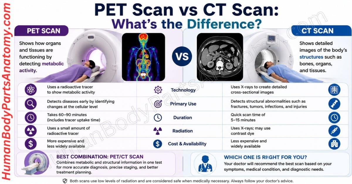
Medical imaging is an important part of modern healthcare, especially when diagnosing, staging, and monitoring cancer. Two of the most commonly used imaging tests are PET (Positron Emission Tomography) scans and CT (Computed Tomography) scans. Although these tests are often performed together, they serve different purposes. A CT scan creates detailed images of the body’s internal structures, while a PET scan shows how tissues and organs are functioning by detecting changes in metabolic activity. Together, they provide doctors with a more complete understanding of a patient’s health.
Why It’s Important to Know the Difference
Understanding the difference between a PET scan and a CT scan can help patients better understand why a specific imaging test is recommended. Each scan provides unique information that supports accurate diagnosis, cancer staging, treatment planning, and follow-up care.[1]
In many cases, healthcare providers combine both scans because the detailed anatomical images from a CT scan and the functional information from a PET scan work together to improve diagnostic accuracy.
Knowing how these imaging tests differ can help patients feel more prepared, reduce uncertainty, and make informed decisions about their healthcare. Because each test offers different insights, learning the basics of pet vs ct scan differences can also help you prepare better questions for your care team.
| Description | CT scan | PET scan |
|---|---|---|
| What it shows | Anatomy and structure [1] | Metabolic and cellular function [3] |
| How it works | Rotating X-ray source and detectors [5] | Radioactive tracer emitting gamma rays [4] |
| Typical duration | A few minutes to about half an hour [1] | About two hours total; ~30 min scanning [2] |
| Main strength | Spatial detail, speed, availability [1] | Detecting functional change early [2] |
| Common use | Trauma, tumors, bleeding, fractures [1] | Cancer staging, treatment response, viability [4] |
What Is a PET Scan?
A PET (Positron Emission Tomography) scan is a nuclear imaging test that shows how well your tissues and organs are working.[2]
Unlike imaging tests that mainly display the body’s structure, a PET scan reveals metabolic activity and body function — information not available through procedures that show anatomy alone.[3] This functional focus is what sets a PET scan apart in the broader pet scan vs ct scan comparison.
During the scan, healthcare providers give the patient a small amount of a radioactive tracer. They usually inject it into a vein, but depending on the area being examined, they may instead ask the patient to swallow or inhale the tracer.[4]
The tracer settles in areas of the body with high levels of chemical activity, which can be a sign of cancer or other diseases. Because chemical changes can appear before symptoms develop, a PET scan can find signs of disease at an early stage, often before the disease is visible on other imaging tests.[2]
Healthcare providers commonly use PET scans to diagnose diseases, determine how far a condition has progressed, monitor treatment response, and detect whether a disease has returned.[2]
An important clarification: a PET scan is a diagnostic and staging tool, not a routine cancer screening test. Healthcare providers use PET scans to evaluate known or suspected medical conditions. Although FDG PET can detect certain diseases, healthcare providers recommend established screening tests, such as colonoscopy, when routine screening is appropriate because FDG PET is not a standard screening tool.[4]
How Does a PET Scan Work?
A PET scan begins with administration of a small amount of radioactive tracer, most often into a vein. The tracer travels through the bloodstream and is absorbed by tissues according to their specific affinity.[4]
The tracer emits positrons, which interact with neighbouring electrons to produce gamma rays. A ring of detectors inside the scanner picks up these gamma rays, and a computer processes the data to generate a three-dimensional image of the tracer’s distribution throughout the body. Areas of higher metabolic activity exhibit increased tracer uptake and appear as brighter spots on the images.[4]
However, increased tracer uptake is not always cancer. Inflammation, infection, recent treatment, and normal physiological uptake in certain tissues can all produce similar findings.[12]
A radiologist, a doctor who specializes in diagnosing conditions using medical imaging, reviews your results and interprets them alongside your medical history and other test results.[2]
Unlike CT or MRI, which show anatomy or body form, PET shows metabolic activity or body function.[3] This is why the two types of imaging answer different clinical questions.
Common Uses of PET Scans
Healthcare providers widely use PET scans to diagnose, stage, and monitor many medical conditions. They often combine PET scans with CT scans (PET/CT) to capture both functional and structural information in a single examination.[1]
1. Cancer Detection and Treatment
Healthcare providers rely on PET imaging in oncology because it can detect metastatic tumors that other imaging techniques may not reveal. PET also helps differentiate malignant from benign growths and accurately assess the spread of malignant tumors.[3] Specifically, PET helps doctors:
- Identify whether a known or suspected abnormality is likely to be cancerous
- Determine whether cancer has spread to other parts of the body (staging)
- Monitor how well treatments such as chemotherapy, radiation therapy, or immunotherapy are working
- Investigate suspected cancer recurrence after treatment.[4]
For many oncologists, this is a key part of the ct scan vs pet scan for cancer decision when planning a patient’s care.
2. Brain and Neurological Disorders
PET scans can evaluate brain activity and help investigate conditions including stroke, epilepsy, Alzheimer’s disease, and Parkinson’s disease. PET is also used to detect recurrent brain tumours.[3]
In dementia evaluation specifically, different PET tracers answer different questions:
- FDG PET measures how brain regions use energy
- Amyloid PET measures abnormal deposits of beta-amyloid — higher levels of which are consistent with amyloid plaques, a hallmark of Alzheimer’s disease.[6]
- In epilepsy, seizure foci typically appear hypometabolic on an interictal FDG PET scan.[4]
3. Heart Disease
PET scans are also used to evaluate heart health. They help doctors measure blood flow to the heart muscle, identify areas of poor circulation caused by coronary artery disease, and assess whether heart muscle that is not contracting well is still viable rather than scarred.[7]
PET imaging combining myocardial perfusion and FDG is considered the gold standard for evaluating myocardial viability.[7]
However, precision matters here: while FDG PET distinguishes hibernating (viable) myocardium from scar in order to identify patients who might benefit from revascularization, several clinical trials — including a substudy of the STICH trial, in which viability assessment did not identify patients with a differential survival benefit from bypass surgery versus medical therapy alone — have raised questions about this concept.[13]
Decisions about bypass surgery or angioplasty are made on the basis of the whole clinical picture, not a PET result alone.
What Is a CT Scan?
A Computed Tomography (CT) scan is a noninvasive imaging procedure that uses special X-ray equipment to create detailed pictures of areas inside the body.[1]
A CT scan uses a computerized X-ray system that directs a narrow beam of X-rays through the body while rotating rapidly around the patient. A computer then processes the signals into detailed cross-sectional images, or “slices.”[5]
This structural emphasis is a core part of the ct vs pet scan comparison that doctors consider before ordering either test.
Each picture shows the organs, bones, and other tissues in a thin slice of the body. The full series is like a sliced loaf of bread: you can look at each slice individually as a two-dimensional picture, or view the whole loaf as a three-dimensional image.
Modern machines take continuous pictures in a helical (spiral) fashion, which is faster and produces better-quality 3-D images that may improve detection of small abnormalities.[1]
Healthcare providers widely use CT scans to diagnose circulatory system diseases, including coronary artery disease, aneurysms, and blood clots. They also use CT scans to detect spinal conditions, kidneys and bladder stones, abscesses, inflammatory diseases, and injuries affecting the head, skeletal system, and internal organs.[1]
How Does a CT Scan Work?
Unlike a conventional X-ray, which uses a fixed X-ray tube, a CT scanner uses a motorised X-ray source that rotates around the circular opening of a doughnut-shaped structure called a gantry.
The patient lies on a bed that moves slowly through the gantry while the tube rotates, and digital detectors positioned directly opposite the source capture the X-rays as they leave the body.
Each time the source completes one full rotation, the computer uses mathematical reconstruction techniques to build a 2-D image slice, with tissue thickness per slice usually ranging from 1 to 10 millimetres. Successive slices can then be digitally stacked to form a three-dimensional image.[5]
During the procedure, the technologist may ask you to hold your breath briefly to prevent motion blur and produce clearer images. In some cases, they administer a contrast agent beforehand to highlight specific structures and improve image quality.
Healthcare providers may give the contrast agent by mouth, inject it into a vein, or administer it as an enema. Iodine- and barium-based contrast agents are the two most commonly used for CT scans.[1]
Healthcare providers widely use CT scans because they are fast, painless, and highly detailed. They rely on CT imaging to diagnose medical conditions, evaluate injuries, plan treatments, and monitor a patient’s response to therapy.[1]
Common Uses of CT Scans in Medical Imaging
Healthcare providers commonly use computed tomography (CT) scans because they produce detailed cross-sectional images of the body’s organs, bones, blood vessels, and soft tissues. CT has many uses in the diagnosis, treatment, and monitoring of cancer.[1]
Recognizing these applications is an important piece of any ct scan vs pet scan comparison for patients trying to understand their imaging options.
1. Tumor Detection and Evaluation
Healthcare providers use CT scans to detect tumors and evaluate their size, shape, and location. This information helps them determine whether the patient needs additional testing or treatment.[1]
2. Emergency and Trauma Assessment
Healthcare providers widely use CT scans to detect trauma, brain bleeds, and injuries to the head, skeletal system, and internal organs, helping them make fast and informed treatment decisions.[1]
3. Detecting Infections and Internal Injuries
Doctors use CT to identify abscesses and inflammatory diseases, such as ulcerative colitis and sinusitis, that may not be apparent during a physical examination.[1] The detailed images help diagnose the underlying cause of symptoms and guide appropriate treatment.
4. Cancer Diagnosis and Treatment Monitoring
CT helps provide information about the stage of a cancer, determine whether a cancer is responding to treatment, and detect recurrence of a tumour.[1]
5. Guiding Medical Procedures
CT determines exactly where to perform a biopsy and guides certain local treatments, including cryotherapy, radiofrequency ablation, and the implantation of radioactive seeds for brachytherapy. It also helps plan external-beam radiation therapy or surgery.[1]
6. Monitoring Chronic Conditions
CT is used to monitor ongoing conditions affecting the lungs, heart, abdomen, blood vessels, and other organs, helping doctors track changes and adjust treatment when necessary.[1]
Key Differences Between PET and CT Scans
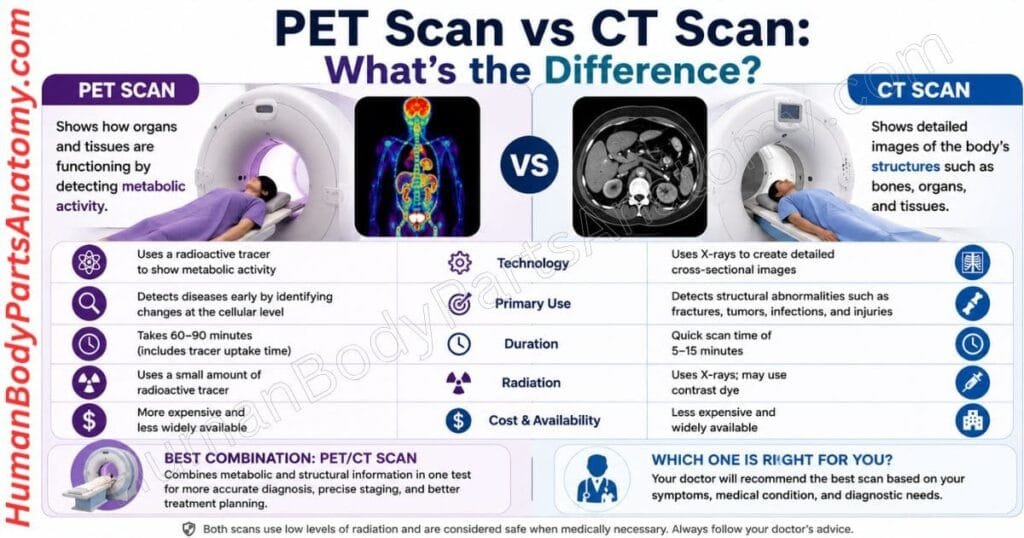
PET and CT scans are both advanced imaging tests, but they serve different purposes. A CT scan creates detailed pictures of the body’s structures.[1] A PET scan shows how organs and tissues are functioning by measuring metabolic activity.[3] Understanding the difference between PET vs CT scan can help patients know why a doctor recommends one test over the other.
1. PET Scan vs CT Scan: Structural Imaging vs Functional Imaging
The biggest difference is the type of information each provides.
- CT produces detailed images of the body’s anatomy, making it well suited to identifying fractures, internal bleeding, infections, tumours, and organ abnormalities.[1]
- PET detects changes in cellular activity using a radioactive tracer. Because chemical changes can show up before symptoms appear, PET can find signs of disease at an early stage, often before it is visible on other imaging tests.[2]
Because they provide different types of information, doctors often combine both tests in a PET/CT scan. During a PET/CT scan, the CT scan is performed first to create detailed anatomical images of the body’s organs and structures. The PET scan then captures functional information by showing the metabolic activity within tissues and cells.[1]
2. Contrast Agents and Radiation Exposure
Both scans involve ionizing radiation, but they use it differently. A CT scan may use an iodine-based contrast dye to highlight blood vessels, organs, and tissues.[1] A PET scan uses a small amount of radioactive tracer that collects in areas of increased metabolic activity.[2]
To put the doses in context: the average person in the United States receives an effective dose of about 3 millisieverts (mSv) per year from natural sources; a typical chest CT is about 6.1 mSv, comparable to two years of natural background radiation, and a low-dose chest CT is about 1.5 mSv.[1]
The PET component alone is approximately 7.5 mSv, while a combined FDG PET/CT typically ranges from about 14 to 30 mSv depending on the CT protocol.[4]
Although exposure to ionizing radiation can increase cancer risk, a single CT scan adds only a small amount of risk. Healthcare providers recommend CT scans when the benefits of diagnosing or evaluating a serious medical condition outweigh the potential radiation risks.[1]
The FDA advises that a CT scan should always be performed if it is medically necessary and other exams using no or less radiation are unsuitable.[8] Children are more sensitive to radiation than adults because their bodies are growing and their cells divide more rapidly, so special considerations apply in paediatric imaging.[1]
Radiation in a PET scan is safe for most adults but can be harmful to a developing baby, so tell your provider if you are or may be pregnant, or if you are breastfeeding.[2]
3. Diagnostic Applications
Healthcare providers commonly use CT scans to diagnose bone fractures and skeletal injuries, internal injuries and bleeding, lung diseases, kidney and bladder stones, tumors, abdominal and inflammatory conditions, and stroke or traumatic brain injuries.[1]
Healthcare providers primarily use PET scans to evaluate how organs and tissues function. They commonly recommend PET scans to stage cancer and assess the extent of disease, monitor treatment response, investigate suspected cancer recurrence,[4] evaluate myocardial perfusion and heart muscle viability,[7] and diagnose certain neurological disorders, including Alzheimer’s disease and other forms of dementia.[2][6]
This split in applications is one of the clearest illustrations of the ct vs pet scan divide in everyday clinical practice.
4. Imaging Technology
CT uses multiple X-ray measurements taken from different angles, which a computer reconstructs into cross-sectional and 3-D views.[5] PET detects gamma rays produced when positrons emitted by the tracer interact with electrons in the body, and reconstructs these signals into a map of metabolic activity.[4]
5. Image Quality and Detail
CT provides excellent spatial resolution and is especially effective for viewing bones, lungs, blood vessels, and internal organs, helping doctors identify the exact size, shape, and location of structural abnormalities.[1]
PET does not produce the same level of anatomical detail, and its lower spatial resolution can limit detection of small lesions — a recognised limitation in, for example, identifying small periesophageal nodes.[4]
Instead, PET reveals areas of abnormal metabolic activity. Combining the two provides both structural and functional information in a single examination and may give a more complete picture of a tumour’s location, growth, or spread than either test alone.[1]
6. Functional vs. Structural Imaging
One of the easiest ways to understand PET scan vs CT scan is by comparing what each measures. CT shows the body’s physical structure — bones, organs, and soft tissues.[1] PET shows how cells and organs are functioning by measuring metabolic activity.[3] This functional imaging allows PET to detect certain disease processes before structural changes become apparent.[2]
7. Procedure Time and Patient Experience
A CT scan is one of the fastest imaging tests available. The length depends on the size of the area being scanned, but it usually lasts only a few minutes to half an hour.[1] Patients lie still while the scanner captures images.
A PET scan takes longer because the tracer needs time to be absorbed. Patients typically wait 45 to 60 minutes after the injection, then remain very still for about 30 minutes of scanning. The full appointment takes about two hours including absorption time.[2]
Both procedures are noninvasive and do not cause pain, though lying in one position may be slightly uncomfortable. Most CT scanners surround only portions of the body rather than enclosing the patient, so claustrophobia is less common than many people expect.[1]
8. Sensitivity and Diagnostic Accuracy
Each test has distinct strengths, and neither is universally superior.
CT is highly accurate for structural abnormalities such as fractures, internal bleeding, organ injuries, tumours, and lung disease.[1]
PET is more sensitive to metabolic change. In non-small cell lung cancer, for example, FDG PET demonstrated higher sensitivity (71% versus 43%) and overall accuracy (76% versus 68%) than CT for detecting N2 mediastinal lymph nodes, allowing more precise staging and better patient selection.[4]
That advantage is not uniform across all cancers, however. For locoregional lymph node assessment in cancers of the oesophagus and gastroesophageal junction, FDG PET showed lower accuracy than the combination of CT and endoscopic ultrasound (48% versus 69%), largely due to reduced sensitivity and spatial resolution for small nodes.[4] The most appropriate test depends on the cancer type, the clinical question, and the stage of disease.
For many cancer patients the combined PET/CT is valuable precisely because it shows anatomy and metabolic activity simultaneously, which may improve the ability to diagnose cancer, determine how far a tumour has spread, plan treatment, and monitor response — and may reduce the number of additional imaging tests needed.[1]
This trade-off is frequently at the center of the ct scan vs pet scan for cancer decision that oncologists weigh for each patient.
9. Role in Medical Procedures
CT plays a direct role in guiding procedures: determining exactly where to perform a biopsy, guiding local treatments such as cryotherapy and radiofrequency ablation, and helping plan external-beam radiation therapy or surgery.[1]
Healthcare providers primarily use PET scans to guide treatment planning by determining the extent of disease and monitoring how well treatment works over time. A positive FDG PET finding generally indicates the need for a tissue biopsy at the corresponding location to confirm the diagnosis.[4]
10. Cost and Availability
CT scans are widely available in hospitals and imaging centers across the United States. For most people, CT is performed on an outpatient basis without an overnight stay.[1]
They are generally less expensive and are often used as the first imaging test because they are fast, accurate, and accessible.
PET requires specialised equipment, radioactive tracers, and trained personnel. As a result it is more expensive and may only be available at larger hospitals or dedicated imaging centers. Doctors usually recommend PET when detailed information about tissue function or cancer activity is needed.[4]
How CT and PET Scans Help Diagnose Cancer
Both scans play an important role in cancer diagnosis but provide different information. CT shows detailed images of organs and tissues[1]; PET shows how cells and tissues are functioning.[2]
Doctors often use both together to diagnose cancer, determine its stage, and create the most effective treatment plan.[1] This shared role in oncology is why the pet scan vs ct scan comparison comes up so often when discussing cancer care.
How a CT Scan Helps Detect Cancer
A CT scan helps doctors diagnose the presence of a tumour, provide information about its stage, determine exactly where to perform a biopsy, help plan radiation therapy or surgery, determine whether a cancer is responding to treatment, and detect recurrence.[1]
How a PET Scan Helps Detect Cancer
A PET scan helps doctors identify areas of increased metabolic activity that may represent active cancer, determine whether cancer has spread, measure how well treatment is working, and investigate suspected recurrence.[4]
Because cancerous tumours usually metabolise glucose more rapidly than normal tissues, they take up more FDG and appear different from other tissues on a PET scan.[1]
PET can also help distinguish active tumour from post-treatment changes such as fibrosis, which can closely resemble recurrence on anatomical imaging alone.[11]
Because PET scans show how tissues function, they help doctors interpret PET scan normal vs cancer findings by highlighting areas with abnormal cellular activity. A bright area is a clue requiring interpretation in context — not a diagnosis in itself.[12]
Benefits of CT and PET Scans for Cancer Detection and Staging
Healthcare providers often use both tests together because each provides unique information. As a result, the answer to the “CT scan vs PET scan for cancer” question depends on the type of cancer, its stage, and the specific clinical situation.
Benefits of a CT Scan
- Produces detailed images of internal organs and tissues.[1]
- Fast and widely available at most hospitals and imaging centers.[1]
- Helps detect tumors and evaluate their size and location.[1]
- Assists in planning surgery, radiation therapy, and biopsies.[1]
- Useful for monitoring changes during follow-up care.[1]
Benefits of a PET Scan
- Detects metabolically active disease throughout the body, including metastatic tumours that other imaging techniques might miss.[3]
- Helps identify spread beyond the original site and evaluate treatment response.[4]
- Can identify recurrence in patients with suspicious findings on conventional imaging.[11]
- Is often combined with CT for more accurate diagnosis and staging.[1]
A note on surveillance: while PET can detect recurrence, routine PET surveillance in patients without symptoms is not automatically beneficial. Researchers analyzed SEER–Medicare data from more than 97,000 patients with lung cancer and 4,400 patients with esophageal cancer. They found that greater use of PET scans to detect cancer recurrence did not improve two-year survival, suggesting that healthcare providers may overuse PET for this specific purpose.[14]
Your oncology team will individualize your follow-up imaging schedule based on your cancer type, treatment, and overall clinical condition.
Risks and Limitations of CT and PET Scans
Although both scans are valuable diagnostic tools, they also have certain limitations.
CT Scan Limitations
- Cannot determine whether a tumour is cancerous without additional testing, such as a biopsy [1]
- Uses ionizing X-ray radiation, although doses are optimised and controlled [1]
- Contrast agents can rarely cause allergic reactions, including mild itching or hives and, in rare cases, more serious symptoms such as shortness of breath or swelling. They can also very rarely affect kidneys function in people with existing kidney disease, so healthcare providers may perform a simple blood test beforehand to check kidneys function.[1]
- May reveal incidental findings that lead to further, possibly invasive, follow-up testing [9]
PET Scan Limitations
- Provides lower spatial resolution and less anatomical detail than CT [4]
- Usually costs more than a standard CT scan and is less widely available
- Involves ionizing radiation, with a combined PET/CT dose typically higher than CT alone [4]
- Some cancers do not show up on a PET scan [2]. Tumours with low FDG affinity include renal cell carcinoma, hepatocellular carcinoma, mucinous tumours of the gastrointestinal tract, certain carcinoid tumours, and low-grade lymphomas [12]
- False-positive results can occur due to inflammation, infection, recent surgery or treatment, and normal physiological uptake [12]
- Accuracy can be affected by high blood glucose, recent strenuous exercise, and caffeine, alcohol, or tobacco use within 24 hours [4]
Weighing these drawbacks against the benefits is part of the larger ct scan vs pet scan comparison patients should discuss with their doctor.
When Do Doctors Recommend a CT Scan for Cancer?
Healthcare providers often use CT scans as one of the first imaging tests when they suspect cancer because CT creates detailed cross-sectional images that help them detect tumors and assess how far the disease has progressed.
Doctors may recommend a CT scan to investigate possible signs of cancer; detect and measure tumours; provide information about the stage of a cancer; determine exactly where to perform a biopsy; help plan external-beam radiation therapy or surgery; determine whether a cancer is responding to treatment; and detect recurrence of a tumour.[1]
Healthcare providers also use CT scans in two established screening settings. They use CT colonography to screen for large colorectal polyps and tumors, and they use low-dose CT to screen people at high risk for lung cancer.
The U.S. Preventive Services Task Force recommends annual lung cancer screening with low-dose CT for people aged 50 to 80 with a 20 pack-year or greater smoking history who currently smoke or quit within the past 15 years.
Notably, whole-body CT has not been shown to be an effective screening method for healthy people, and most doctors recommend against it for those without signs or symptoms of disease.[1] This is often where the pet vs ct scan conversation begins during a cancer workup.
When Do Doctors Recommend a PET Scan for Cancer?
A PET scan helps doctors see how metabolically active cells are inside the body. Because cancerous tumours usually metabolise glucose more rapidly than normal tissues, they take up more FDG and appear different from surrounding tissue [1] — an effect related to the Warburg phenomenon, in which cancer cells show increased glucose uptake and glycolysis even when oxygen is adequate.[4]
Doctors may recommend a PET scan to:
- Determine whether a suspicious lump or mass is cancerous, especially when CT or MRI results are unclear.[3]
- Stage cancer accurately by identifying whether it has spread to nearby lymph nodes or distant organs.[4][11]
- Evaluate how aggressive a tumor is, as highly active tumors usually have increased metabolic activity.[4][11]
- Monitor treatment response by showing whether cancer cells are becoming less active after chemotherapy, radiation therapy, immunotherapy, or targeted therapy.[4][11]
- Detect cancer recurrence early by identifying active cancer cells before structural changes become visible on other imaging tests.[4][11]
Beyond FDG, other PET agents provide information about tissue oxygen levels, new blood vessel formation, bone growth, and whether tumour cells are actively dividing. For example, PSMA-targeted imaging drugs are used for PET/CT imaging of PSMA-positive lesions in men with suspected prostate cancer metastases.[1]
Healthcare providers often combine PET and CT into a single PET/CT examination to obtain both detailed anatomical images and metabolic information, improving diagnostic accuracy and disease staging.[1] This is typically the stage where the ct vs pet scan comparison becomes most relevant for staging and monitoring decisions.
How to Prepare for CT and PET Scans
Preparing for a PET Scan
Healthcare providers may ask you to fast (avoid eating or drinking) for four to six hours before the test. They may also advise you to avoid tobacco products and foods or drinks containing caffeine or alcohol for 24 hours before the scan.[2] In many cases, they also recommend following a low-carbohydrate, sugar-free diet during the 24 hours leading up to the scan.[4]
If you have diabetes and use insulin, you may need to change the timing of your regular dose, because people with diabetes may not absorb the sugar in the tracer, which can affect results. Your provider will give you specific instructions — do not stop or adjust any medicine unless told to do so [2].
Avoid strenuous exercise before your appointment because it can significantly increase radiotracer uptake in your tissues and affect the accuracy of the scan.[4]
Tell your healthcare provider if you have claustrophobia so they can arrange medication to help you relax. You should also inform them if you are pregnant, think you may be pregnant, or are breastfeeding before undergoing the scan.[2]
Preparing for a CT Scan
Your healthcare provider will give you preparation instructions based on the part of your body being examined. If your CT scan requires contrast, they may ask you not to eat or drink for four to six hours before the test.[10]
Tell your provider if you have allergies — particularly to iodine or contrast dye — if you have kidney disease or diabetes, or if there is any possibility you are pregnant. Depending on the area being scanned, your provider may reduce the radiation dose or use an alternative imaging method.[1]
What to Expect During the Scan
Both scans are noninvasive and painless, though lying in one position may be slightly uncomfortable. You must remain still to avoid blurred images, and during a CT you may be asked to hold your breath briefly.[1]
PET scans use only a small amount of radioactive tracer. Your body eliminates most of the tracer through urine and stool within 2 to 10 hours, and drinking plenty of water after the scan helps flush out the remaining tracer more quickly. Allergic reactions to the tracer are rare and usually mild.[2]
Final Thoughts
CT scans and PET scans serve different purposes, and the right choice depends on your symptoms, medical history, and your doctor’s recommendation. A CT scan is excellent for viewing detailed body structures, while a PET scan helps evaluate how organs and tissues are functioning at the cellular level.
Healthcare providers choose a PET scan, a CT scan, or both based on whether they need structural detail, functional information, or a combination of both to guide your care. In many cases, combining PET and CT gives them the most complete picture for diagnosis and treatment planning.[1]
If you have been advised to have an imaging test, three useful questions to ask your provider are: Why is the test needed? Will the results change the treatment decisions? Is there an alternative test that doesn’t involve radiation? [1]
FAQ’s-
Neither scan is better in every situation. A CT scan shows the size, shape, and location of a tumor, while a PET scan detects active cancer cells. Doctors often recommend a PET-CT scan because it combines both tests for a more complete evaluation.
It depends on the purpose. A PET scan is better for detecting active cancer and checking if it has spread, while a CT scan is better for showing the exact location and structure of a tumor.
In many cases, yes. Using both scans together helps doctors diagnose cancer more accurately, determine its stage, and plan the most effective treatment.
Yes. Some cancers may not cause clear structural changes on a CT scan but can be detected on a PET scan because it identifies areas of increased metabolic activity.
A PET scan, especially a PET-CT scan, is commonly used to detect whether cancer has spread to other parts of the body and to help determine the cancer stage.
References –
- National Cancer Institute (NCI), National Institutes of Health. Computed Tomography (CT) Scans and Cancer Fact Sheet. Reviewed February 8, 2024. https://www.cancer.gov/about-cancer/diagnosis-staging/ct-scans-fact-sheet
- MedlinePlus, National Library of Medicine, National Institutes of Health. PET Scan. Last updated September 10, 2024. https://medlineplus.gov/lab-tests/pet-scan/
- NIH Clinical Center, National Institutes of Health. Positron Emission Tomography (PET): About PET.https://www.cc.nih.gov/pet/about
- Kapoor M, Heston TF, Kasi A. PET Scanning. In: StatPearls [Internet]. NCBI Bookshelf, National Library of Medicine, National Institutes of Health. Last updated February 26, 2025. PMID: 32644515. https://www.ncbi.nlm.nih.gov/books/NBK559089/
- National Institute of Biomedical Imaging and Bioengineering (NIBIB), National Institutes of Health. Computed Tomography (CT) Fact Sheet.https://www.nibib.nih.gov/sites/default/files/Computed_Tomography_Fact_Sheet.pdf
- National Institute on Aging (NIA), National Institutes of Health. How Biomarkers Help Diagnose Dementia. Updated January 23, 2026. https://www.nia.nih.gov/health/alzheimers-symptoms-and-diagnosis/how-biomarkers-help-diagnose-dementia
- Ahmed I, Devulapally P. Nuclear Medicine PET Scan Cardiovascular Assessment, Protocols, and Interpretation. In: StatPearls [Internet]. NCBI Bookshelf, National Library of Medicine, National Institutes of Health. Last updated July 30, 2023. PMID: 34033393. https://www.ncbi.nlm.nih.gov/books/NBK570631/
- U.S. Food and Drug Administration (FDA). Computed Tomography (CT).https://www.fda.gov/radiation-emitting-products/medical-x-ray-imaging/computed-tomography-ct
- U.S. Food and Drug Administration (FDA). What Are the Radiation Risks from CT?https://www.fda.gov/radiation-emitting-products/medical-x-ray-imaging/what-are-radiation-risks-ct
- MedlinePlus, National Library of Medicine, National Institutes of Health. Abdominal CT Scan. Medical Encyclopedia. https://medlineplus.gov/ency/article/003789.htm
- Volpi S, Ali JM, Tasker A, Peryt A, Aresu G, Coonar AS. The role of positron emission tomography in the diagnosis, staging and response assessment of non-small cell lung cancer. Annals of Translational Medicine. 2018;6(5):95. doi:10.21037/atm.2018.01.25. PMID: 29666818. PMCID: PMC5890043.
- Long NM, Smith CS. Causes and imaging features of false positives and false negatives on 18F-PET/CT in oncologic imaging. Insights into Imaging. 2011;2(6):679–698. doi:10.1007/s13244-010-0062-3. PMID: 22347986. PMCID: PMC3259390.
- Chen K, Miller EJ, Sadeghi MM. PET-based imaging of ischemic heart disease. PET Clinics. 2019;14(2):211–221. doi:10.1016/j.cpet.2018.12.003. PMID: 30826019. PMCID: PMC6426655.
- Healy MA, Yin H, Reddy RM, Wong SL. Use of positron emission tomography to detect recurrence and associations with survival in patients with lung and esophageal cancers. JNCI: Journal of the National Cancer Institute. 2016;108(7):djv429. doi:10.1093/jnci/djv429. PMID: 26903519. PMCID: PMC4948569.
Read More-
Human Body-
- Human Anatomy: Guide to Bones, Muscles, Organs, Systems, Functions & Diagram
- Human Skeleton Anatomy: All 206 Bones Explained with Functions & Diagrams
- Human Muscle Anatomy: Validated Guide to Every Major Muscles & Functions
Head, Face & Senses-
- Nose Anatomy: Parts of the Nose, Structure, Nasal Cavity & Sinuses Explained
- Skull Anatomy: Parts of the Skull, Structure, Cranial, Facial Bones & Functions
- Mouth Anatomy: Guide on Parts of Mouth, Lips, Palate, Gums & Oral Cavity
- Eye Anatomy: Parts of the Eye, Cornea, Lens, Retina, Optic Nerve & Diagram
- Ear Anatomy: Parts of the Ear, Outer, Middle & Inner Ear & Structures
Brain & Nervous System-
- Brain Anatomy: Parts of the Brain, Structure, Functions & Regions Explained
- Brainstem Anatomy: Complete Guide with Midbrain, Pons & Medulla Explained
- Midbrain Anatomy: Complete Guide with Parts, Structure & Key Functions
- Cerebrum Anatomy: Guide with Parts, Lobes, Hemispheres, Functions & Diagram
- Cerebellum Anatomy: Guide with Parts, Structure, Functions & Diagram
- Basal Ganglia Anatomy: Guide with Parts, Structure & Role in Movement
- The 4 Lobes of the Brain: Complete Guide with Locations & Functions
Spine & Back-
- Cervical Spine Anatomy: C1–C7 Vertebrae, Muscles & Nerves Explained
- Spine Anatomy: Parts of the Spine, Vertebrae, Curves, Spinal Cord & Diagram
- Neck Muscle Anatomy: Guide with Key Muscles, Groups, Functions & Diagrams
- Rib Cage Anatomy: Ribs, Sternum, Thoracic, Vertebrae & Functions Explained
Organs-
- Pancreas Anatomy: Parts of Pancreas, Structure, Location, Functions & Role
- Thyroid Anatomy: Guide on Key Parts, Location, Structure & Functions
- Stomach Anatomy: Parts of Stomach, Regions, Layers & Digestive Function
- Heart Anatomy: Guide on Parts of Heart, Chambers, Valves & Blood Flow
- Liver Anatomy: Key Parts of Liver, Functions, Lobes, Segments & Diagram
- Kidney Anatomy: Guide on Parts of Kidney, Structure, Functions & Diagram
Upper Limb-
- Forearm Anatomy: Parts of the Forearm, Radius, Ulna, Muscles & Diagram
- Shoulder Anatomy: Parts of the Shoulder, Bones, Joint Structure & Diagram
- Wrist Anatomy: Parts of the Wrist, 8 Carpal Bones, Tendons & Diagram
- Hand Anatomy: Parts of the Hand, Bones, Muscles with Functions & Diagram
- Arm Anatomy: Parts of Arm, Bones, Muscles & Joints with Functions & Diagram
Lower Limb-
- Leg Anatomy: Parts of the Leg, Bones, Muscles & Lower Leg with Functions
- Knee Anatomy: Parts of Knee, Bones, Ligaments, Cartilage & Joint Structure
- Thigh Muscle Anatomy: Key Muscle Groups, Names, Functions & Diagram
Medical Disclaimer
All content on HumanBodyPartsAnatomy.com is educational and based on verified, peer-reviewed medical sources. Articles are authored or reviewed by qualified medical or biomedical professionals to ensure accuracy.
This website does not provide medical advice, diagnosis, or treatment. Always consult a licensed healthcare professional for personal medical guidance.
No commercial or promotional interests influence the medical content published on this site.