
📅 Published on August 22, 2026 | 🕒 Last Reviewed & Updated on October 5, 2026
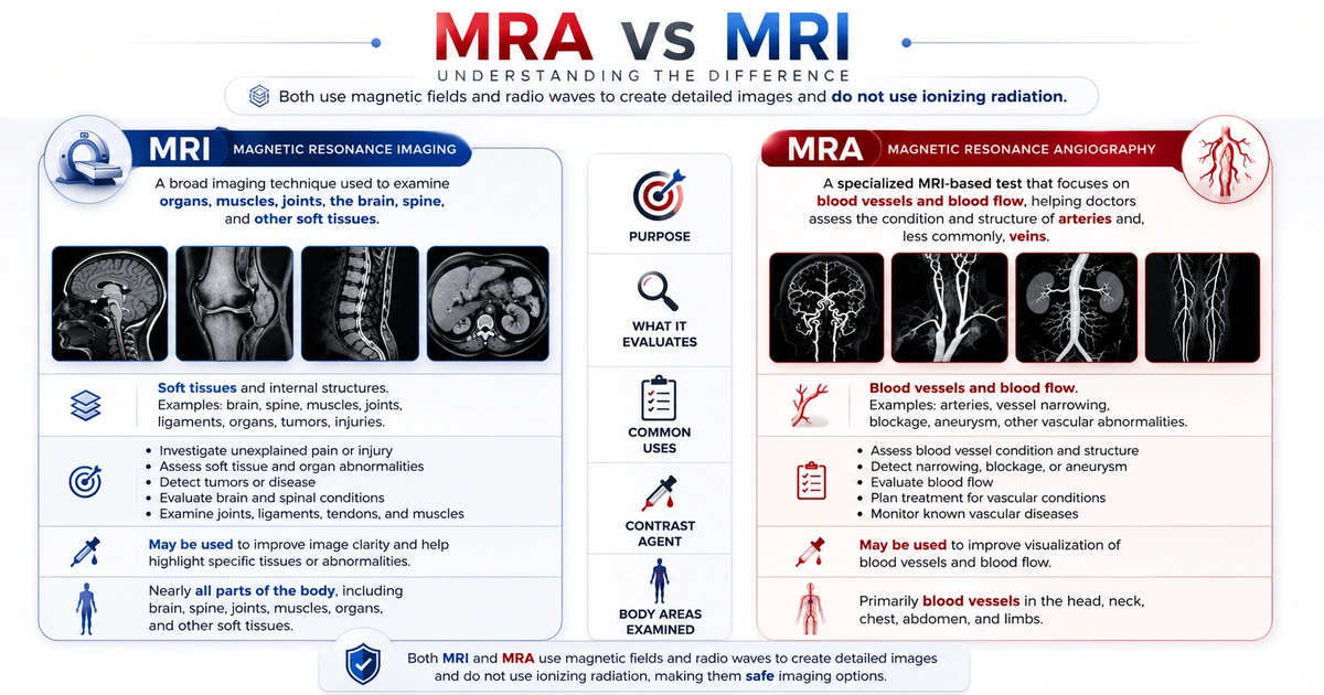
The key difference between MRI and MRA is the part of the body each test is intended to evaluate. Magnetic resonance imaging (MRI) is a broad imaging technique used to examine organs, muscles, joints, the brain, spine, and other soft tissues.[1] Magnetic resonance angiography (MRA) is a specialized MRI-based test that focuses on blood vessels and blood flow. It helps doctors evaluate the structure and condition of arteries and, less commonly, veins. When doctors need to examine veins specifically, they generally use a related technique called magnetic resonance venography (MRV).[1][2]
Both tests use magnetic fields and radio waves to create detailed images and do not use ionizing radiation.[1][2] Your doctor will recommend the test based on your symptoms, medical history, and the area they need to examine.
Overview of MRA vs MRI
As we know, MRI and MRA have different diagnostic goals, but they are closely related technologies. Understanding MRA vs MRI can help explain why a provider may choose one scan over the other. MRA is derived from MRI technology and uses the same basic principles to create detailed images of internal structures.[2] This makes both examinations useful when doctors need clear, high-resolution information without exposing the patient to X-ray radiation.[1][2]
Healthcare providers may recommend these two tests for different clinical questions. For example, a healthcare provider may choose an MRI to investigate unexplained pain, an injury, changes in an organ, or soft-tissue abnormalities.[1] An MRA may be appropriate when a healthcare provider needs to evaluate the condition or structure of a blood vessel, such as possible narrowing, blockage, an aneurysm, or another vascular abnormality.[2]
The exact imaging approach can also vary depending on the body part being studied and the information the doctor needs. In some cases, additional techniques or a contrast agent may be used to improve image clarity.[1][2]
What Is an MRI?
A clear MRA vs MRI comparison starts with understanding what an MRI examines.
Magnetic resonance imaging (MRI) is a noninvasive diagnostic scan that uses magnets and radio waves to create highly detailed images of areas inside your body. It does not use X-ray radiation.[1] MRI can show soft tissues especially well, making it useful for examining the brain, spine joints, muscles, ligaments, tendons, organs, and other internal structures.[1][3]
Your doctor may order an MRI to investigate a wide range of health problems, including sports injuries, ligament or tendon damage, brain and spinal conditions, tumors, abnormal tissue changes, and certain diseases affecting internal organs.[1][8] Some MRI examinations use a contrast material to make specific tissues or blood vessels easier to distinguish. [1]
How Should You Prepare for an MRI?
Your preparation depends on the type of MRI you are having. The imaging center will provide instructions before your appointment. In general, you can prepare in the following ways:
Choose Clothing Without Metal
Wear loose, comfortable clothing that does not contain metal zippers, snaps, hooks, or decorative fasteners. You may need to change into a medical gown before the scan.[1]
Before entering the MRI room, remove jewelry, watches, piercings, hair accessories, belts, and other metal objects. Keep valuables at home whenever possible.[1]
Tell the MRI Team About Implants
Make sure your doctor and MRI technologist know if you have any implanted medical device, pacemaker, cochlear implant, surgical clip, metal fragment, artificial joint, or other object containing metal.[1]
Some implants are safe to scan under specific conditions, while others may not be MRI-compatible. Providing complete information about previous surgeries and implants helps the medical team determine the safest approach.[3]
Ask About Food, Drinks, and Medications
Many MRI scans do not require changes to your normal diet or medications. However, certain examinations may require you to fast or follow special eating and drinking instructions.[11]
Do not stop prescription medications unless your healthcare provider specifically tells you to. Carefully follow any instructions given by your imaging center.
Tell Your Doctor If You Have Claustrophobia
During an MRI, you typically lie on a table that moves into a large, tunnel-shaped scanner. You must remain still while the images are being captured.[1]
If you feel uncomfortable in enclosed spaces, tell your healthcare provider before the appointment. Depending on your situation, they may discuss ways to make the experience easier, such as relaxation techniques, an appropriate open MRI system, or medication to help you relax.[1]
What Happens During an MRI?
MRI scans are generally painless. The machine can produce loud tapping, knocking, or buzzing sounds that may damage your hearing. To protect your hearing, you will receive earplugs or headphones.[1][3]
The technologist will monitor you throughout the examination and can communicate with you during the scan. The length of the test varies depending on the body area being examined and the type of images needed.[1]
What Is an MRA?
Magnetic resonance angiography (MRA) is a noninvasive imaging test that produces detailed images of blood vessels and the way blood moves through them. It uses a strong magnetic field and radio waves rather than X-rays. Because of this, MRA can provide useful information about the circulatory system without the need for a catheter-based procedure.[2]
Doctors may recommend an MRA when they need to look for problems such as narrowed arteries, blocked or occluded blood vessels, aneurysms, abnormal blood vessel development, or changes related to a stroke. Depending on the reason for the exam, doctors can use MRA to examine blood vessels in the brain, neck, chest, abdomen, kidneys, pelvis, or legs.[2]
Some MRA scans use no contrast agent. In other cases, doctors inject a contrast agent into a vein to make certain blood vessels appear more clearly on the images.[2]
How Should You Prepare for an MRA?
MRA preparation depends on the type of scan and the part of the body being examined. In many cases, preparation is straightforward.
Follow MRI Safety Instructions
Wear comfortable clothing and avoid metal whenever possible. Before the scan, you may need to remove jewelry, watches, piercings, glasses, or other metal items.[2]
Tell your healthcare provider about any implants, medical devices, or metal fragments in your body. This is especially important if you have a pacemaker, surgical clips, cochlear implant, insulin pump, or a history of metal-related injuries. Your medical team will determine whether the device is safe for an MRI examination.[2][3]
Tell Your Doctor About Kidney Problems or Previous Reactions
Some MRA examinations use a gadolinium-based contrast agent to provide clearer images of blood vessels. Before receiving contrast, tell your doctor if you have kidney disease or have experienced a previous reaction to MRI contrast.[2]
Gadolinium can harm people with kidney failure and cause a rare condition called nephrogenic systemic fibrosis. For this reason, healthcare providers generally avoid contrast-enhanced MRA when kidney function is poor.[4][7] Because many MRA studies do not require contrast, your healthcare team may choose a non-contrast approach. They will determine whether you need contrast and whether you require any additional precautions.[7]
Check Whether You Need to Avoid Food or Caffeine
Many MRA exams do not require major changes to your normal routine. You may usually eat, drink, and take your medications as directed unless your healthcare provider tells you otherwise.[2]
Certain specialized exams may have different instructions. Caffeine restriction, for example, is not a requirement of MRA itself. It applies to a different heart study called vasodilator stress perfusion cardiac MRI, which uses medications such as adenosine or regadenoson to widen the coronary arteries. Because caffeine blocks the same receptors that medication acts on, standardized cardiac MR protocols advise avoiding caffeine and related substances for 12 to 24 hours before that test.[14] Always follow the instructions provided by your imaging center.
What Happens During an MRA?
For the examination, you will lie on a movable table that slides into the MRI scanner. It is important to stay as still as possible while the images are being captured. The machine makes loud tapping or knocking sounds, so earplugs or headphones are provided to protect your hearing.[2][3]
An MRA does not normally cause pain, although some people may feel uncomfortable in the enclosed scanner. The test time can vary depending on the area being examined and whether contrast is required.[2]
How are MRAs and MRIs similar?
MRA and MRI are closely related medical imaging techniques. Both use advanced magnetic technology to create detailed images of structures inside the body. Although an MRA focuses mainly on blood vessels, the two examinations have many similarities.
- Both use magnets and radio waves — MRI and MRA scanners use a strong magnetic field and radiofrequency waves to produce images. Unlike X-rays and CT scans, these tests do not use ionizing radiation.[1][2]
- Both are noninvasive procedures — You do not need surgery or an incision for either examination. You typically lie on a padded table that moves into the scanner. The scan itself is usually painless. The machine may produce loud clicking, tapping, or humming noises, so you will receive earplugs or headphones to protect your hearing.[1][2]
- Both can be performed with contrast —
In some cases, a healthcare provider may recommend a gadolinium-based contrast agent to help highlight certain tissues or blood vessels. The provider usually injects the contrast through a small IV. Your healthcare team will review your medical history and kidney function when determining whether contrast is appropriate.[1][2] - Both require you to remain still — Movement can make MRI and MRA images blurry. You may need to stay in the same position for the duration of the scan and, for certain exams, briefly hold your breath.[1][2]
- Both require MRI safety screening — Because the scanner contains a powerful magnet, medical staff will ask you about metal objects, implants, medical devices, or metal fragments in your body before the examination. This screening helps protect you and ensures that you can undergo the scan safely.[3]
The main difference is their purpose.
MRI scans a wide range of tissues and organs, while MRA focuses specifically on producing detailed images of blood vessels and blood flow.[1][2]
How Are MRAs and MRIs Different?
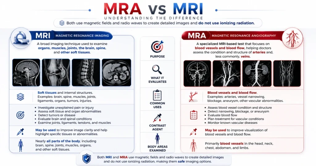
MRI and MRA are advanced imaging tests that use magnets and radio waves to create detailed images inside the body. Although both use similar technology, they serve different purposes. MRI evaluates organs, tissues, and other body structures, while MRA specifically examines blood vessels and blood flow.[1][2]
MRI Looks at Organs and Body Structures
An MRI (magnetic resonance imaging) provides detailed images of many parts of the body, including the brain, spine, joints, muscles, and internal organs. It is especially useful for viewing soft tissues that may not be clearly seen with other imaging methods.[3][11]
Doctors may recommend an MRI to investigate problems such as joint or ligament injuries, spinal disorders, brain tumors, inflammation, or abnormalities affecting internal organs. The exact area scanned depends on the symptoms and the medical condition being evaluated.[8][11]
MRA Examines Blood Vessels
An MRA (magnetic resonance angiography) is a specialized type of MRI that is used to visualize arteries and veins. It can help doctors assess the size, shape, and condition of blood vessels and, in some cases, how blood moves through them.[2][7]
An MRA may help doctors identify conditions such as aneurysms, narrowed arteries, blocked blood vessels, and other abnormalities that affect circulation. Depending on the reason for the test, doctors may examine blood vessels in the brain, neck, heart, kidneys, abdomen, or legs.[2][7]
What About Contrast?
Both MRI and MRA can use a contrast agent. Doctors sometimes inject contrast through an IV to make certain tissues or blood vessels appear more clearly in the images.[1][2]
For an MRA, contrast may help the radiologist see blood vessels and areas of abnormal blood flow more clearly. However, MRA does not always require contrast, and doctors can perform many MRI examinations without it. The decision depends on the body area the doctor examines, the suspected condition, and the information the doctor needs.[2]
MRA vs. MRI: What Each Test Is Used For
This MRA vs MRI comparison shows how each examination is selected for a different diagnostic purpose.
MRI and MRA are advanced imaging procedures that use magnets and radio waves to create detailed images of the body. Although both use similar technology, they serve different purposes. MRI primarily examines organs, muscles, joints, and other tissues, while MRA focuses on blood vessels and blood flow.[1][2]
When Is an MRI Recommended?
An MRI provides highly detailed images of soft tissues and is useful for investigating many different health conditions. Doctors may order an MRI to examine:
- Brain and spine: It can help detect tumors, multiple sclerosis, spinal cord abnormalities, herniated discs, and other conditions affecting the brain or spine.[8][11]
- Muscles, joints, and soft tissues: MRI can reveal ligament and tendon tears, cartilage injuries, muscle damage, and other problems involving joints such as the knee, shoulder, or hip.[3]
- Internal organs: MRI can provide detailed views of organs such as the liver, kidneys, heart, and pancreas and may help identify abnormalities or disease.[1][3]
- Tumors and other growths: MRI may also be used to locate a tumor, determine its size and position, or monitor changes during treatment.[1]
When Is an MRA Recommended?
An MRA, or magnetic resonance angiography, is a specialized MRI technique that focuses on blood vessels. It helps healthcare providers examine arteries and, in some situations, veins without the need for a conventional invasive angiogram. MRA may be used to check for:[2]
- Blocked or narrowed arteries: It can show areas where blood vessels have become narrowed or obstructed, reducing normal blood flow.[2]
- Brain and neck blood vessels: MRA can assess arteries supplying the brain and help doctors look for conditions such as aneurysms, narrowing, or other vessel abnormalities.[5]
- Aortic and other vascular problems: It can examine the aorta and major blood vessels for aneurysms, narrowing, or structural changes.[2]
- Blood flow problems in the legs or kidneys: MRA can help identify vascular disease affecting the legs, including peripheral artery disease, as well as problems involving the renal arteries.[6][7]
MRI vs. MRA Results
MRI and MRA use magnets and radio waves to create detailed images of the body without using radiation. Although they use similar technology, doctors use them for different purposes. MRI helps examine organs, tissues, muscles, joints, and other internal structures, while MRA focuses specifically on blood vessels and blood flow.[1][2]
Knowing what each test can reveal makes it easier to understand why your doctor may recommend one test instead of the other.
What Can an MRA Show?
A magnetic resonance angiogram (MRA) produces detailed images of blood vessels and helps doctors evaluate the movement of blood through them. Depending on the area being examined, doctors may use MRA to study arteries and veins in the brain, neck, chest, abdomen, pelvis, or limbs.[2]
An MRA may help detect:
- Narrowed arteries: Reduced vessel width can interfere with normal blood circulation.[2]
- Blocked arteries or vessels: A significant blockage can restrict blood supply to tissues.[2]
- Brain aneurysms: These occur when a weakened section of a blood vessel expands or bulges. MRA can show the size, location, and shape of an aneurysm.[5]
- Abnormal blood vessel development: Some people have unusual vessel patterns or malformations that may need further evaluation.[2]
- Changes in circulation: MRA can help doctors identify areas where blood flow is reduced, disrupted, or unusual.[2]
Depending on the situation, an MRA may be performed with or without contrast material. Contrast can make certain blood vessels more visible and help the radiologist evaluate them in greater detail.[2]
What Can an MRI Show?
An MRI (magnetic resonance imaging) provides detailed views of many of the body’s soft tissues and internal structures. It is commonly used when doctors need a closer look at areas that may not be clearly visible with other imaging methods.[3][11]
MRI can help identify:
- Muscle, tendon, and ligament injuries, including damage from exercise, sports, or accidents.[3]
- Brain abnormalities, including tumors, cysts, inflammation, and other structural changes.[8]
- Spinal conditions, such as disc problems, spinal stenosis, and abnormalities of the spinal cord.[11]
- Joint disorders, including cartilage damage and arthritis-related changes.[3]
- Abnormal growths or tumors affecting organs or soft tissues.[1][9]
- Certain bone and bone marrow problems that require detailed imaging, such as bone infection or tumor.[11]
Because MRI can distinguish between different types of soft tissue, it is particularly useful for examining the brain, spine, joints, muscles, and internal organs.[1][3]
MRI vs. MRA – What Is the Difference?
For a practical MRA vs MRI comparison, the most important difference is the area and type of anatomy each test is designed to evaluate.
| Feature | MRI (Magnetic Resonance Imaging) | MRA (Magnetic Resonance Angiography) |
|---|---|---|
| Primary focus | Organs, soft tissues, bones, joints, and other body structures | Arteries, veins, and blood flow |
| What it examines | Brain, spine, muscles, joints, abdominal organs, and other tissues | Blood vessels in the brain, neck, heart, abdomen, legs, and other areas |
| Contrast dye | May be used when more detailed images are needed | May be used to make certain blood vessels easier to see, but some MRA techniques do not require contrast |
| Type of detail | Provides detailed information about tissue structure and anatomy | Shows the structure of blood vessels and can help evaluate blood flow |
| Common uses | Tumors, joint injuries, spinal conditions, multiple sclerosis, and organ abnormalities | Aneurysms, narrowed or blocked vessels, blood clots, vascular malformations, and other circulation problems |
| Imaging technique | Uses standard MRI sequences tailored to the body part being examined | Uses specialized MRI techniques designed to highlight blood vessels |
| Preparation | Usually involves removing metal objects and following specific instructions based on the scan; fasting may be required in some situations | Similar preparation; additional instructions may apply if contrast is planned |
| Best for | Evaluating organs, tissues, and overall structural abnormalities | Evaluating blood vessels and suspected circulation problems |
| Main limitation | Not specifically designed for detailed blood-vessel evaluation | Focuses on vascular structures rather than providing the same level of detail for general soft-tissue conditions |
Sources for the comparison above: MRI and MRA scope and preparation [1][2]; MRI soft-tissue advantage and the CT comparison for bone [3][11]; MRA indications [2][5][6][7].
Are there any risks with MRI or MRA scans?
MRI and MRA exams are considered safe for most people and do not involve the radiation used in X-rays or CT scans. Even so, certain precautions are important before the test.[1][2][3]
- Implants and medical devices: MRI scanners produce a powerful magnetic field that can affect some implanted devices or metal objects in the body. Before your scan, tell the imaging team about any pacemaker, implantable pump, cochlear implant, metal clips, artificial joints, or other implanted device. The team will verify that the equipment is MRI-safe.[1][3]
- Possible effects from contrast material: Some MRA and MRI examinations require a gadolinium-based contrast agent to provide clearer images of blood vessels and tissues. Most people have no problems, although temporary symptoms such as nausea, headache, or a mild skin reaction can occur. Serious reactions are uncommon. The FDA requires a class warning that gadolinium can remain in the body, including the brain, for months to years after administration. However, studies have not linked this retention to adverse health effects in people with normal kidney function.[4] People with significant kidney problems may not be able to receive gadolinium contrast, so clinicians should evaluate kidney function before administering it.[11]
- Heating from radiofrequency energy: The radiofrequency energy used during the scan can warm body tissue, and the potential for heating is greater during longer examinations. Heating and burns are the most commonly reported MRI adverse events, so tell the technologist immediately if you feel any part of your body getting hot or burning.[3]
- Claustrophobia and anxiety: The MRI scanner is a narrow, tunnel-shaped machine, and some people may feel nervous or uncomfortable inside it. Inform your healthcare provider if you are prone to claustrophobia. Depending on the situation, they may suggest relaxation techniques or medication to help you remain comfortable during the examination.[1][3]
- Noise and movement: MRI machines produce loud knocking and buzzing sounds that can damage hearing without adequate ear protection. Earplugs or headphones are provided to reduce the noise. You will also need to stay as still as possible so the images are clear.[1][3]
- Pregnancy: Tell your healthcare provider if you are or may be pregnant.[1] The American College of Obstetricians and Gynecologists and the American College of Radiology state that doctors can perform MRI scans during any trimester of pregnancy when medically indicated.[12] Gadolinium contrast is the exception because it crosses the placenta. Doctors should use gadolinium during pregnancy only when it is essential and when the expected benefits outweigh the uncertain risks.[13]
Overall, most patients tolerate MRI and MRA scans well. Proper safety screening before the examination helps healthcare providers identify potential risks and perform the scan safely.[3]
FAQ’s –
MRI (magnetic resonance imaging) and MRA (magnetic resonance angiography) both use a strong magnetic field and radio waves to create detailed images without X-rays. The main difference is what they focus on.[1][2]
MRI is mainly used to examine organs, muscles, bones, joints, the brain, spine, and other soft tissues.[1] MRA is a specialized MRI technique that focuses on blood vessels and blood flow. It can help doctors identify narrowed or blocked blood vessels, aneurysms, and other vascular abnormalities.[2][5]
No. MRA is not necessarily better than MRI, and MRI is not necessarily better than MRA. Each test is designed for a different purpose.[1][2]
Doctors may recommend an MRI to evaluate tissues, organs, joints, or the brain, while an MRA may be used when they need detailed images of arteries, veins, or blood flow. In some cases, both tests may be performed during the same examination.[10]
No. An MRA scan is generally painless. You lie still inside the MRI scanner while it creates images of your blood vessels using magnetic fields and radio waves.[2]
Some MRA exams require contrast. In that case, you may feel a brief pinch when an IV is inserted. Most people do not feel the scan itself.[2]
No. Some MRA scans can be performed without contrast. However, your doctor may recommend a contrast-enhanced MRA when it can provide clearer or more detailed images of certain blood vessels.[2]
The type of MRA used depends on the blood vessels being examined, your kidney function, and the medical question your doctor is trying to answer.[2][7]
Yes, MRI can detect and evaluate many tumors and can provide detailed images of soft tissues. It is commonly used to examine areas such as the brain, liver, breast, prostate, and other parts of the body.[1]
MRA is primarily used to examine blood vessels rather than cancer itself. It may help show whether a tumor is near, pressing on, or affecting nearby blood vessels.[2]
An imaging test alone may not confirm whether a growth is cancerous. Your doctor may recommend additional tests, including follow-up imaging or a biopsy, to make a definite diagnosis.[9]
References –
- U.S. National Library of Medicine, NIH. MRI. MedlinePlus Medical Encyclopedia. Review date 15 July 2024. https://medlineplus.gov/ency/article/003335.htm
- U.S. National Library of Medicine, NIH. Magnetic resonance angiography. MedlinePlus Medical Encyclopedia. Review date 10 May 2024. https://medlineplus.gov/ency/article/007269.htm
- U.S. Food and Drug Administration, Center for Devices and Radiological Health. MRI (Magnetic Resonance Imaging): Benefits and Risks. Content current as of 9 December 2017. https://www.fda.gov/radiation-emitting-products/mri-magnetic-resonance-imaging/benefits-and-risks
- U.S. Food and Drug Administration. FDA Drug Safety Communication: FDA warns that gadolinium-based contrast agents (GBCAs) are retained in the body; requires new class warnings. 19 December 2017; updated 16 May 2018. https://www.fda.gov/drugs/drug-safety-and-availability/fda-drug-safety-communication-fda-warns-gadolinium-based-contrast-agents-gbcas-are-retained-body
- National Institute of Neurological Disorders and Stroke (NINDS), NIH. Cerebral Aneurysms. NIH publication, 2025. https://www.ninds.nih.gov/sites/default/files/2025-05/cerebral-aneurysms.pdf
- National Heart, Lung, and Blood Institute (NHLBI), NIH. Peripheral Artery Disease — Diagnosis. Accessed 20 August 2026. https://www.nhlbi.nih.gov/health/peripheral-artery-disease/diagnosis
- National Institute of Diabetes and Digestive and Kidney Diseases (NIDDK), NIH. Renal Artery Stenosis. Last reviewed July 2014. https://www.niddk.nih.gov/health-information/kidney-disease/renal-artery-stenosis
- NINDS, NIH. Neurological Diagnostic Tests and Procedures. NIH publication, December 2024. https://www.ninds.nih.gov/health-information/disorders/neurological-diagnostic-tests-and-procedures
- National Cancer Institute (NCI), NIH. Tests and Procedures Used to Diagnose Cancer. Accessed 20 August 2026. https://www.cancer.gov/about-cancer/diagnosis-staging/diagnosis
- U.S. National Library of Medicine, NIH. Head MRI. MedlinePlus Medical Encyclopedia. Accessed 20 August 2026. https://medlineplus.gov/ency/article/003791.htm
- U.S. National Library of Medicine, NIH. Lumbar MRI scan. MedlinePlus Medical Encyclopedia. Accessed 20 August 2026. https://medlineplus.gov/ency/article/007352.htm
- Organization of Teratology Information Specialists (OTIS). Magnetic Resonance Imaging (MRI). MotherToBaby Fact Sheets. Brentwood (TN); March 2024. NCBI Bookshelf ID NBK582539. PMID: 35951802. https://www.ncbi.nlm.nih.gov/books/NBK582539/
- Committee on Obstetric Practice, American College of Obstetricians and Gynecologists. Committee Opinion No. 723: Guidelines for Diagnostic Imaging During Pregnancy and Lactation. Obstet Gynecol. 2017; PMID: 28937575.
- Kramer CM, Barkhausen J, Bucciarelli-Ducci C, Flamm SD, Kim RJ, Nagel E. Standardized cardiovascular magnetic resonance imaging (CMR) protocols: 2020 update. J Cardiovasc Magn Reson. 2020;22(1):17. PMID: 32089132. DOI: 10.1186/s12968-020-00607-1
Read More-
Pain Management –
- Lower Back Pain: An Overview of Types, Risk Factors, and When to Seek Medical Care
- Lower Back Pain in Women: Why It Affects Women Differently — Causes, Symptoms, Treatment & Prevention
- Lower Back Pain Treatments: Effective Relief Options, Tests & Expert Tips
- Lower Back Pain Causes & Symptoms: Common Reasons & Risk Factors
- RICE Method For Injuries – Rest, Ice, Compression & Elevation
- 13 Best Exercises for Knee Pain: Step-by-Step Guide with Images
- 15 Effective Home Remedies for Knee Pain with Pictures That Actually Work
Diagnostic Testing –
- PET Scan vs CT Scan: What’s the Difference and Which One Do You Need?
- CT Scan vs. MRI Scan: Key Differences, Uses, and Which One You Need
Medical Disclaimer
All content on HumanBodyPartsAnatomy.com is educational and based on verified, peer-reviewed medical sources. Articles are authored or reviewed by qualified medical or biomedical professionals to ensure accuracy.
This website does not provide medical advice, diagnosis, or treatment. Always consult a licensed healthcare professional for personal medical guidance.
No commercial or promotional interests influence the medical content published on this site.