📅 Published on July 11, 2025 | 🕒 Last updated on May 20, 2026
Overview of Midbrain Anatomy
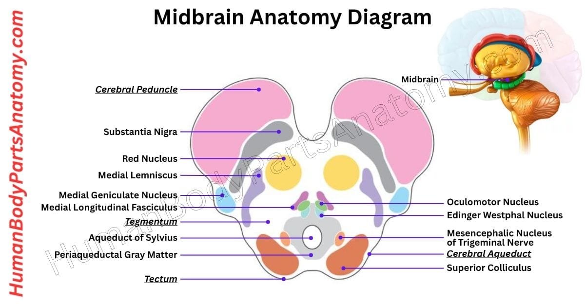
The midbrain, also known as the mesencephalon, is the topmost part of the brainstem.[1][2] It serves as a bridge between the cerebrum and diencephalon above and the pons below.[1] Despite of being 1.5-2 cm, it plays a vital role in several important functions, including vision, hearing, body movement, alertness, sleep cycles, and temperature regulation.[1][3] Anatomically, the midbrain is mostly found within the posterior cranial fossa, with its upper section extending above a membrane called the tentorial notch.[1][2] Midbrain Anatomy is made up of four key parts: Tectum, Cerebral aqueduct, Tegmentum & Cerebral peduncles.[1]
Towards the front (rostrally), the midbrain connects to the diencephalon, which includes structures like the thalamus and hypothalamus.[1][2]
At the back (caudally), it links with parts of the hindbrain such as the pons, medulla oblongata, and cerebellum. Interestingly, the midbrain spreads outward slightly as it moves toward the front of the brain.[1]
When studying the midbrain, scientists often look at cross-sections at two levels—superior colliculi or inferior colliculi.[1]
Midbrain Anatomy Diagram

Anatomy of the Midbrain
- Tectum
- Cerebral aqueduct
- Tegmentum
- Cerebral peduncles
Midbrain Anatomy
Tectum
The tectum is the upper part of the midbrain, positioned just behind a narrow channel called the cerebral aqueduct.[1] It lies opposite the tegmentum, which forms the floor of the midbrain.[1] The tectum plays a key role in reflexes related to vision and hearing.[1][2]
A major structure within the tectum is the corpora quadrigemina, made up of four rounded bumps called colliculi.[1]
They are arranged in two pairs:
- The superior colliculi are involved in visual processing.[1][3] They help guide quick eye movements known as saccades, which allow both eyes to move rapidly in the same direction.[1] These structures also help process information from the optic nerves.[1][3] Some nerve fibers cross to the opposite side of the brain, while others remain on the same side.[1] From here, the tectospinal tract begins, sending signals to neck muscles to coordinate head and eye movements.[1][3] Each superior colliculus is linked to the lateral geniculate nucleus, an important relay center in the visual system.[1]
- The inferior colliculi are located just above the origin of the trochlear nerve.[1] They are involved in processing sound.[1][3] Each one connects to the medial geniculate nucleus, which helps relay auditory information to the brain.[1]
The reticulospinal tract influences alertness and body movements and also receives signals from the tectum. This pathway runs both upward and downward through the brainstem and spinal cord, helping coordinate responses to sensory input.[1][3]
Cerebral aqueduct
The cerebral aqueduct is a narrow channel within the brain that connects the third ventricle (in the upper midbrain) to the fourth ventricle (lower in the brainstem).[1]
It plays a key role in moving cerebrospinal fluid (CSF) through the ventricular system, helping protect and nourish the brain.[1]
This aqueduct runs between two important midbrain structures: the tectum (behind) and the tegmentum (in front).[1]
The aqueduct is a region called the periaqueductal gray (PAG), which is involved in reducing pain (analgesia), calming the body (quiescence), and supporting social bonding.[4]
Just in front of the PAG, near the level of the inferior colliculus, lies the dorsal raphe nucleus.[1] This cluster of neurons releases serotonin, a chemical that influences mood and alertness.[1][3]
Two key pairs of cranial nerve nuclei are found nearby:
- The oculomotor nuclei involved in eyelid lifting and most eye movements are located at the level of the superior colliculus.[1][6]
- The trochlear nuclei help move the eyes for near vision.[1][6] They are found just below, next to the dorsal raphe, at the level of the inferior colliculus.[1]
These nerves exit the brain in different ways. The oculomotor nerve travels forward through the tegmentum, while the trochlear nerve is unique. It is the only cranial nerve that exits the brainstem from the back (dorsally), just below the inferior colliculus.[1][6]
Also nearby is the Edinger-Westphal nucleus, which sits between the oculomotor nucleus and the cerebral aqueduct.[1][6] It helps control pupil size and lens shape, supporting visual focus.[1]
Tegmentum
The midbrain tegmentum is the lower part of the midbrain. It is located in front of the cerebral aqueduct and much larger than the tectum above it.[1]
It connects to the cerebellum through two thick bundles of nerve fibers called the superior cerebellar peduncles.[1]
These bundles enter from the lower back part of the tegmentum and cross over each other at the level of the inferior colliculus.[1] Between them lies the median raphe nucleus, which helps with memory formation.[1][3]
The tegmentum is full of nerve cells and connections that help control basic body functions like breathing, heart rate, and reflexes.[1][3] It includes part of the reticular formation, which helps keep us awake and alert.[1]
Many important nerve pathways also run through this area.
- One of these pathways is the medial lemniscus, a thin band of fibers that carries touch and position signals to the brain.[1][3] It runs along the front side of the tegmentum and stays in a fairly steady position.[1]
- Another pathway, the spinothalamic tract, carries pain and temperature signals.[1][3] It lies just behind the medial lemniscus at lower levels and moves to the side as it goes upward.[1]
In the upper part of the tegmentum are two rounds. The reddish areas are called the red nuclei, which help control movement.[8] They send signals down to the spinal cord through the rubrospinal tract, mainly to the neck and upper body.[8]
Just in front of the red nuclei is the ventral tegmental area (VTA).[1][9] This part makes dopamine, a brain chemical linked to reward, motivation, and pleasure.[1]
The VTA connects with areas in the forebrain, like the hypothalamus and mammillary bodies, helping to control emotions and behavior.[1][9]
Cerebral Peduncles
The cerebral peduncles are two thick bands of nerve fibers located at the front of the midbrain, below a part called the tegmentum.[1] The space these two peduncles are called the interpeduncular fossa, which holds cerebrospinal fluid.[1]
Each cerebral peduncle contains a large bundle of nerve fibers called the cerebral crus (or crus cerebri).[1] These fibers carry important signals from the thalamus and cerebral cortex down to the lower parts of the brain and spinal cord.[1]
The middle part of each crus carries signals that help control muscles, while the outer parts connect the brain to the pons, a part of the brainstem.[1]
In the past, the whole cerebral peduncle was sometimes called the crus cerebri. But today, scientists use cerebral peduncle to mean all the nerve fibers connecting the cerebrum, including those in the tegmentum.[1]
The dark area called the substantia nigra is attached to the cerebral peduncle, whichmeans black substance because it contains a lot of melanin.[1][5] This area is part of the basal ganglia, a group of brain structures that help control movement, learning, and emotions.[5]
The substantia nigra has two parts:
- The pars compacta, which makes dopamine, a chemical important for movement and motivation.[5]
- The pars reticulata helps control signals going out of the basal ganglia.[5]
FAQ’s
The midbrain, or mesencephalon, is the uppermost part of the brainstem located between the pons and the thalamus.[1][2] It acts as a crucial link between the spinal cord and higher brain centers, coordinating motor, sensory, and reflex pathways.[1]
The midbrain has three major regions:
Tectum – contains the superior and inferior colliculi for visual and auditory reflexes.[1][3]
Tegmentum – includes nuclei like the red nucleus and substantia nigra, important for movement and alertness.[1][5][8]
Cerebral peduncles – carry motor signals from the cerebral cortex to the brainstem and spinal cord.[1]
The midbrain controls eye movement, hearing, body movement, arousal, and pain modulation.[1][3][4] It helps process visual and auditory information and transmits motor commands between the brain and the body.[1]
It receives blood from the posterior cerebral, superior cerebellar, and basilar arteries.[1][7] Blockage in these vessels can lead to midbrain strokes, causing symptoms like eye paralysis, weakness, or coordination loss.[7]
Two cranial nerves originate here:
Oculomotor nerve (CN III) – controls most eye movements and pupil constriction.[6]
Trochlear nerve (CN IV) – controls the superior oblique muscle for precise eye motion.[6]
Midbrain-related disorders include:
Parkinson’s disease – due to degeneration of the substantia nigra.[5]
Weber’s and Benedikt’s syndromes – caused by midbrain stroke.[7]
Lesions affecting the periaqueductal gray may alter pain and consciousness levels.[4]
During embryonic growth, the mesencephalon develops into the midbrain and remains undivided. It forms essential neural tracts and nuclei that control movement, reflexes, and sensory integration.[1]
The midbrain helps you react quickly to visual and auditory cues, maintain posture, and control eye and body coordination.[1][3] Without it, reflexive actions like turning your head toward sound or tracking moving objects wouldn’t be possible.[1]
References-
- National Institutes of Health (NIH) / StatPearls Publishing. 2024. Neuroanatomy, Mesencephalon Midbrain. https://www.ncbi.nlm.nih.gov/books/NBK551509. PMID: 31855353.
- Johns Hopkins Medicine. 2024. Brain Anatomy and How the Brain Works. https://www.hopkinsmedicine.org/health/conditions-and-diseases/anatomy-of-the-brain.
- National Institutes of Health (NIH) / StatPearls Publishing. 2024. Neuroanatomy, Superior Colliculus. https://www.ncbi.nlm.nih.gov/books/NBK544224. PMID: 31536279.
- National Institutes of Health (NIH) / StatPearls Publishing. 2023. Neuroanatomy, Periaqueductal Gray. https://www.ncbi.nlm.nih.gov/books/NBK554391. PMID: 32119337.
- National Institutes of Health (NIH) / StatPearls Publishing. 2024. Neuroanatomy, Substantia Nigra. https://www.ncbi.nlm.nih.gov/books/NBK536995. PMID: 30725680.
- National Institutes of Health (NIH) / StatPearls Publishing. 2023. Neuroanatomy, Cranial Nerve 3 (Oculomotor). https://www.ncbi.nlm.nih.gov/books/NBK537126. PMID: 30725680.
- NIH / StatPearls Publishing. 2024. Brainstem Stroke. https://www.ncbi.nlm.nih.gov/books/NBK560896.
- National Institutes of Health (NIH) / StatPearls Publishing. 2023. Neuroanatomy, Red Nucleus. https://www.ncbi.nlm.nih.gov/books/NBK551628. PMID: 31869092.
- NIH / PMC. 2024. The Formation and Function of the VTA Dopamine System. https://pmc.ncbi.nlm.nih.gov/articles/PMC11011984. PMID: 38585560.
Read More-
Human Head
- Skull Anatomy: Complete Guide with Parts, Names, Functions & Diagram
- Ultimate Guide to Eye Anatomy: Parts, Structure, Functions & Diagram
- Tongue Anatomy: Complete Guide with Parts, Names, Functions & Diagram
- Mouth Anatomy: Complete Guide with Parts, Names, Functions & Diagram
- Complete Guide to Tooth Anatomy: Learn Parts, Names & Diagram
- Ultimate Guide to Ear Anatomy: Parts, Structure, Functions & Diagram
- Nose Anatomy: Complete Guide with Parts, Names, Functions & Diagram
Brain
- Basal Ganglia Anatomy: Complete Guide with Names, Functions & Diagram
- Lobes of the Brain: Complete Guide with Names, Functions & Diagram
- Parts of the Cerebrum Anatomy: Complete Guide with Names, Functions & Diagram
Organs
- Kidney Anatomy: Complete Guide with Parts, Names, Functions & Diagram
- Liver Anatomy: Complete Guide with Parts, Names, Functions & Diagram
- Heart Anatomy: Complete Guide with Parts, Names, Functions & Diagram
External Sources-
- Wikipedia
- KenHub
- Optometrists
- Cleveland Clinic
- American Academy of Ophthalmology
Medical Disclaimer
All content on HumanBodyPartsAnatomy.com is educational and based on verified, peer-reviewed medical sources. Articles are authored or reviewed by qualified medical or biomedical professionals to ensure accuracy.
This website does not provide medical advice, diagnosis, or treatment. Always consult a licensed healthcare professional for personal medical guidance.
No commercial or promotional interests influence the medical content published on this site.