📅 Published on March 16, 2024 | 🕒 Last updated on July 6, 2026
Overview of Eye Anatomy
An eye is a part of an organism that helps it see things. It takes in light and turns it into signals for the brain.[1] In fancy terms, it is an optical system that collects light, adjusts its intensity, focuses it to form an image, and sends signals to the brain.[1] Eye anatomy consists of various parts that make it a prime and important human body part. There are two main types of eyes: compound and non-compound.[1] Compound eyes, found in insects, have many small visual units. Non-compound eyes, like those in mammals, including humans, have one lens and form a single image on the retina.[1]
You are born with two eyes that work together to give you a wide field of view, depth perception, and color vision. If the focus is right, things look clear. But your eye muscles can adjust to get the focus spot-on.[1]
When light hits your retinas, special cells send messages to your brain about what they see — the color, brightness, and details. Your brain then decodes these messages to create the images you see.[1]
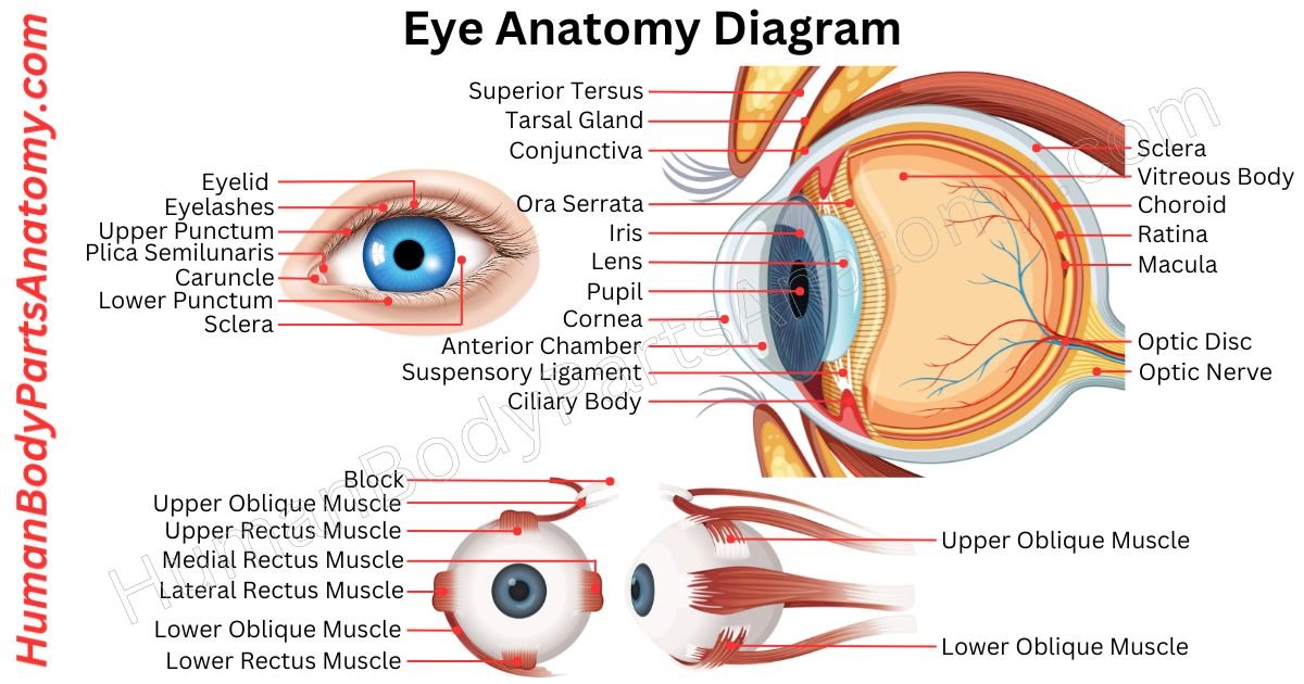
Eye Anatomy Diagram
Parts of Eye
- Cornea
- Iris
- Pupil
- Lens
- Ciliary Body
- Retina
- Fovea Centralis
- Optic Nerve
- Sclera
- Choroid
- Aqueous Humor
- Vitreous Humor
- Conjunctiva
- Eyelids
- Lacrimal Glands
- Eyelashes
- Meibomian Glands
- Optic Disk
- Optic Chiasm
- Optic Tracts
- Macula
- Anterior Chamber
- Posterior Chamber
- Suspensory Ligaments
- Scleral Venous Sinus (Canal of Schlemm)
- Visual Cortex
- Superior and Inferior Lateral Geniculate Nuclei (LGN)
- Ocular Adnexa
- Tenon’s Capsule
- Fornix
Eye Muscles
- Eyelid Muscles
- Extraocular Muscles
- Medial Rectus
- Superior Rectus
- Inferior Rectus
- Superior Oblique
- Inferior Oblique
- Intrinsic Muscles
- Iris Sphincter Muscle
- Iris Dilator Muscle
- Ciliary Muscle
Eye Anatomy: Parts, Names & Functions
Cornea
The cornea is like a clear shield covering the front of your eye.[2] It protects against dirt, germs, and UV rays while also helping to focus light for vision.[2][3]
It sits in front of a watery chamber in your eye.[2] If it gets damaged, it can heal quickly.[3] However, it is important to protect it since it is vulnerable to injury.[2][3] It has six layers, each with a specific job.[6]
- Epithelium: It is the outermost layer and serves as a barrier against the outside world. It is very sensitive to pain, helping you react quickly to protect your eyes.[6]
- Bowman’s layer: Made mostly of collagen, this tough layer provides structure and helps the cornea maintain its shape.[6]
- Stroma: This is the thickest layer and strengthens the cornea while also helping to focus light onto the retina.[6]
- Pre-Descemet’s layer (PDL): Also known as “Dua’s layer,” it is airtight, creating a strong barrier between the inside of the eye and the outside environment.[6][38]
- Descemet’s layer: This thin but strong layer protects the inside of the eye from injury and infection.[6]
- Endothelium: Responsible for maintaining fluid balance within the cornea and the eye, ensuring proper function.[6]
Together, these layers work like laminated glass in car windshields, providing strength and protection to the eye.[6]
The laminated glass consists of layers of glass with a plastic sheet in between. The layers of the cornea work together to make it resilient and efficient.[6]
Iris
The iris is the colorful part of your eye that controls the black opening called the pupil. This pupil lets light into your eye. The color of your iris is unique to you, like a fingerprint.[4]
The iris has two layers: one at the front with pigment that gives it color and one underneath with muscles.[4]
Two main muscles govern pupil size: the sphincter pupillae, which constricts the pupil by circular contraction, and the dilator pupillae, which dilates the pupil by radial contraction.[4][5]
The iris adjusts the size of your pupil.[4] When it is wider, more light gets in, and when it is narrower, less light gets in.[4] It helps you see better in different lighting conditions, like going from bright sunlight to indoors.[5]
Your iris automatically adjusts your pupil size to help you see clearly, a process called the pupillary light reflex.[4][5]
The iris is good at blocking light because of its pigmented layers, ensuring that only the right amount of light reaches the retina at the back of the eye.[4]
Some people are born without an iris, which can blur their vision, but their eyes still work.[4]
Pupil
The pupil is the circular aperture in the center of the iris through which light enters the eye. In bright light, it constricts; in dim light, it dilates. This response is called the pupillary light reflex.[5]
The size of the pupil is controlled by two muscles in the iris: the iris dilator (widens the pupil) and the iris sphincter (narrows the pupil).[4][5] Light enters through the pupil, is focused by the lens, and strikes the retina.[5]
The retina converts light into neural signals for the brain, which then forms the perceived image.[12] Neuronal pathways connecting the pupil to the brainstem and midbrain control its size via the autonomic nervous system.[5]
The pupil also provides a pathway for aqueous humor — a nutrient-rich fluid — to flow from the posterior chamber to the anterior chamber of the eye.[7]
Humans have round pupils, while many other species (e.g., cats, goats) have slit-shaped or rectangular pupils adapted to their environments.[5]
Lens
The crystalline lens is a transparent, biconvex structure located behind the iris and in front of the vitreous humor.[8] In adults, it measures approximately 10 mm in diameter and 4 mm in axial depth.[8]
Composed predominantly of crystallin proteins, the lens is transparent and can change shape to focus light on the retina — a process called accommodation.[8] This change in curvature allows clear vision at varying distances.[8]
When light enters the eye, the lens refracts it to create a focused image on the retina.[8] This image is initially inverted; the visual cortex of the brain interprets and corrects it.[8]
Ciliary muscles surrounding the lens alter its curvature, while zonular fibers (suspensory ligaments) maintain its position.[8][10] The lens contributes approximately 25–35% of the total refractive power of the eye.[8]
The lens is avascular and relies entirely on the aqueous humor for nutrients and metabolic waste removal.[7][8]
Aqueous humor flows through the eye and drains into the bloodstream via the trabecular meshwork and Canal of Schlemm.[7]
Ciliary Body
The ciliary body is a component of the eye that sits underneath the colorful iris.[10] It resembles a ring with folds called ciliary processes, creating clear fluid.[10] This fluid covers the gap between your cornea and iris, supplying pressure and nutrition to your eye.[7][10]
The ciliary body has two primary functions: aqueous humor secretion and lens accommodation.[10] It connects to the lens via zonular fibers and modifies lens curvature through contraction and relaxation of the ciliary muscle.[10]
When viewing a near object, the ciliary muscle contracts, zonular fibers relax, and the lens becomes more spherical.[10] When viewing a distant object, the ciliary muscle relaxes, zonular fibers tighten, and the lens flattens.[10]
Presbyopia makes it difficult to focus on nearby things as we age. The changes in the ciliary body cause it.[9][11]
Overall, the ciliary body, iris, and choroid are part of the uvea, working together to support vision by controlling light and lens shape.[10]
Retina
The retina is like the camera film in your eye. It is a layer at the back of your eyeball that catches the light.[12]
Inside the retina, There are different types of cells. The main ones are rods and cones. Rods work in dim light and give us black-and-white vision. Cones work in bright sunlight and help us see colors and details.[12]
Some special cells called photoreceptors turn light into messages for your brain.[12] These messages travel through the optic nerve to your brain, becoming the pictures you see.[12][14]
If something happens to your retina, like damage or missing, your eye can still take in light, but your brain won’t get all the information it needs to make clear pictures.[12]
The retina is like the screen of a camera that captures images and sends them to our brain for us to see.[12]
Fovea Centralis
The fovea centralis is like the high-definition center of our eye, located right in the middle of the back of the eye.[13]
Surrounding the fovea are two concentric zones: the parafovea (inner ring, densely packed with cones) and the perifovea (outer ring, with more rods).[13]
The fovea itself contains only cone photoreceptors — approximately 199,000 per mm² — with no rods, blood vessels, or overlying neurons, which maximizes its resolving power.[13]
Rods, which predominate in the peripheral retina, provide low-light (scotopic) vision and are particularly sensitive to motion and faint objects in the dark.[13]
When we wish to examine something in detail — such as reading — the eyes move reflexively (via saccades) to project the image onto the fovea.[13]
Optic Nerve
The optic nerve is like a conduit that carries visual information from your eyes to your brain.[14] It starts at the back of your eye, where the retina is, and travels to your brain.[14]
Unlike other nerves in your body, it is wrapped in layers of tissue called meninges instead of the usual nerve coverings.[14]
It is part of the central nervous system, not the peripheral nervous system, because it comes from a different part of the brain during development.[14]
Depending on the person, the optic nerve has around 770,000 to 1.7 million tiny nerve fibers.[14] These fibers connect to cells in your retina, with some connecting to only a few cells for detailed vision and others to thousands for broader vision.[14]
After leaving the eye, the optic nerve travels toward the brain through a small hole called the optic canal. Along the way, some fibers cross over at a spot called the optic chiasm, while others keep going straight back.[24][25]
Post-chiasmal fibers form the optic tracts, which relay visual information primarily to the lateral geniculate nucleus (LGN) of the thalamus.[15][25] From there, signals are projected via the optic radiation to the primary visual cortex (V1) in the occipital lobe.[15]
A subset of optic nerve fibers projects to the superior colliculus (reflexive eye movements), the pretectal nucleus (pupillary reflex), and the suprachiasmatic nucleus (circadian rhythms).[14]
Sclera
The sclera — commonly called the white of the eye — is a dense, fibrous outer coat composed of collagen and elastic fibers.[16] It is derived embryologically from neural crest cells.[16]
In children, the sclera is thin and may appear slightly bluish (due to underlying choroidal pigment); in older adults, it can acquire a yellowish hue from lipid deposition.[16] Individuals with darker skin may have naturally darker sclerae due to increased melanin concentration.[16]
The sclera covers approximately 83% of the eyeball’s outer surface, merging anteriorly with the cornea at the limbus and posteriorly with the dural sheath of the optic nerve.[16]
It provides structural rigidity, maintains globe shape, and serves as the attachment point for the six extraocular muscles.[16][36]
Surface blood vessels and the adjacent conjunctiva become engorged during inflammation, producing the characteristic redness of the eye.[16]
Human eyes are distinctive among primates in that the sclera is prominently visible, partly because the human iris is proportionally smaller relative to the visible eye.[16]
Choroid
The choroid is a highly vascular, pigmented layer located between the sclera and retina.[17] It is the main blood supply for the outer retinal layers, including the photoreceptors.[17]
It comprises five distinct layers:[17]
- Bruch’s membrane: The innermost layer; interfaces with the retinal pigment epithelium and regulates nutrient and waste exchange.[17]
- Choriocapillaris: A dense capillary network that supplies oxygen and nutrients to the outer retina and removes metabolic waste; also helps regulate ocular temperature.[17]
- Sattler’s layer: Medium-caliber blood vessels.[17]
- Haller’s layer: Large-caliber blood vessels embedded in a stroma of fibers, melanocytes, and smooth muscle cells.[17]
- Suprachoroidea: The outermost layer; positioned between the choroid and sclera; facilitates fluid drainage and helps maintain intraocular pressure.[17]
Choroidal blood is supplied by the posterior ciliary arteries and drains via the vortex veins.[17] Autonomic innervation regulates blood flow and contributes to pressure homeostasis.[17]
Aqueous Humor
Humor in your eye is a transparent fluid that helps keep your eye healthy. There are two types: aqueous humor and vitreous humor.[7]
Aqueous humor is like a nourishing drink for your eye. It keeps your eye inflated and provides nutrients. It flows in and out, maintaining the right pressure inside your eye.[7]
Your eye has two chambers: the anterior chamber (between the lens and cornea) and the posterior chamber (between the lens and iris).[28][30]
Aqueous humor is produced primarily by the non-pigmented epithelium of the ciliary body’s ciliary processes via active secretion, ultrafiltration, and diffusion.[7][10]
It flows through the pupil into the anterior chamber, where it drains primarily via the trabecular meshwork into the Canal of Schlemm and then into episcleral veins.[7][32]
Vitreous Humor
The vitreous humor is a transparent, gel-like substance filling the vitreous chamber — the large space between the lens and the retina.[18]
It constitutes approximately 80% of the volume of the eye.[18] It maintains the shape of the globe and provides mechanical support to the retina.[18]
In forensic medicine, vitreous humor is valuable for toxicological analysis because its protected location makes it less susceptible to post-mortem contamination than blood.[18]
Biochemically, the vitreous is composed of water (~99%), collagen fibrils, hyaluronic acid, salts, sugars, ascorbic acid, and trace proteins.[18] Specialized cells called hyalocytes (phagocytic cells) maintain its clarity by removing debris.[18]
Conjunctiva
The conjunctiva is a thin, clear membrane that covers the inside of your eyelids and the white part of your eye. It helps protect your eye by creating a mucus layer in your tears.[19]
This layer lubricates your eye and acts as a barrier against irritants.[19] Working in concert with the lacrimal and meibomian glands, the conjunctiva supports a three-layer tear film: mucin (conjunctiva), aqueous (lacrimal glands), and lipid (meibomian glands).[19][21][22]
It is like protecting your eye, keeping it safe and moist. Problems with the conjunctiva can lead to dry eyes or infections.[19]
Eyelids
The eyelids are mobile folds of skin and muscle that protect the anterior eye surface.[20] Eyelid opening is primarily accomplished by the levator palpebrae superioris muscle, innervated by cranial nerve III, and by Müller’s muscle, innervated by sympathetic fibers.[20]
The primary function of the eyelids is to spread tears uniformly across the ocular surface during blinking, maintaining corneal health and moisture.[20]
The blink reflex also protects the eye from foreign particles, with eyelashes serving as the first mechanical barrier to debris.[20]
Lid morphology varies between populations due to differences in orbital fat distribution and skin fold anatomy.[20]
Lacrimal Gland
The lacrimal glands are like tear factories for our eyes. They sit near the top and outer sides of our eye sockets. When they get inflamed, it is called dacryoadenitis.[21]
These glands produce tears, which travel across our eyes and eventually drain into our noses. The gland has two parts: one closer to the eye and one deeper inside the eye socket.[21]
The part closer to the eye is called the palpebral lobe; you can see it if you turn your upper eyelid inside out. The deeper part is the orbital lobe, which connects to the palpebral lobe with small tubes.[21]
Lacrimal secretions pass through 6–12 excretory ducts into the superior conjunctival fornix, spread across the eye surface via blinking, and drain medially through the lacrimal puncta, canaliculi, lacrimal sac, and nasolacrimal duct into the nasal cavity.[21]
Eyelashes
Eyelashes are the hairs on our eyelids that help protect our eyes by catching debris and triggering the blink reflex when something gets too close.[20]
They are super sensitive, with many nerve endings, helping us sense even light touches. Besides guarding our eyes, they keep the tear film intact, preventing excessive evaporation. Plus, they are a big part of what makes eyes attractive![20]
Meibomian Glands
Meibomian glands are like tiny oil factories located along the edges of our eyelids. They make an oily meibum, which stops tears from drying up too quickly. It keeps our eyes moist and comfortable.[22]
Meibomian glands keep tears where they are supposed to be, preventing them from spilling onto our cheeks. When we blink, the oily edge helps trap tears against our eyeballs and seals our eyelids tight.[22]
Usually, we have about 25 of these glands on the top eyelid and 20 on the bottom. However, if these glands aren’t working right, it can lead to dry eyes and posterior blepharitis.[22]
Optic Disc
The optic disc (optic nerve head) is the site where all retinal ganglion cell axons converge and exit the eye to form the optic nerve.[23] Because it contains no photoreceptors, it corresponds to the physiological blind spot in each eye’s visual field.[23]
A typical human optic disc contains approximately 1.0–1.2 million nerve fiber axons. It is located approximately 3–4 mm nasal to the fovea. Its shape is slightly oval, measuring on average about 1.76 mm horizontally and 1.92 mm vertically.[23]
Within the disc is a central depression called the optic cup. The cup-to-disc ratio (C/D ratio) is a clinically important metric: an elevated ratio raises suspicion for glaucomatous optic neuropathy.[23][29]
The central retinal artery and vein enter and exit the eye through the optic disc, making it the hub of the retinal vascular supply.[23]
Optic Chiasm
The optic chiasm is an X-shaped structure at the base of the brain, situated immediately anterior to the pituitary stalk and above the pituitary gland.[24]
At the chiasm, visual fibers from the nasal half of each retina cross (decussate) to the opposite optic tract, while fibers from the temporal retina remain ipsilateral.[24]
This arrangement ensures that the left visual cortex processes information from the right visual field (of both eyes), and vice versa.[24]
The optic chiasm receives blood supply primarily from branches of the anterior cerebral artery, the anterior communicating artery, and small perforators from the internal carotid artery.[24]
Optic Tract
The optic tracts are paired fiber bundles that arise from the posterior aspect of the optic chiasm and carry visual information toward the brain.[25]
Each optic tract contains:
- Ipsilateral temporal retinal fibers (carrying contralateral visual field information from that eye).[25]
- Contralateral nasal retinal fibers (carrying contralateral visual field information from the opposite eye).[25]
Most optic tract fibers terminate in the lateral geniculate nucleus of the thalamus; a minority project to the superior colliculus, pretectum, and hypothalamus for non-image-forming visual functions.[15][25]
Macula
The macula is a crucial part of your eye that helps you see things directly in front of you, like small details. It is like the central hub of your retina, which is at the back of your eyeball.[26]
When light enters your eye, it hits the retina, where cells called photoreceptors turn it into electrical signals.[26] These signals travel to your brain through the optic nerve, creating the images you see.[14]
The macula specializes in processing this information, allowing you to focus on specific aspects of what you are looking at.[26]
Damage to the macula — as in age-related macular degeneration (AMD), the leading cause of central vision loss in adults over 50 — results in blurring or a central scotoma even though peripheral vision may be preserved.[26][27]
Anterior Chamber
The front of the eye has a space filled with fluid called the anterior chamber (AC). It sits between the colored The anterior chamber (AC) is the fluid-filled space between the posterior surface of the cornea and the anterior surface of the iris and lens.[28]
Three clinically significant conditions affect the anterior chamber:
- Hyphema: Accumulation of blood in the anterior chamber, typically following blunt ocular trauma. Blood settles inferiorly due to gravity.[28]
- Anterior Uveitis: Inflammation of the iris and ciliary body (the anterior uvea), causing pain, photophobia, and a cellular inflammatory infiltrate visible in the AC.[28]
- Glaucoma: Inadequate drainage of aqueous humor through the trabecular meshwork elevates intraocular pressure, damaging the optic nerve and causing progressive vision loss.[28][29]
The axial depth of the anterior chamber normally ranges from 1.5 to 4.0 mm, with an average of approximately 3.0 mm.[28]
The chamber becomes shallower with age and in hyperopia.[28] A depth below 2.5 mm significantly increases risk of angle-closure glaucoma.[28][29]
Posterior Chamber
The posterior chamber is a narrow, ring-shaped space located behind the iris and in front of the anterior surface of the lens and zonular apparatus.[30] It is the primary site of aqueous humor production by the ciliary processes.[7][30]
Obstruction of aqueous flow from the posterior to the anterior chamber — known as pupillary block — can occur with a dense mature cataract or a malpositioned intraocular lens implant.[30]
Pupillary block causes pressure to accumulate in the posterior chamber, bowing the iris anteriorly (iris bombé), obstructing the trabecular meshwork, and precipitating acute angle-closure glaucoma.[29][30]
Suspensory Ligament
The suspensory ligament of the eyeball is also known as Lockwood’s ligament. It acts like a hammock under the eye. It stretches between two points on the sides of the eye and holds up the muscles below it.[33]
Its primary role is to maintain the vertical position of the eyeball within the orbit and prevent inferior displacement (enophthalmos or hypoglobus) following orbital floor fractures or loss of inferior muscular support.[33]
Canal of Schlemm (Scleral Venous Sinus)
Schlemm’s canal is a circumferential venous channel located in the corneoscleral junction (limbus), serving as the primary drainage route for aqueous humor.[31]
Adjacent anatomical structures include:
- Internal Scleral Sulcus: A circumferential groove at the corneoscleral junction that houses the canal.[31]
- Scleral Spur: A wedge-shaped projection of scleral tissue at the posterior border of the sulcus; the attachment point for the trabecular meshwork and ciliary muscle.[31]
- Trabecular Meshwork: A porous connective tissue network spanning the internal scleral sulcus; aqueous humor percolates through its pores before entering Schlemm’s canal.[32]
Schlemm’s canal is lined by a continuous layer of specialized endothelial cells that create transcellular pores allowing bulk flow of aqueous humor into collector channels and then into episcleral veins.[31]
Dysfunction of this drainage system is the primary mechanism in open-angle glaucoma.[29]
Lateral Geniculate Nucleus
The lateral geniculate nucleus (LGN) is a layered, ovoid nucleus located in the posterior thalamus on each side of the brain.[15] It serves as the primary thalamic relay station for visual information route to the cortex.[15]
The LGN comprises six neuronal layers:
- Layers 1 and 2 (magnocellular): Receive input from M-type retinal ganglion cells; process motion and low-contrast stimuli.[15]
- Layers 3–6 (parvocellular): Receive input from P-type retinal ganglion cells; process fine detail and color.[15]
The LGN transmits processed visual signals to the primary visual cortex (area V1) via the optic radiation.[15] It also receives significant feedback projections from V1, enabling top-down modulation of visual processing.[15]
Tenon’s Capsule
Tenon’s capsule is like a thin covering around the eyeball, stretching from the optic nerve to the cornea’s edge. It keeps the eyeball separate from the fatty tissue around it and helps it move smoothly.[33]
A gap separates it from the eye’s outer layer by a space for lymph fluid. This fluid space connects to areas around the brain.[33]
The capsule has holes at the back for blood vessels and nerves. It joins the optic nerve’s covering and the eye’s outer layer.[33]
At the front, it sticks to the conjunctiva, which covers the eye’s surface. Tenon’s capsule and the conjunctiva are linked to the eye’s center region.[33]
Fornix
The fornix of the eye is like a stretchy connection between the inner surface of the eyelids and the eyeball. Each eye has two fornix: one on top and one on the bottom.[34]
This connection lets the eyelids move smoothly. The fornix comprises three sides of tissue that attach to the eyeball and a flexible fold on the fourth side.[34]
This setup lets the eyelids and eyeballs move without getting in each other’s way. The fornix is a protective layer that’s about 3 to 5 layers thick.[34]
FAQ’s
The human eye integrates multiple structures for vision. Key components include the cornea (primary focusing element, ~65–75% of refractive power),[2][6] the iris (controls pupil aperture and light entry),[4] the lens (accommodation, fine-tuning focus),[8] the retina (photoreception and phototransduction),[12] and the optic nerve (transmits encoded signals to the visual cortex).[14]
The eye is housed within the bony orbital cavity (orbit), which is formed by seven bones: the frontal, zygomatic, maxilla, sphenoid, ethmoid, lacrimal, and palatine bones.[35] Together they provide structural protection against mechanical trauma and serve as attachment sites for the extraocular muscles.[35]
Each eye is moved by six extraocular muscles: the superior, inferior, medial, and lateral rectus muscles, and the superior and inferior oblique muscles.[36] These allow precise, conjugate eye movements — including saccades, smooth pursuit, and vergence — in all directions of gaze.[36]
The eye does not contain true synovial joints.[36] The globe rests within the orbital cavity surrounded by fatty tissue, fascia, and the six extraocular muscles, permitting rotational movement that is biomechanically analogous to a ball-and-socket joint, but without a formal joint capsule or articular cartilage.[36]
Ocular pain (ophthalmalgia) arises from one of the most densely innervated surfaces in the body — the corneal epithelium contains approximately 7,000 free nerve endings per mm².[3][6] Surface causes include dry eye, corneal abrasion, foreign bodies, and keratitis.[3] Deeper causes include elevated intraocular pressure (glaucoma), uveitis, scleritis, and optic neuritis.[29]
The optic nerve acts as the principal data cable between the retina and the brain.[14] It carries approximately 1.0–1.7 million axons encoding luminance, color, contrast, and motion information from retinal ganglion cells to the lateral geniculate nucleus and other visual centers.[15][14] Optic nerve damage — from glaucoma, ischemia, or demyelination — can cause irreversible partial or complete vision loss.[14][29]
The crystalline lens fine-tunes retinal focus through accommodation — changing shape via ciliary muscle contraction and relaxation.[10][8] It contributes approximately 25–35% of the eye’s total refractive power (complementing the cornea’s ~65–75%). Lens transparency is maintained by its avascularity and specialized aqueous humor supply; opacity (cataract) is the world’s leading cause of reversible blindness.[8]
Tears form a stratified film over the cornea and conjunctiva that lubricates, nourishes, and immunologically protects the ocular surface.[19][21] The tear film has three layers: mucin (conjunctival goblet cells), aqueous (lacrimal glands), and lipid (meibomian glands).[22][19] Inadequate tear volume or composition leads to dry eye disease, corneal irregularity, and pain.[19][22]
The white of the eye is the sclera — a tough, collagen-rich fibrous coat covering approximately 83% of the globe’s outer surface.[16] It maintains ocular shape, protects intraocular structures, and anchors the extraocular muscles.[36][16] Changes in scleral color (icterus, blue sclerae in osteogenesis imperfecta) signal systemic conditions.[16]
Yes. The eyeball grows from approximately 16–17 mm at birth to an adult axial length averaging 24 mm. The majority of growth occurs in the first 2 years of life, with slower elongation continuing until approximately ages 13–15.[37] After adolescence the eye typically stabilizes, although conditions such as high axial myopia may cause continued pathological elongation.[37]
Human visual function encompasses central acuity, peripheral vision, color discrimination (trichromacy), depth perception (stereopsis), and scotopic (night) vision.[1][12] Common refractive errors — myopia (nearsightedness), hyperopia (farsightedness), and astigmatism — affect an estimated 150 million Americans and represent the most frequent correctable cause of vision impairment in the United States.[1]
References-
- National Eye Institute (NEI), National Institutes of Health (NIH). (2023). How the Eyes Work. U.S. Department of Health & Human Services, Bethesda, MD.
https://www.nei.nih.gov/learn-about-eye-health/healthy-vision/how-eyes-work - MedlinePlus, National Library of Medicine (NLM), National Institutes of Health (NIH). (2022). Cornea. U.S. National Library of Medicine, Bethesda, MD.
https://medlineplus.gov/cornea.html - National Eye Institute (NEI), NIH. (2023). Corneal Conditions. U.S. Department of Health & Human Services.
https://www.nei.nih.gov/learn-about-eye-health/eye-conditions-and-diseases/corneal-conditions - Tadi P, Lui F. (2023). Anatomy, Head and Neck: Eye Iris Sphincter Muscle. In: StatPearls. Treasure Island (FL): StatPearls Publishing; NCBI Bookshelf, National Library of Medicine (NLM), NIH.
https://www.ncbi.nlm.nih.gov/books/NBK532252/
NCBI Bookshelf ID: NBK532252 - Kardon RH, et al. (2023). Physiology, Pupillary Light Reflex. In: StatPearls. Treasure Island (FL): StatPearls Publishing; NCBI Bookshelf, NLM, NIH.
https://www.ncbi.nlm.nih.gov/books/NBK554379/
NCBI Bookshelf ID: NBK554379 - Wilson ME, et al. (2023). Anatomy, Head and Neck: Eye Cornea. In: StatPearls. Treasure Island (FL): StatPearls Publishing; NCBI Bookshelf, NLM, NIH.
https://www.ncbi.nlm.nih.gov/books/NBK470340/
NCBI Bookshelf ID: NBK470340 - Goel M, Picciani RG. (2023). Physiology, Aqueous Humor Circulation. In: StatPearls. Treasure Island (FL): StatPearls Publishing; NCBI Bookshelf, NLM, NIH.
https://www.ncbi.nlm.nih.gov/books/NBK519007/
NCBI Bookshelf ID: NBK519007 - Rehman I, Hazhirkarzar B, Patel BC. (2023). Anatomy, Head and Neck, Eye. In: StatPearls. Treasure Island (FL): StatPearls Publishing; NCBI Bookshelf, NLM, NIH.
https://www.ncbi.nlm.nih.gov/books/NBK482428/
NCBI Bookshelf ID: NBK482428 - National Eye Institute (NEI), NIH. (2023). Presbyopia. U.S. Department of Health & Human Services.
https://www.nei.nih.gov/learn-about-eye-health/eye-conditions-and-diseases/presbyopia - Sadun AA, et al. (2023). Anatomy, Head and Neck: Ciliary Body. In: StatPearls. Treasure Island (FL): StatPearls Publishing; NCBI Bookshelf, NLM, NIH.
https://www.ncbi.nlm.nih.gov/books/NBK526110/
NCBI Bookshelf ID: NBK526110 - Mayo Clinic Staff. Mayo Foundation for Medical Education and Research (MFMER). (2023). Presbyopia — Symptoms and Causes. Rochester, MN.
https://www.mayoclinic.org/diseases-conditions/presbyopia/symptoms-causes/syc-20376504 - Remington LA, et al. (2023). Anatomy, Head and Neck: Eye Retina. In: StatPearls. Treasure Island (FL): StatPearls Publishing; NCBI Bookshelf, NLM, NIH.
https://www.ncbi.nlm.nih.gov/books/NBK545159/
NCBI Bookshelf ID: NBK545159 - Balaratnasingam C, et al. (2023). Anatomy, Head and Neck: Eye Fovea Centralis. In: StatPearls. Treasure Island (FL): StatPearls Publishing; NCBI Bookshelf, NLM, NIH.
https://www.ncbi.nlm.nih.gov/books/NBK554514/
NCBI Bookshelf ID: NBK554514 - Patel BC, et al. (2023). Anatomy, Head and Neck: Eye Optic Nerve. In: StatPearls. Treasure Island (FL): StatPearls Publishing; NCBI Bookshelf, NLM, NIH.
https://www.ncbi.nlm.nih.gov/books/NBK542252/
NCBI Bookshelf ID: NBK542252 - StatPearls Publishing. (2023). Anatomy, Head and Neck: Lateral Geniculate Nucleus. Treasure Island (FL): StatPearls Publishing; NCBI Bookshelf, NLM, NIH.
https://www.ncbi.nlm.nih.gov/books/NBK526127/
NCBI Bookshelf ID: NBK526127 - Cioffi GA, et al. (2023). Anatomy, Head and Neck: Eye Sclera. In: StatPearls. Treasure Island (FL): StatPearls Publishing; NCBI Bookshelf, NLM, NIH.
https://www.ncbi.nlm.nih.gov/books/NBK554382/
NCBI Bookshelf ID: NBK554382 - Nickla DL, Wallman J. (2023). Anatomy, Head and Neck: Choroid. In: StatPearls. Treasure Island (FL): StatPearls Publishing; NCBI Bookshelf, NLM, NIH.
https://www.ncbi.nlm.nih.gov/books/NBK519014/
NCBI Bookshelf ID: NBK519014 - Sebag J. (2023). Anatomy, Head and Neck: Eye Vitreous. In: StatPearls. Treasure Island (FL): StatPearls Publishing; NCBI Bookshelf, NLM, NIH.
https://www.ncbi.nlm.nih.gov/books/NBK507807/
NCBI Bookshelf ID: NBK507807 - Azari AA, Barney NP. (2023). Anatomy, Head and Neck: Conjunctiva. In: StatPearls. Treasure Island (FL): StatPearls Publishing; NCBI Bookshelf, NLM, NIH.
https://www.ncbi.nlm.nih.gov/books/NBK553190/
NCBI Bookshelf ID: NBK553190 - Knop E, Knop N. (2023). Anatomy, Head and Neck: Eyelid. In: StatPearls. Treasure Island (FL): StatPearls Publishing; NCBI Bookshelf, NLM, NIH.
https://www.ncbi.nlm.nih.gov/books/NBK519100/
NCBI Bookshelf ID: NBK519100 - Dartt DA. (2023). Anatomy, Head and Neck: Lacrimal Gland. In: StatPearls. Treasure Island (FL): StatPearls Publishing; NCBI Bookshelf, NLM, NIH.
https://www.ncbi.nlm.nih.gov/books/NBK532934/
NCBI Bookshelf ID: NBK532934 - Nichols KK, et al. (2023). Anatomy, Head and Neck: Meibomian Glands. In: StatPearls. Treasure Island (FL): StatPearls Publishing; NCBI Bookshelf, NLM, NIH.
https://www.ncbi.nlm.nih.gov/books/NBK470417/
NCBI Bookshelf ID: NBK470417 - Jonas JB, et al. (2023). Anatomy, Head and Neck: Optic Disc. In: StatPearls. Treasure Island (FL): StatPearls Publishing; NCBI Bookshelf, NLM, NIH.
https://www.ncbi.nlm.nih.gov/books/NBK542278/
NCBI Bookshelf ID: NBK542278 - Horton JC. (2023). Anatomy, Head and Neck: Optic Chiasm. In: StatPearls. Treasure Island (FL): StatPearls Publishing; NCBI Bookshelf, NLM, NIH.
https://www.ncbi.nlm.nih.gov/books/NBK526115/
NCBI Bookshelf ID: NBK526115 - Kupersmith MJ. (2023). Anatomy, Head and Neck: Optic Tract. In: StatPearls. Treasure Island (FL): StatPearls Publishing; NCBI Bookshelf, NLM, NIH.
https://www.ncbi.nlm.nih.gov/books/NBK545141/
NCBI Bookshelf ID: NBK545141 - StatPearls Publishing. (2023). Anatomy, Head and Neck: Macula. Treasure Island (FL): StatPearls Publishing; NCBI Bookshelf, NLM, NIH.
https://www.ncbi.nlm.nih.gov/books/NBK554440/
NCBI Bookshelf ID: NBK554440 - National Eye Institute (NEI), NIH. (2023). Age-Related Macular Degeneration.
https://www.nei.nih.gov/learn-about-eye-health/eye-conditions-and-diseases/age-related-macular-degeneration - Allingham RR, et al. (2023). Anatomy, Head and Neck: Eye Anterior Chamber. In: StatPearls. Treasure Island (FL): StatPearls Publishing; NCBI Bookshelf, NLM, NIH.
https://www.ncbi.nlm.nih.gov/books/NBK554416/
NCBI Bookshelf ID: NBK554416 - National Eye Institute (NEI), NIH. (2023). Glaucoma.
https://www.nei.nih.gov/learn-about-eye-health/eye-conditions-and-diseases/glaucoma - Weinreb RN, et al. (2023). Anatomy, Head and Neck: Eye Posterior Chamber. In: StatPearls. Treasure Island (FL): StatPearls Publishing; NCBI Bookshelf, NLM, NIH.
https://www.ncbi.nlm.nih.gov/books/NBK559096/
NCBI Bookshelf ID: NBK559096 - Rhee DJ. (2023). Anatomy, Head and Neck: Canal of Schlemm. In: StatPearls. Treasure Island (FL): StatPearls Publishing; NCBI Bookshelf, NLM, NIH.
https://www.ncbi.nlm.nih.gov/books/NBK559101/
NCBI Bookshelf ID: NBK559101 - Tamm ER. (2023). Anatomy, Head and Neck: Trabecular Meshwork. In: StatPearls. Treasure Island (FL): StatPearls Publishing; NCBI Bookshelf, NLM, NIH.
https://www.ncbi.nlm.nih.gov/books/NBK538141/
NCBI Bookshelf ID: NBK538141 - StatPearls Publishing. (2023). Anatomy, Head and Neck: Tenon Capsule. Treasure Island (FL): StatPearls Publishing; NCBI Bookshelf, NLM, NIH.
https://www.ncbi.nlm.nih.gov/books/NBK545221/
NCBI Bookshelf ID: NBK545221 - Bron AJ, et al. (2023). Anatomy, Head and Neck: Conjunctival Fornix. In: StatPearls. Treasure Island (FL): StatPearls Publishing; NCBI Bookshelf, NLM, NIH.
https://www.ncbi.nlm.nih.gov/books/NBK563177/
NCBI Bookshelf ID: NBK563177 - Rootman J. (2023). Anatomy, Head and Neck: Orbit. In: StatPearls. Treasure Island (FL): StatPearls Publishing; NCBI Bookshelf, NLM, NIH.
https://www.ncbi.nlm.nih.gov/books/NBK519520/
NCBI Bookshelf ID: NBK519520 - Demer JL. (2023). Anatomy, Head and Neck: Extraocular Muscles. In: StatPearls. Treasure Island (FL): StatPearls Publishing; NCBI Bookshelf, NLM, NIH.
https://www.ncbi.nlm.nih.gov/books/NBK519536/
NCBI Bookshelf ID: NBK519536 - Levin LA, Nilsson SFE. (2023). Physiology, Eye. In: StatPearls. Treasure Island (FL): StatPearls Publishing; NCBI Bookshelf, NLM, NIH.
https://www.ncbi.nlm.nih.gov/books/NBK556093/
NCBI Bookshelf ID: NBK556093 - Dua HS, Faraj LA, Said DG, Gray T, Lowe J. (2013). Human Corneal Anatomy Redefined: A Novel Pre-Descemet’s Layer (Dua’s Layer). Ophthalmology. Elsevier Inc.
https://doi.org/10.1016/j.ophtha.2013.01.018
PMID: 23714320
Medical Disclaimer
All content on HumanBodyPartsAnatomy.com is educational and based on verified, peer-reviewed medical sources. Articles are authored or reviewed by qualified medical or biomedical professionals to ensure accuracy.
This website does not provide medical advice, diagnosis, or treatment. Always consult a licensed healthcare professional for personal medical guidance.
No commercial or promotional interests influence the medical content published on this site.