📅 Published on January 13, 2024 | 🕒 Last updated on March 9, 2026
Overview of Arm Anatomy
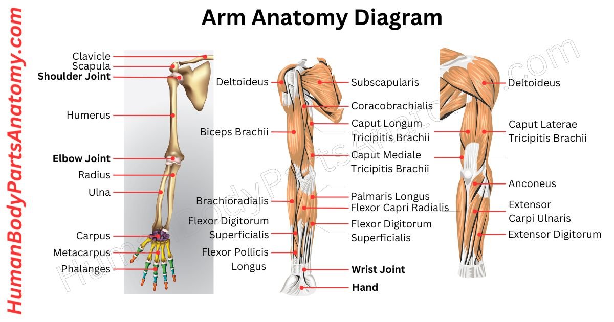
The upper extremity, or arm, is a vital part of the human body that enables us to lift, hold, and move objects.[1] The human arm anatomy consists of three main parts: the upper arm, forearm, and hand. It stretches from the shoulder to the fingers and includes 30 bones in the upper extremity, along with muscles, blood vessels, and nerves.[1] The brachial plexus is the group of nerves that controls movement and feeling in the arm.[12]
The shoulder joint is shaped like a ball-and-socket joint, which allows the arm to move in many directions.[3] But because the socket is shallow, it is not as stable as the hip joint.[3] The elbow works mostly like a hinge and helps the arm bend and straighten.[8] It also features a special joint that allows the forearm to rotate the hand up or down (this is known as pronation and supination).[8]
The wrist joint is shaped to allow back-and-forth and side-to-side movement, while the small joints between the wrist bones allow only a little motion.[9] The finger joints work like hinges and let the fingers bend and straighten.[11]
In this article, we will look at all the parts of the arm, along with their names, functions, and locations, using simple diagrams.
Arm Anatomy Diagram

Anatomy of Arm
- Shoulder
- Biceps
- Triceps
- Forearm
- Hand
- Thumb
- Fingers
- Nails
- Joints
- Shoulder Joint
- Elbow Joint
- Wrist Joint
- Metacarpophalangeal (MCP) Joints
- Interphalangeal (IP) Joints
Arm Muscle Anatomy
- Anterior Compartment Muscles:
- Biceps Brachii
- Brachialis
- Coracobrachialis
- Posterior Compartment Muscles
- Triceps brachii
- Anconeus
- Intrinsic Muscles
Arm Anatomy: Parts & Functions
Shoulder
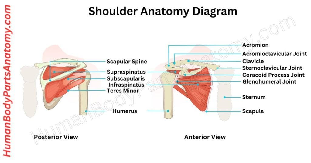
The human shoulder comprises 3 bones: the clavicle (collarbone), scapula (shoulder blade), and humerus (upper arm bone).[1]
These remarkable skeletal elements connect with an intricate network of muscles, ligaments, and tendons. This combination creates a very effective shoulder joint design.[1]
Within this system, the glenohumeral joint with the acromioclavicular joint strengthen the overall splendor. The shoulder joint allows an inseparable connection between the humerus and the scapula.[3]
Functionally, the shoulder joint is a good ball and socket splendor, releasing the great potential of circular rotations. A hinge-like extension elevates the arm to move freely.[3]
Read More – Shoulder Anatomy: Ultimate Guide to Parts, Names, Functions & Diagram
- What are the Shoulder Injuries and Disorders?
- What is a Dislocated Shoulder?
- How to do Shoulder replacement surgery?
- What is Shoulder pain?
- How to perform a Shoulder CT scan?
- How to perform a Shoulder MRI scan?
- What is Frozen shoulder? How to take aftercare?
- What is a dislocated shoulder? How to take aftercare?
Biceps
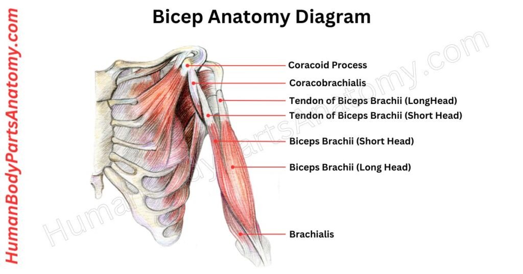
The biceps brachii muscle is located in the anterior compartment of the upper arm. What sets it apart is its dual-headed origin.[1]
The short head originates from the coracoid process, while the long head arises from the supraglenoid tubercle of the scapula.[1]
Unlike typical muscles, the biceps brachii display an exceptional capacity to traverse not one but two joints – the shoulder joint and the elbow joint. It facilitates interesting forearm flexion and supination actions.[1]
The corkscrew is twisted into the cork with force by the biceps. The biceps attach to the brachialis and coracobrachialis muscles to form a trio within the anterior compartment of the upper arm.[1]
It shares nerve supply, forming a collaboration that enhances their collective strength.[1]
Read More – Ultimate Guide to Bicep Anatomy: Parts, Names, Functions & Diagram
- Long head of the biceps tendon pain: differential diagnosis and treatment.
- Diagnosis and treatment of distal biceps and anterior elbow pain in throwing athletes.
- Does bicep pathology affect rotator cuff repair outcomes?
- Left neck and right biceps muscle vibrations have similar effects on perceived body orientation.
Triceps
The triceps brachii is a strong muscle at the back of your upper arm. It helps straighten your elbow and has three parts: a long head, a lateral head, and a medial head. These parts come together into one tendon near your elbow.[1]
The long head begins from a spot on your shoulder blade, while the lateral and medial heads start from your upper arm bone. They all connect to a bony part on your elbow called the olecranon process.[1]
Its main job is to extend your forearm at the elbow. When your arm is slightly bent, the biceps usually have more power than the triceps.[1]
Apart from straightening your arm, the triceps also help keep your elbow stable when your hand is doing delicate tasks like writing.[1]
The triceps hang out in the back of your arm, while muscles like the biceps and brachialis live in the front. A barrier called the lateral intermuscular septum separates these muscle groups.[1]
Forearm
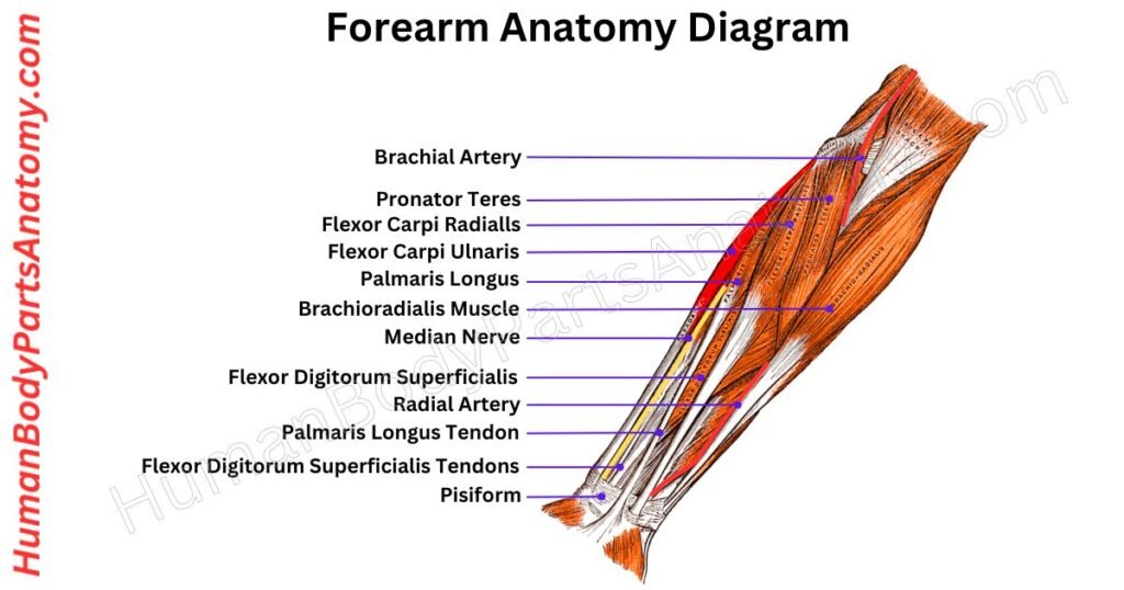
The forearm is the part between your elbow and wrist, comprising the radius and ulna bones. It is packed with muscles—20 in total—that help you do delicate movements with your arm, wrist, and fingers.[2]
The forearm has two main muscle groups: the front (for flexing) and the back (for extending). Tough layers of tissue split these groups. They are like compartments that keep the muscles in place.[2]
These muscles are a big deal for making your arm, wrist, and fingers move smoothly. They are split into two types: ones that do their job in the forearm (intrinsic) and others that handle finger movement (extrinsic).[2]
The intrinsic muscles help rotate your forearm, while the extrinsic ones deal with bending and straightening your fingers.[2]
One more muscle, the brachioradialis, goes from your arm to your wrist and helps to bend your elbow.
Read More – Complete Guide to Forearm Anatomy: Parts, Names, Functions & Diagram
- How a forearm fracture X-ray looks like?
- Effects of forearm rotation on wrist flexor and extensor muscle activities.
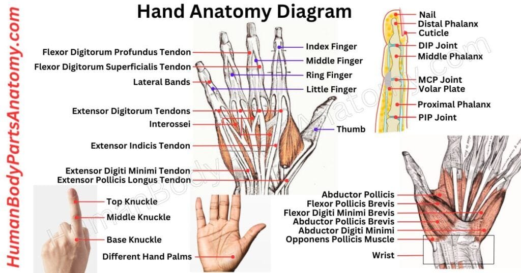
Hand
The hand is what we use to grab and hold things. Humans and some animals, like monkeys and koalas, have hands with fingers and a thumb.[5]
Usually, our hand has five parts: four fingers and one thumb. Inside, 27 bones help the hand move.[6] Some people might have a different number of tiny bones.[5]
The bigger bones in the middle connect the fingers to the wrist bones. Our hands are made up of small and big bones that help us do many different things![5]
Read More – Comprehensive Guide to Hand Anatomy: Parts, Functions & Diagram
- What is Hand Injuries and Disorders?
- What is Claw Hand?
- How to perform a hand X-ray?
- What is Chapped hands?
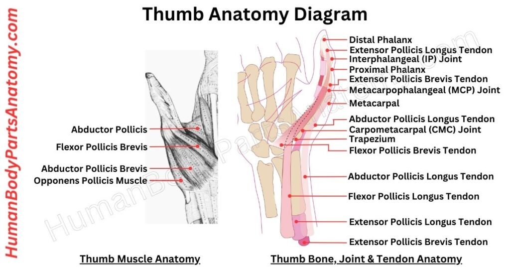
Thumb
The human thumb is the most important part of our arm anatomy. It underwent millions of years of evolution. The first digit of the hand sits beside the index finger when the palm faces forward.[5]
The thumb is one of the fingers of the five fingers and is positioned as the outermost finger.[5] It has two phalanges and an incredibly flexible joint, giving a wide range of movements and precise control.[15]
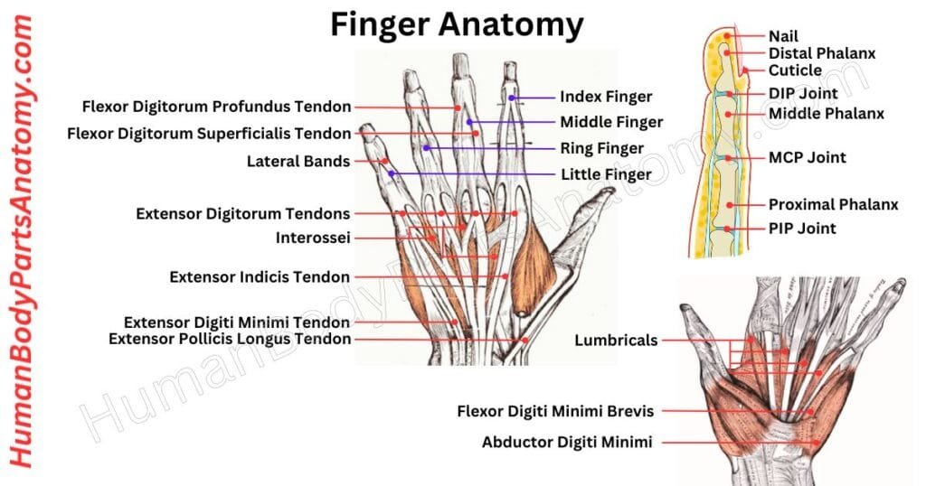
Fingers
Fingers are standout digits found on the front limbs of most four-legged vertebrates, particularly those with handy limbs like humans and other primates.[5]
Most animals with four limbs have five fingers, and if they are shorter than the metacarpals (the bones in the palm), we call them toes.[5] On the flip side, we call them fingers if they are long.
These fingers can bend and are opposable in humans, making them essential for feeling things and pulling off delicate movements. This flexibility is a big deal for the hands, helping us grab and handle stuff with finesse.[5]
Read More – Complete Guide to Finger Anatomy with Parts, Names, Functions & Diagram
- What are the Finger Injuries and Disorders?
- What is a Trigger Finger?
- Different Finger pain? Causes & Care.
- What is Smashed fingers?
- What is webbing of the fingers or toes?
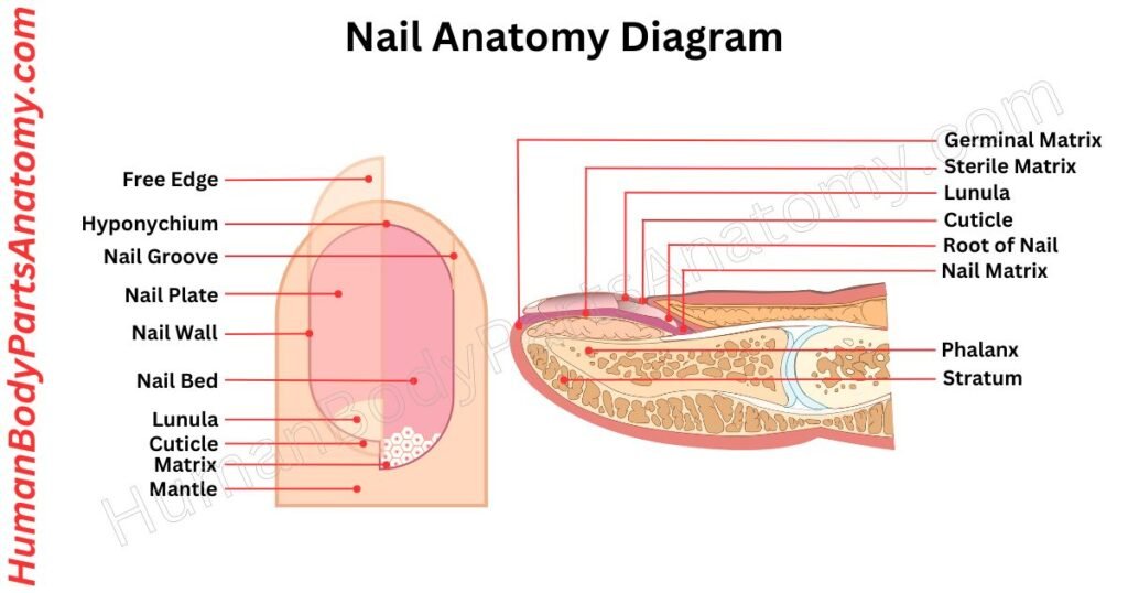
Nails
Primates, including humans, have nails on their fingers and toes. These nails are like claws but made of a tough stuff called keratin. They are about 0.5 millimeters thick and a little curved.[7]
Nails stick to the nail bed but separate at the tips so we can scratch stuff. They are also important for feeling things.[7]
Nails have folds of skin around them: side ones called lateral nail folds and a bottom one called the proximal nail fold. A thin skin layer, the cuticle, protects the nail’s lower part.[7]
Read More – Complete Guide to Nail Anatomy with all Parts, Names & Diagrams
Joints
The arm contains five main joints. Let’s go through each of them, understand their functions, and look at a labeled diagram for better clarity.
Shoulder Joint
The shoulder joint connects your arm to your body and can move in many ways. It is like a ball fitting into a socket.[3]
This joint lets you move your arm in many directions: forward, backward, away from your body, toward it, and even in circles. It is super flexible but unstable because the bones don’t support it much.[3]
The shoulder relies on muscles, ligaments, and tendons to keep it steady. But it is easy to hurt this joint because it is so flexible. That is why shoulder injuries are common.[3]
Read More – Shoulder Anatomy: Ultimate Guide to Parts, Names, Functions & Diagram
Elbow Joint
The elbow joint is where your upper arm bone (humerus) meets the bones in your lower arm (radius and ulna). It is a special joint that lets you move your arm differently.[8]
Inside, a smooth surface on the bones is covered with a slippery fluid that helps them glide smoothly. This fluid keeps things moving without any trouble.[8]
Around the joint, there’s a tough covering like a capsule, and inside, there’s a lining that keeps everything running smoothly. This joint is like a hinge—it moves back and forth, like a door opening and closing.[8]
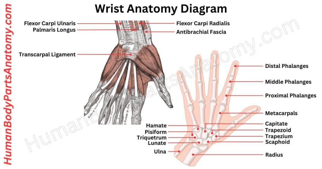
Wrist Joint
The wrist joint is called the radiocarpal joint. It links the lower part of the forearm bone (radius) with three small wrist bones. It is the main part of the wrist but includes nearby joints, too.[9]
In this joint, the end of the radius bone smoothly connects with the scaphoid and lunate bones. However, the link with the triquetral bone involves a disk between them.[9]
The radiocarpal joint lets your wrist move in different ways: bending forward (flexion), backward (extension), moving sideways away from your body (abduction), and towards your body (adduction).[9]
This joint’s structure and job are super important for wrist movement and keeping it steady.
Read More – Wrist Anatomy: Ultimate Guide to Parts, Names, Functions & Diagram
Metacarpophalangeal (MCP) Joints
The MCP joints link the bones of the palm to the fingers. There are five joints, one for each finger, linking a big hand bone to the first finger bone.[10]
The hand bones’ spherical tops fit into the finger bones’ curving bottoms to form these joints. They allow your fingers to flex, extend, separate, come together, create circles, and spin slightly.[10]
MCP joints are critical for hand usage because they provide strength and flexibility to your fingers. Ligaments, joint capsules, surrounding muscles, and tendons contribute to smooth and easy-to-control motions.[10]
Interphalangeal (IP) Joints
The interphalangeal joints in your hand are like hinges between your finger bones (phalanges). They let your fingers bend towards your palm.[11]
Each finger, except the thumb, has two sets of these joints. The “proximal interphalangeal joints” are between the first two bones of your fingers. The “distal interphalangeal joints” are between the last two bones.[11]
These joints are similar in structure, but there are some tiny differences. For instance, the way certain parts are attached and the flexibility of the joints vary slightly. The distal joints are smaller and don’t move as much as the proximal ones.[11]
Arm Muscle Anatomy
Your upper arm and forearm have over twenty muscles. These muscles help you to move your arms, hands, fingers, and thumbs.[1]
Some muscles are for tasks like sewing, while others help with bigger actions like throwing a ball or doing push-ups.[1]
Some muscles are deep inside your arm, while others are closer to your skin. You can see these muscles when you flex.[1]
Tendons are soft tissues that connect these muscles to your arm and shoulder bones. They let you straighten your elbow, lift your arms, and do all sorts of movements.[1]
Anterior Compartment Muscles
The upper arm houses three muscles in its front area. The biceps brachii have two parts, the long and short heads, found at the top. Below them, there are the coracobrachialis and brachialis muscles nestled under the biceps.[1]
Biceps Brachii
The biceps brachii is a front-arm muscle made of two parts. One part starts near the shoulder blade, and the other shares its start with another muscle near the same spot.[1]
Both parts join and attach to the radius bone in the forearm. It gets signals from the musculocutaneous nerve and blood from the brachial artery.[1]
The biceps brachii is mainly responsible for bending the elbow and turning the palm up. It also helps bring the arm forward. The longer part helps keep the shoulder steady.[1]
Brachialis
The brachialis muscle is in the front of your arm and helps you bend your elbow. It connects the upper arm bone to the forearm bone.[1]
It gets messages from nerves in your arm and is supplied with blood from certain arteries. This muscle is key to bending your arm at the elbow.[1]
Coracobrachialis
The coracobrachialis muscle sits on the inner side of your upper arm. It is attached to the shoulder blade and the upper arm bone.[1]
This muscle is good at pulling your arm closer to your body and bending your arm at the shoulder. The nerves that make it work come from the neck area, and it gets its blood supply from certain branches of an artery in the arm.[1]
Posterior Compartment Muscles
The back part of the upper arm houses a special muscle called the triceps brachii and the Anconeus. These are the 2 muscles found in this particular compartment.[1]
Triceps Brachii
The triceps brachii is a big muscle in the back of your arm. It has three parts, each starting from a different place but joining together at the same endpoint.[1]
One part starts from the shoulder blade, another from the lower part of your upper arm bone, and the third from the upper part of your upper arm bone. It all comes together and connects to the back of your elbow and the covering of your lower arm.[1]
Anconeus
The anconeus muscle is a little helper at the back of your elbow. It links the bumps on your upper arm and forearm. You assist the larger triceps muscle in straightening your elbow and keeping the joint stable. Nerves from your neck (C7-C8) run the show for this muscle, which gets blood from an arm artery.[1]
FAQ’s
The human arm is divided into the upper arm, forearm, wrist, and hand. Major bones include the humerus (upper arm), radius and ulna (forearm), along with muscles, nerves, and blood vessels that support movement and strength.[1]
Each arm contains 3 major bones: the humerus in the upper arm, and the radius and ulna in the forearm.[4] Together, they form joints that allow bending, rotation, and lifting.[1]
Key muscles include the biceps brachii, triceps brachii, brachialis, and brachioradialis. These muscles help with flexion, extension, and rotation of the arm and forearm.[1]
The arm technically refers only to the upper arm (shoulder to elbow), while the forearm refers to the section between the elbow and wrist. Many people use “arm” to describe the entire limb, but anatomically, they are distinct.[1]
The arm is mainly controlled by the brachial plexus, a network of nerves that includes the median, radial, and ulnar nerves. These nerves control sensation, reflexes, and movement.[1]
The humerus is the largest bone in the arm, connecting the shoulder to the elbow. It plays a critical role in lifting, throwing, pushing, and stabilizing the arm.[1]
Frequent arm injuries include fractures, muscle strains, tennis elbow, and nerve compression. These may result from sports, repetitive use, or accidents.[14]
Blood is supplied by the brachial artery, which branches into the radial and ulnar arteries in the forearm.[13] Veins such as the cephalic and basilic veins return blood to the heart.[1]
References-
- Sharma, S. et al. (2023) Anatomy, Shoulder and Upper Limb, Arm Structure and Function. StatPearls. NCBI Bookshelf. Available at: https://www.ncbi.nlm.nih.gov/books/NBK507841/
- Simons, S.M. et al. (2023) Anatomy, Shoulder and Upper Limb, Forearm Muscles. StatPearls. NCBI Bookshelf. Available at: https://www.ncbi.nlm.nih.gov/books/NBK536975/
- Fadaei, R. et al. (2024) Anatomy, Shoulder and Upper Limb, Glenohumeral Joint. StatPearls. NCBI Bookshelf. Available at: https://www.ncbi.nlm.nih.gov/books/NBK537018/
- Mayo Clinic Staff (2023). Arm bones. Mayo Clinic. Available at: https://www.mayoclinic.org/diseases-conditions/broken-arm/multimedia/arm-bones/img-20007018
- Caggiano, S. et al. (2023) Anatomy, Shoulder and Upper Limb, Hand Bones. StatPearls. NCBI Bookshelf. Available at: https://www.ncbi.nlm.nih.gov/books/NBK547684/
- InformedHealth.org (2024). In brief: How do hands work? Institute for Quality and Efficiency in Health Care (IQWiG). NCBI Bookshelf. Available at: https://www.ncbi.nlm.nih.gov/books/NBK279362/
- Khan, W.S. et al. (2023) Anatomy, Shoulder and Upper Limb, Nails. StatPearls. NCBI Bookshelf. Available at: https://www.ncbi.nlm.nih.gov/books/NBK534769/
- Poudel, R.R. et al. (2023) Anatomy, Shoulder and Upper Limb, Elbow Joint. StatPearls. NCBI Bookshelf. Available at: https://www.ncbi.nlm.nih.gov/books/NBK532948/
- Laskowski, E.R. et al. (2023) Anatomy, Shoulder and Upper Limb, Hand Radiocarpal Joint. StatPearls. NCBI Bookshelf. Available at: https://www.ncbi.nlm.nih.gov/books/NBK539744/
- Shakeri, A. et al. (2023) Anatomy, Shoulder and Upper Limb, Metacarpophalangeal Joints. StatPearls. NCBI Bookshelf. Available at: https://www.ncbi.nlm.nih.gov/books/NBK538428/
- Caggiano, S. et al. (2023) Anatomy, Shoulder and Upper Limb, Hand Bones. StatPearls. NCBI Bookshelf. Available at: https://www.ncbi.nlm.nih.gov/books/NBK547684/
- MedlinePlus (NLM/NIH) (2025) Brachial plexus. Reviewed 11 February 2025. Available at: https://medlineplus.gov/ency/article/002239.htm
- Brown, S.M. et al. (2023) Anatomy, Shoulder and Upper Limb, Brachial Artery. StatPearls. NCBI Bookshelf. Available at: https://www.ncbi.nlm.nih.gov/books/NBK537145/
- MedlinePlus (NLM/NIH) (2023) Arm injuries and disorders. Updated 2023. Available at: https://medlineplus.gov/arminjuriesanddisorders.html
- Mayo Clinic Staff. (2023). Hand and wrist bones. Mayo Clinic. Available at: https://www.mayoclinic.org/bones-of-the-wrist-and-hand/img-20006951
Read More-
Lower Limb
- Hip Bone Anatomy – Complete Guide with Parts, Names, Functions & Diagram
- Complete Guide on Leg Anatomy with Parts, Functions & Diagram
- Complete Guide to Thigh Muscle Anatomy: Learn Parts, Names & Diagram
- Knee Anatomy: Complete Guide to Parts, Names, Functions & Diagram
- Femur Anatomy: Complete Guide with Parts, Names, Functions & Diagram
- Hip Muscle Anatomy – Complete Guide with Parts, Names, Functions & Diagram
Upper Limb
- Complete Guide to Finger Anatomy with Parts, Names, Functions & Diagram
- Complete Guide to Forearm Anatomy: Parts, Names, Functions & Diagram
- Comprehensive Guide to Arm Anatomy: Parts, Names & Diagram
- Comprehensive Guide to Hand Anatomy: Parts, Functions & Diagram
- Ultimate Guide to Bicep Anatomy: Parts, Names, Functions & Diagram
- Shoulder Anatomy: Ultimate Guide to Parts, Names, Functions & Diagram
- Wrist Anatomy: Ultimate Guide to Parts, Names, Functions & Diagram
- Complete Guide to Nail Anatomy with all Parts, Names & Diagrams
- Spine Anatomy: Complete Guide with Parts, Names, Functions & Diagram
External Sources-
- Wikipedia
- KenHub
- Optometrists
- Cleveland Clinic
- American Academy of Ophthalmology
Medical Disclaimer
All content on HumanBodyPartsAnatomy.com is educational and based on verified, peer-reviewed medical sources. Articles are authored or reviewed by qualified medical or biomedical professionals to ensure accuracy.
This website does not provide medical advice, diagnosis, or treatment. Always consult a licensed healthcare professional for personal medical guidance.
No commercial or promotional interests influence the medical content published on this site.