📅 Published on July 12, 2025 | 🕒 Last updated on January 31, 2026
Overview of Brainstem Anatomy
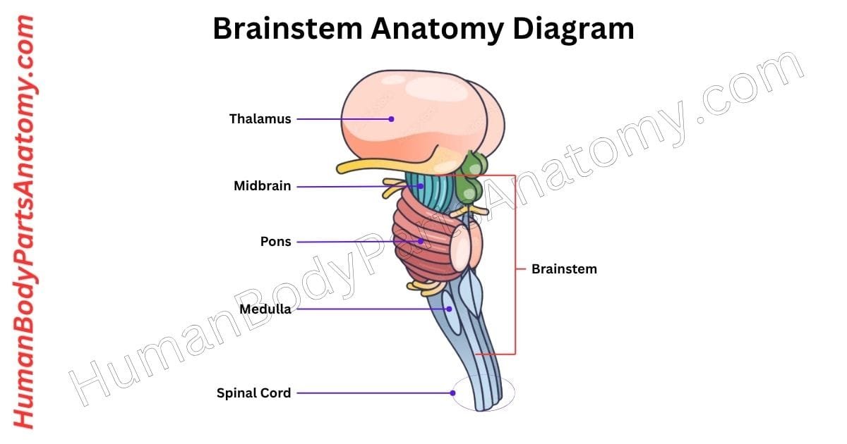
The brainstem is a small yet vital part of the brain. It is located at its base and connects the cerebrum (the largest part of the brain) to the spinal cord.[1] The brainstem anatomy consists of three main structures: the midbrain, the pons, and the medulla oblongata.[1] The brainstem anatomy consists solely of the midbrain, pons, and medulla oblongata; the thalamus is in the forebrain and connects via pathways.[2] Although the brainstem accounts for only about 2 to 3% of the brain’s total weight, it plays a critical role in sustaining life.[3] It helps regulate essential functions such as heart rate, breathing, and blood pressure.[1] It also manages the sleep-wake cycle and maintains alertness.[4]
The brainstem serves as a communication hub, transmitting motor commands from the brain to the body and sensory information from the body back to the brain.[1] It contains key neural pathways like:
- The corticospinal tract (for voluntary muscle movements),[1]
- The dorsal column-medial lemniscus pathway (for fine touch, vibration, and body position awareness),[1]
- The spinothalamic tract (for pain, temperature, and crude touch).[1]
Additionally, the brainstem is the origin point for 10 of the 12 cranial nerves, which are responsible for many functions of the face, head, and neck, including movement and sensation.[1]
Anatomy of Brainstem Diagram

Brainstem Anatomy
1. Midbrain
- Tectum (posterior part)
- Superior colliculi
- Inferior colliculi
- Tegmentum (central part)
- Red nucleus
- Substantia nigra
- Reticular formation
- Cerebral aqueduct
- Cerebral peduncles (crura cerebri)
2. Pons
- Basilar (ventral) part
- Corticospinal and corticobulbar tracts
- Pontine nuclei
- Tegmentum (dorsal part)
- Reticular formation
- Cranial nerve nuclei
- Medial lemniscus
- Middle cerebellar peduncles
3. Medulla Oblongata
- Pyramids
- Olives (olivary bodies)
- Gracile and cuneate nuclei
- Reticular formation
- Vital centers
- Cranial nerve nuclei
Cranial Nerves Originating in the Brainstem
- Midbrain: CN III (Oculomotor), CN IV (Trochlear)
- Pons: CN V (Trigeminal), CN VI (Abducens), CN VII (Facial), CN VIII (Vestibulocochlear)
- Medulla: CN IX (Glossopharyngeal), CN X (Vagus), CN XI (Accessory), CN XII (Hypoglossal)
Midbrain
The midbrain, also known as the mesencephalon, is the topmost part of the brainstem, positioned between the diencephalon (which includes the thalamus and hypothalamus) and the pons.[5]
Despite being less than 2 cm long, it plays a crucial role in multiple vital functions, including vision, hearing, motor control, sleep-wake cycles, alertness, and temperature regulation.[5]
One important part of the midbrain is the substantia nigra. It is closely linked to the basal ganglia and is essential for controlling movement. It makes a chemical called dopamine, which helps with movement and motivation.[6]
Anatomically, the midbrain sits mainly in the posterior cranial fossa, with its upper portion extending above the tentorial notch.[5] It has four main parts:
- Tectum – helps with eye and ear reflexes,[5]
- Tegmentum – carries signals related to movement and feelings,[5]
- Cerebral aqueduct – a small canal for brain fluid,[5]
- Cerebral peduncles – large nerve bundles that connect the brain to the spinal cord.[5]
The midbrain connects forward to the forebrain (specifically the diencephalon) and backward to the hindbrain (pons, medulla, and cerebellum). Toward the front, it broadens slightly on either side.[5]
In anatomical studies, the midbrain is often examined through cross-sections at two levels—either at the superior colliculi (involved in visual processing) or the inferior colliculi (involved in auditory processing).[5]

Read More – Midbrain Anatomy: Complete Guide with Parts, Names, Functions & Diagram
Pons
The pons are a central part of the brainstem located between the medulla oblongata below and the midbrain above.[1]
On its front side, the pons have a smooth, rounded shape that bulges outward. Along this surface runs a shallow groove called the basilar groove, where the basilar artery rests.[1]
On the back side of the pons, thick bundles of nerve fibers called the middle cerebellar peduncles extend to the cerebellum. These are the largest connections between the pons and cerebellum.[1]
They are essential for coordinating movement. The upper floor of the fourth ventricle is a fluid-filled space in the brain also formed by the back of the pons.[1]
At the point where the pons meets the medulla, a small groove gives rise to three cranial nerves:
- Cranial nerve VI (abducens)[1]
- Cranial nerve VII (facial)[1]
- Cranial nerve VIII (vestibulocochlear)[1]
These nerves emerge in order from the middle to the side. Higher up, from the side of the pons, cranial nerve V (trigeminal)—the largest cranial nerve—comes out. It carries both sensory and motor signals.[1]
Inside the pons are several key clusters of nerve cells (called nuclei) that perform vital roles:
- Cranial nerve nuclei handle functions like facial sensation, hearing, eye movement, and facial expression.[1]
- The locus coeruleus is located toward the back side near the periaqueductal gray. It is the main source of norepinephrine, a chemical that helps keep you alert and awake. This area is part of the reticular activating system and is one of the first regions affected by Alzheimer’s disease.[7]
- The pontine nuclei found at the front of the pons help control movement. They send information to the cerebellum through the middle cerebellar peduncles and also play a role in regulating breathing patterns.[1]
Medulla Oblongata
The medulla oblongata is the lowest part of the brainstem. It connects the pons above to the spinal cord below and passes through an opening in the skull called the foramen magnum.[8]
Front (Ventral) Surface:
On its front side, the medulla has two long ridges called pyramids. These contain nerve fibers that carry movement commands from the brain to the body. Most of these fibers cross over to the opposite side at the lower end of the medulla in a process called pyramidal decussation.[8]
- Fibers that cross form the lateral corticospinal tract, which controls limb muscles on the opposite side.[8]
- Fibers that don’t cross continue in the anterior corticospinal tract, helping to control trunk muscles.[8]
Between the pyramids is a groove called the anterior median fissure, which continues into the spinal cord.[8]
Next to each pyramid is a rounded swelling called the olive (or olivary body). This structure sends signals to the cerebellum, helping coordinate movement and timing.[8]
The hypoglossal nerve (cranial nerve XII), which controls tongue movements, exits the medulla between the pyramid and the olive.[8] Behind the olive is a groove called the postolivary sulcus, where three important cranial nerves emerge:
- Glossopharyngeal (IX) – helps with taste and swallowing,[8]
- Vagus (X) – manages heart rate, breathing, and digestion,[8]
- Accessory (XI) – controls muscles in the neck and shoulders.[8]
Back (Dorsal) Surface:
At the back of the medulla, the inferior cerebellar peduncles link it to the cerebellum, supporting balance and coordination. In the middle is a narrow groove called the posterior median sulcus.[8]
On either side of this groove are two visible bumps:
- Gracile tubercles – carry touch information from the legs and lower body,[8]
- Cuneate tubercles – carry touch information from the upper body above spinal level T6.[8]
These structures are part of the dorsal column–medial lemniscus pathway, which helps the brain detect fine touch, pressure, and body position.[8]
The upper back portion of the medulla also helps form the floor of the fourth ventricle, a cavity filled with cerebrospinal fluid (CSF). This fluid cushions the brain and maintains a stable environment.[8]
FAQ’s
The brainstem is the lower part of the brain that connects the brain to the spinal cord. It controls vital life-support functions such as breathing, heart rate, blood pressure, swallowing, and sleep cycles. The brainstem also serves as a major pathway for nerve signals traveling between the brain and the body, making it essential for movement, sensation, consciousness, and survival.[1]
The brainstem has three primary parts: the midbrain, pons, and medulla oblongata.[1] The midbrain regulates eye movement and motor control.[5] The pons helps control breathing, sleep, and facial sensation.[1] The medulla oblongata manages critical autonomic functions like heart rate, respiration, blood pressure, and reflexes such as coughing and vomiting.[8]
The brainstem is responsible for involuntary functions that keep you alive, such as breathing, swallowing, heart rhythm, and sleep cycles. It also serves as a relay center, transmitting signals between the brain and the rest of the body.[1]
Brainstem damage can cause serious or life-threatening problems because it controls vital functions like breathing, heart rate, consciousness, and motor coordination. Symptoms may include paralysis, difficulty swallowing, speech problems, coma, or respiratory failure. Causes include stroke, trauma, tumors, or neurodegenerative diseases. Brainstem injuries often require urgent medical care.[1]
The brainstem contains respiratory and cardiovascular control centers that automatically regulate breathing rhythm and heart rate. The medulla oblongata adjusts oxygen intake and blood pressure based on body needs, while the pons fine-tunes breathing patterns. These automatic processes occur without conscious effort, ensuring continuous oxygen supply and stable circulation essential for survival.[1]
The brainstem regulates sleep-wake cycles through the reticular activating system (RAS), which controls alertness and consciousness. It helps manage transitions between sleep stages and maintains wakefulness. Damage to this system can lead to sleep disorders, reduced awareness, or coma, highlighting the brainstem’s key role in maintaining consciousness and brain activity.[4]
The brainstem is located at the base of the brain, just above the spinal cord and in front of the cerebellum. It acts as the central communication bridge between the brain and the rest of the body. Its strategic position allows it to regulate vital bodily functions and transmit nerve signals efficiently.[1]
References-
- National Center for Biotechnology Information (NCBI), U.S. National Library of Medicine (NLM). (2023). Neuroanatomy, Brainstem. URL: https://www.ncbi.nlm.nih.gov/books/NBK544297/. PMID: 31335017.
- National Institutes of Health (NIH). (2024). Anatomy, Central Nervous System. URL: https://www.ncbi.nlm.nih.gov/books/NBK542179/.
- National Institutes of Health (NIH). (2023). Brainstem Anatomy: A Study on the Basis of the Pattern of Fiber Organization. URL: https://pubmed.ncbi.nlm.nih.gov/31715404/. DOI: 10.1016/j.wneu.2019.11.016. PMID: 31715404.
- National Institutes of Health (NIH). (2011). Reassessment of the Structural Basis of the Ascending Arousal System. URL: https://pmc.ncbi.nlm.nih.gov/articles/PMC3119596/. PMID: 21176049. DOI: 10.1002/cne.22559.
- National Center for Biotechnology Information (NCBI), U.S. National Library of Medicine (NLM). (2024). Neuroanatomy, Mesencephalon Midbrain. URL: https://www.ncbi.nlm.nih.gov/books/NBK551509/.
- National Center for Biotechnology Information (NCBI), U.S. National Library of Medicine (NLM). (2024). Neuroanatomy, Substantia Nigra. URL: https://www.ncbi.nlm.nih.gov/books/NBK536995/.
- National Institutes of Health (NIH). (2021). The Mechanistic Link Between Selective Vulnerability of the Locus Coeruleus and Neurodegeneration in Alzheimer’s Disease. URL: https://pubmed.ncbi.nlm.nih.gov/33427939/. DOI: 10.1007/s00401-020-02248-1. PMID: 33427939.
- National Center for Biotechnology Information (NCBI), U.S. National Library of Medicine (NLM). (2023). Neuroanatomy, Medulla Oblongata. URL: https://www.ncbi.nlm.nih.gov/books/NBK551589/.
Read More-
Human Head
- Skull Anatomy: Complete Guide with Parts, Names, Functions & Diagram
- Ultimate Guide to Eye Anatomy: Parts, Structure, Functions & Diagram
- Tongue Anatomy: Complete Guide with Parts, Names, Functions & Diagram
- Mouth Anatomy: Complete Guide with Parts, Names, Functions & Diagram
- Complete Guide to Tooth Anatomy: Learn Parts, Names & Diagram
- Ultimate Guide to Ear Anatomy: Parts, Structure, Functions & Diagram
- Nose Anatomy: Complete Guide with Parts, Names, Functions & Diagram
Brain
- Basal Ganglia Anatomy: Complete Guide with Names, Functions & Diagram
- Lobes of the Brain: Complete Guide with Names, Functions & Diagram
- Parts of the Cerebrum Anatomy: Complete Guide with Names, Functions & Diagram
- Midbrain Anatomy: Complete Guide with Parts, Names, Functions & Diagram
- The Cerebellum Anatomy: Complete Guide with Names, Functions & Diagram
Organs
- Kidney Anatomy: Complete Guide with Parts, Names, Functions & Diagram
- Liver Anatomy: Complete Guide with Parts, Names, Functions & Diagram
- Heart Anatomy: Complete Guide with Parts, Names, Functions & Diagram
External Sources-
- Wikipedia
- KenHub
- Optometrists
- Cleveland Clinic
- American Academy of Ophthalmology
Medical Disclaimer
All content on HumanBodyPartsAnatomy.com is educational and based on verified, peer-reviewed medical sources. Articles are authored or reviewed by qualified medical or biomedical professionals to ensure accuracy.
This website does not provide medical advice, diagnosis, or treatment. Always consult a licensed healthcare professional for personal medical guidance.
No commercial or promotional interests influence the medical content published on this site.