📅 Published on August 3, 2024 | 🕒 Last updated on February 18, 2026
Overview of Forearm Anatomy
The forearm is the part of your arm between your elbow and wrist and it is the lower part of your arm.[1] Forearm anatomy consists of various bones, muscles, joints, and nerves.[1] Inside the forearm are two bones called the radius and ulna,[2][3] connected by an elastic tissue called the interosseous membrane.[4] On the outside, your forearm has muscles that help you move your wrist, fingers, and elbow.[1] Some muscles bend your wrist and fingers (flexors), some straighten them out (extensors), and others help you turn your hand palm up or palm down.[1][8] In the middle of your forearm, a line divides it into two sections.[8] The back part holds the muscles that straighten your fingers and wrist, and the radial nerve controls it.[9][8] The front part has the muscles that bend your fingers and wrist, mainly controlled by the median nerve.[8][10]
The ulnar nerve also goes through your forearm.[14] Your forearm muscles are bigger on the front because they fight against gravity when you lift things.[1]
In this article, we will see all the different parts of forearm anatomy to understand their functions and names.
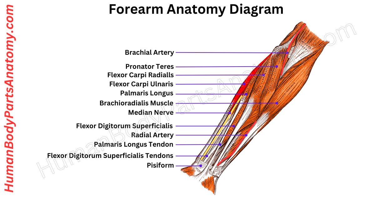
Forearm Anatomy Diagram

Parts of Forearm
Muscles
- Flexor Muscles
- Flexor Digitorum Profundus
- Flexor Digitorum Superficialis
- Flexor Carpi Ulnaris
- Flexor Carpi Radialis
- Extensor Muscles
- Extensor Carpi Radialis Longus
- Extensor Carpi Radialis Brevis
- Extensor Digitorum
- Extensor Carpi Ulnaris
- Extensor Digiti Minimi
- Pronator Muscles
- Pronator Teres
- Pronator Quadratus
- Supinator Muscle
- Other Muscles
- Brachioradialis
- Anconeus
- Palmaris Longus
- Extensor Indicis
Bones
- Ulna
- Radius
Joints
- Elbow Joint
- Radioulnar Joints
Nerves
- Median Nerve
- Radial Nerve
- Ulnar Nerve
Blood Vessels
- Brachial Artery
- Ulnar Artery
- Radial Artery
Forearm Muscle Anatomy – Flexor
The forearm has two bones: the radius on the outer side and the ulna on the inner side.[2][3] There are two muscle groups: one in the front for bending your arm and one in the back for straightening it.[1][8] Together, these muscles are in total twenty. They help move your elbow, wrist, and fingers. They are crucial for making precise movements with your upper limb.[1]
Flexor Digitorum Profundus
It is a strong muscle in your forearm that helps you grip things tightly. It is called the flexor digitorum profundus. You can feel it as a big bump on the back of your forearm near the inner side.[5]
It is the main muscle responsible for gripping because its tendons start lower down your arm than other muscles in the forearm.[5]
Flexor Digitorum Superficialis
The flexor digitorum superficialis is a big muscle in the front of your forearm. It has two parts: one starts from your elbow and the other from your radius bone. These parts join together to form four tendons that connect to your fingers.[6]
This muscle helps you bend your fingers at the middle and first knuckles. It also helps bend your wrist. It gets signals from the median nerve and blood supply from arteries in your arm.[6]
Flexor Carpi Ulnaris
The flexor carpi ulnaris is a muscle on the inner side of your forearm. It has two parts: one starts from the inner elbow and the other from the upper part of the inner forearm.[7]
These parts come together to form a strong tendon that attaches to your little finger’s wrist bones and base.[7]
This muscle is unique because it is controlled by the ulnar nerve, which runs down the inner side of your arm.[7][14] It gets its blood supply from a nearby artery.[7]
Its main job is to bend your hand at the wrist. It also helps move your hand sideways toward your metacarpal bone 5.[7]
Flexor Carpi Radialis
The flexor carpi radialis is a forearm muscle that helps move your hand and wrist. It starts from the inside of your elbow and runs down your forearm to a tendon in your wrist.[8]
From there, it connects to the base of your second and third metacarpal bones. The median nerve controls it. It gets its blood supply from branches of the ulnar and radial arteries.[8]
When it tightens, it bends your wrist downward (flexion) and moves it sideways towards your thumb side (abduction).[8] It also helps turn your palm down (pronation), but not as much as other muscles in the forearm.[1][8]
Palmaris Longus
The palmaris longus muscle is one of the outer muscles on the front side of your forearm. It starts from the medial epicondyle of the humerus. It runs down the middle of your forearm, turning into a tendon.[8]
This tendon then travels toward your wrist, passing over a flexor retinaculum, and attaches to a palmar aponeurosis.[8]
It gets its signals to move from the median nerve, and its blood supply comes from the anterior ulnar recurrent artery.[8]
The main job of the palmaris longus muscle is to work with other muscles in the front of your forearm to help you bend your hand at your wrist. It also helps steady your elbow and gives a little strength to bend your fingers at the joints.[8]
Forearm Muscle Anatomy – Extensor
Extensor Carpi Radialis Longus
The extensor carpi radialis longus is a long muscle in your forearm on the side of your thumb. It starts from a lateral supracondylar ridge of the humerus and the lateral intermuscular septum of the arm. Then, it narrows down into a tendon that connects to the base of your second finger’s hand bone.[1]
This muscle gets signals from the radial nerve in your neck and upper back. Blood comes mainly from a big artery in your arm, with a little help from another artery.[9]
It helps pull your hand back and away from your body and also helps with bending your elbow. It is important for things like grabbing and holding objects.[1]
Extensor Carpi Radialis Brevis
The extensor carpi radialis brevis is a muscle that starts from the lateral epicondyle of the humerus on the outer side of your elbow. Its fibers run down towards your wrist, and the tendon attaches to the back of the bone at the base of your third finger.[1]
This muscle gets its signals from a nerve called the deep branch of the radial nerve. It relies mainly on the radial artery in your arm for energy and nutrients.[9]
The main job of the extensor carpi radialis brevis is to help straighten and move your hand away from your body at the wrist joint.[1]
Extensor Digitorum
The extensor digitorum is a long muscle in your forearm that helps you straighten your fingers and wrist. It starts from the lateral epicondyle of the humerus via the common extensor tendon on your elbow. It runs down to your second to fifth fingers.[1]
Its main job is to straighten your fingers at the middle and end joints, and it also helps straighten your wrist.[1]
Extensor Carpi Ulnaris
The extensor carpi ulnaris muscle is located on the inside of your forearm. It starts from the lateral epicondyle of the humerus via the common extensor tendon on your elbow and runs down to your wrist.[1]
Further, it attaches to the metacarpal bone of the 5th digit.[1] A nerve from your spine powers it[9] and gets blood supply from nearby arteries.[8]
The extensor carpi ulnaris muscle’s main job is to move your hand sideways toward your metacarpal bone (ulnar deviation).[1] It also helps with stretching your hand out, working alongside other muscles.[8]
Extensor Digiti Minimi
The extensor digiti minimi is a thin muscle near the outer side of your forearm. It starts from the lateral epicondyle of the humerus via the common extensor tendon. It stretches down to connect to your digit 5 finger.[1]
This muscle helps straighten your pinky finger at the joint closest to your hand, but it can also help with other pinky movements.[1]
It gets power from a nerve in your arm[9] and blood from nearby arteries.[8] Overall, it plays a role in straightening your pinky finger and extending your wrist.[1][8]
Brachioradialis
The brachioradialis muscle in your forearm is important for bending your elbow and stabilizing during hammering. It connects from the bottom end of your radius bone to a ridge on the outer part of your upper arm bone (humerus).[12]
Depending on your forearm position, this muscle can rotate your forearm inward (pronation) and outward (supination).[12]
The brachioradialis works best when you bend your elbow with your forearm halfway between fully turned inward and outward.[12]
When you bend your forearm inward, the brachioradialis muscle helps to turn it outward (supinate) as it bends.[12]
When your forearm is already turned outward (supinated), it is easier to bend it by turning it inward (pronate). It allows quick movements when lifting weights slowly.[12]
Forearm Anatomy – Pronator Muscles
Pronator Teres
The pronator teres muscle is one of the outer forearm muscles. It has two parts: the humeral head starts from the medial supracondylar ridge of the humerus (inside of the upper arm bone), and the ulnar head from a coronoid process of the ulna(lower arm bone).[13]
These parts come together to form a tendon that attaches to a pronator tuberosity on the lateral side of the radius bone.[13]
This muscle gets signals from the median nerve and blood from the brachial, radial, and ulnar arteries. Its main job is turning the palm downward by rotating the forearm at the top joint near the elbow. It also helps in bending the forearm at the elbow.[13]
Pronator Quadratus
The pronator quadratus is a square-shaped muscle in the lower part of the forearm. It helps to turn the hand so the palm faces downwards.[8]
Its fibers run across the arm and connect from the front of the ulna’s lower quarter to the radius’s lower quarter.[4][8]
This muscle has two heads: the superficial head starts from the front lower part of the ulna’s shaft and attaches to the front lower part of the radius’s shaft and its front section. The deep head also begins from here but attaches closer to the notch of the ulna.[8]
It is unique because it is the only muscle that connects solely from the ulna to the radius. Blood comes to it from the anterior interosseous artery.[8]
When the pronator quadratus tightens, it pulls the radius outer side towards the ulna and rotates the hand downward. Its deep fibers help keep the bones of the forearm together.[8]
Supinator Muscle
The supinator is a muscle in the back of your forearm. It works with other muscles like the brachioradialis and the forearm extensors.[1]
This muscle wraps around the top part of the radius bone, connecting it to the ulna bone. Because it crosses the forearm, the supinator helps turn your hand’s palm up, a movement called supination.[1]
When you turn your hand the other way, with the palm facing down, the pronator quadratus muscle does the work called pronation.[8]
The supinator’s job is to rotate the radius bone laterally at the elbow, paralleling the radius and ulna bones and turning your hand palm up.[1]
Anatomy Forearm Muscles – Other
Anconeus
The anconeus is a small, triangular muscle located at the back of the elbow. It runs from the lower end of the humerus (the upper arm bone) to the upper part of the ulna (one of the forearm bones).[1]
This muscle is part of the group that helps extend the wrist and fingers, along with muscles like the brachioradialis, extensor carpi radialis longus and brevis, extensor digitorum, extensor digiti minimi, and extensor carpi ulnaris.[1]
The anconeus helps straighten the arm at the elbow. It stabilizes the elbow joint by supporting the back of the joint and the ulna.[8]
Extensor Indicis
The extensor indicis is a muscle in the back of the forearm. It is part of a group of muscles that help move the wrist and fingers.[1]
You can feel it by pressing firmly on the bone on the pinky side of your wrist while straightening your index finger.[1]
Its main job is to straighten the index finger at the joints closest to the hand. Unlike other fingers that share muscles for movement, the index finger has its muscle dedicated to this action.[1]
It also plays a minor role in bending the wrist backward.[8]
Forearm Anatomy – Bones
Ulna
The ulna is one of the two long bones in the forearm. It is positioned on the inner side opposite the thumb, running from the elbow to the wrist. It is longer and thinner than the radius.[3]
At the elbow, the ulna has a prominent bony projection called the olecranon process. It fits into the humerus to form a hinge joint and prevent the arm from overextending.[3]
Towards the wrist, the ulna tapers and features a notch that accommodates the head of the radius. Additionally, there is a small bump called the ulnar tuberosity, where various muscles attach.[3]
Radius
The radius is one of the bones in your forearm. It plays a critical role in moving your arm and wrist. It also supports important muscles, tendons, ligaments, and blood vessels.[2]
If you break your radius, you might need surgery to fix it and physical therapy to regain strength and movement.[2]
Because the radius is connected to many muscles and nerves, an injury can also impact these.[2]
Your radius has several important functions:
- Helping your forearm and wrist move, bend, and rotate.[2]
- Supporting seven muscles.[2]
- Stabilizing your arm, wrist, and hand.[2]
Forearm Anatomy – Joints
Elbow Joint
The elbow is the portion where the upper arm meets the forearm and forms a joint crucial for bending and straightening the arm. It comprises bones like the humerus, radius, and ulna.[11][17]
This joint acts like a hinge, enabling us to move our forearm and hand towards and away from our body. Muscles like the biceps and triceps help with bending and straightening the arm.[11][17]
In humans and other primates, this area is called the elbow. However, it is referred to as the forelimb joint in different animals.[11][17] The elbow’s range of motion goes from completely straight (0 degrees) to bent (150 degrees).[15]
Overall, the function of the elbow is to position our hand accurately by adjusting the length of our upper limb.[11][17]
Radioulnar Joints
The proximal radioulnar joint (PRUJ) is a synovial pivot joint in the forearm. It is the joint where the radius’s rounded head fits into the ulna’s radial notch.[2]
This unique setup allows the radius to pivot around the ulna and enables the forearm to rotate.[2] It is essential for twisting motions like turning your palm up or down.[4]
The distal radioulnar joint (DRUJ) is another synovial pivot joint located at the opposite end of the radius and ulna.[2] It features an articular disc that provides cushioning between the bones and is secured by the palmar and dorsal radioulnar ligaments.[4][16]
The DRUJ also allows for rotation and movement of the forearm.[2] It works with the PRUJ to support various hand and wrist activities.[4][16]
The forearm contains flexor muscles on the palm side (like flexor carpi radialis) and extensor muscles on the back side (like extensor digitorum).[1][8] These muscles control wrist movement, finger motion, and grip strength.[1]
The forearm contains two bones: the radius (on the thumb side, allowing rotation) and the ulna (on the pinky side, providing stability). These bones articulate at the elbow and wrist joints.[2][3]
There are typically 20 muscles in the forearm, divided into anterior (flexor) and posterior (extensor) compartments. These muscles control wrist, finger, and forearm movements.[1]
The forearm enables hand rotation, wrist movement, finger control, and gripping actions.[1][4] It also supports lifting, pushing, pulling, and fine motor tasks used in daily activities.[1]
Common causes include muscle strain, tendonitis (tennis elbow or golfer’s elbow), nerve compression, repetitive motion, or injury. Poor ergonomics and overuse are frequent contributors.[11][17]
The radius rotates around the ulna to turn the palm up or down,[2] while the ulna provides stability to the forearm.[3] Both bones are essential for arm strength and flexibility.[2][3][4]
References-
- Mitchell B, Whited L. (2023). Anatomy, Shoulder and Upper Limb, Forearm Muscles. In: StatPearls [Internet]. Treasure Island (FL): StatPearls Publishing; 2025 Jan–. Updated June 5, 2023. PMID: 30725660.
https://www.ncbi.nlm.nih.gov/books/NBK536975/
— Comprehensive review of anterior and posterior forearm muscle groups, innervation, blood supply, and clinical correlations. - Bair MM, Zafar Gondal A. (2023). Anatomy, Shoulder and Upper Limb, Forearm Radius. In: StatPearls [Internet]. Treasure Island (FL): StatPearls Publishing; 2025 Jan–. Updated July 31, 2023. PMID: 31356039.
https://www.ncbi.nlm.nih.gov/books/NBK544512/
— Detailed anatomical description of the radius, ossification, articulations, and fracture relevance. - Baig MA, Byerly DW. (2023). Anatomy, Shoulder and Upper Limb, Forearm Ulna. In: StatPearls [Internet]. Treasure Island (FL): StatPearls Publishing; 2025 Jan–. Updated July 24, 2023. PMID: 31613529.
https://www.ncbi.nlm.nih.gov/books/NBK547749/
— Covers ulna anatomy, proximal and distal articulations, and clinical significance in elbow injuries. - Bandovic I, Holme MR, Black AC, Futterman B. (2023). Anatomy, Shoulder and Upper Limb, Forearm Bones. In: StatPearls [Internet]. Treasure Island (FL): StatPearls Publishing; 2025 Jan–. Updated June 20, 2023.
https://www.ncbi.nlm.nih.gov/books/NBK545260/
— Integrated overview of radius and ulna biomechanics and structural relationships. - Forearm Anatomy Overview. Mayo Clinic; Reviewed by medical professionals.
https://www.mayoclinic.org/diseases-conditions/arm-pain/symptoms-causes/syc-20373146
— Patient-centered explanation of forearm structure, causes of arm pain, and clinical context. - Okafor L, Varacallo MA. (2022). Anatomy, Shoulder and Upper Limb, Hand Flexor Digitorum Superficialis Muscle. In: StatPearls [Internet]. Treasure Island (FL): StatPearls Publishing; 2025 Jan–. Updated October 17, 2022.
https://www.ncbi.nlm.nih.gov/books/NBK539723/
— Functional anatomy, innervation (median nerve), and clinical testing relevance. - Lung BE, Siwiec RM. (2024). Anatomy, Shoulder and Upper Limb, Forearm Flexor Carpi Ulnaris Muscle. In: StatPearls [Internet]. Treasure Island (FL): StatPearls Publishing; 2025 Jan–. Updated January 30, 2024.
https://www.ncbi.nlm.nih.gov/books/NBK526051/
— Discusses ulnar nerve innervation and role in wrist flexion and adduction. - Khan SA, Hafeez AM, Sinkler MA, Arain A. (2023). Anatomy, Shoulder and Upper Limb, Forearm Compartments. In: StatPearls [Internet]. Treasure Island (FL): StatPearls Publishing; 2025 Jan–. Updated July 31, 2023.
https://www.ncbi.nlm.nih.gov/books/NBK539784/
— Describes fascial compartments and compartment syndrome clinical implications. - Glover NM, Black AC, Murphy PB. (2023). Anatomy, Shoulder and Upper Limb, Radial Nerve. In: StatPearls [Internet]. Treasure Island (FL): StatPearls Publishing; 2025 Jan–. Updated November 5, 2023.
https://www.ncbi.nlm.nih.gov/books/NBK534840/
— Radial nerve course, motor/sensory supply, and wrist drop pathology. - Sevy JO, Sinha P, Varacallo M. (2024). Anatomy, Shoulder and Upper Limb, Median Nerve. In: StatPearls [Internet]. Treasure Island (FL): StatPearls Publishing; 2024 Jan–.
https://www.ncbi.nlm.nih.gov/books/NBK448084
— Clinical anatomy of carpal tunnel syndrome and forearm innervation. - Card RK, Lowe JB, Launico MV. (2025). Anatomy, Shoulder and Upper Limb, Elbow Joint. In: StatPearls [Internet]. Treasure Island (FL): StatPearls Publishing; 2025 Jan–. Updated December 9, 2025.
https://www.ncbi.nlm.nih.gov/books/NBK532948/
— Detailed elbow joint structure, ligaments, ROM, and common pathologies. - Lung BE, Ekblad J, Bisogno M. (2024). Anatomy, Shoulder and Upper Limb, Forearm Brachioradialis Muscle. In: StatPearls [Internet]. Treasure Island (FL): StatPearls Publishing; 2025 Jan–. Updated January 30, 2024.
https://www.ncbi.nlm.nih.gov/books/NBK526110/
— Functional anatomy in elbow flexion and clinical reflex testing. - Luxenburg D, Rizzo MG. (2023). Anatomy, Shoulder and Upper Limb, Pronator Teres. In: StatPearls [Internet]. Treasure Island (FL): StatPearls Publishing; 2025 Jan–. Updated May 1, 2023. PMID: 35593871.
https://www.ncbi.nlm.nih.gov/books/NBK580564/
— Anatomical basis of pronator syndrome and median nerve compression. - Byju A, Sinkler MA, Lopez-Ojeda W. (2023). Anatomy, Shoulder and Upper Limb, Ulnar Nerve. In: StatPearls [Internet]. Treasure Island (FL): StatPearls Publishing; 2025 Jan–. Updated July 24, 2023.
https://www.ncbi.nlm.nih.gov/books/NBK499892/
— Ulnar nerve anatomy, cubital tunnel syndrome, and motor deficits. - Normative Values for Elbow Range of Motion. (2019). Published in Journal of Hand and Microsurgery. PMCID: PMC6555111.
https://pmc.ncbi.nlm.nih.gov/articles/PMC6555111/
— Evidence-based elbow flexion-extension and pronation-supination reference ranges. - Arias DG, Varacallo MA. (2025). Anatomy, Shoulder and Upper Limb, Distal Radioulnar Joint. In: StatPearls [Internet]. Treasure Island (FL): StatPearls Publishing; 2025 Jan–. Updated February 26, 2025.
https://www.ncbi.nlm.nih.gov/books/NBK547720/
— DRUJ stability, biomechanics, and triangular fibrocartilage complex relevance. - Elbow Joint Overview. Cleveland Clinic; Medically reviewed content.
https://my.clevelandclinic.org/health/body/elbow-joint
— Patient-friendly explanation of elbow anatomy, ligaments, and injury causes.
Read More-
Lower Limb
- Complete Guide on Leg Anatomy with Parts, Functions & Diagram
- Complete Guide to Thigh Muscle Anatomy: Learn Parts, Names & Diagram
- Knee Anatomy: Complete Guide to Parts, Names, Functions & Diagram
- Femur Anatomy: Complete Guide with Parts, Names, Functions & Diagram
Upper Limb
- Complete Guide to Finger Anatomy with Parts, Names, Functions & Diagram
- Comprehensive Guide to Arm Anatomy: Parts, Names & Diagram
- Comprehensive Guide to Hand Anatomy: Parts, Functions & Diagram
- Ultimate Guide to Bicep Anatomy: Parts, Names, Functions & Diagram
- Shoulder Anatomy: Ultimate Guide to Parts, Names, Functions & Diagram
- Wrist Anatomy: Ultimate Guide to Parts, Names, Functions & Diagram
- Complete Guide to Nail Anatomy with all Parts, Names & Diagrams
Human Head
- Ultimate Guide to Eye Anatomy: Parts, Structure, Functions & Diagram
- Tongue Anatomy: Complete Guide with Parts, Names, Functions & Diagram
- Mouth Anatomy: Complete Guide with Parts, Names, Functions & Diagram
- Complete Guide to Tooth Anatomy: Learn Parts, Names & Diagram
- Ultimate Guide to Ear Anatomy: Parts, Structure, Functions & Diagram
Medical Disclaimer
All content on HumanBodyPartsAnatomy.com is educational and based on verified, peer-reviewed medical sources. Articles are authored or reviewed by qualified medical or biomedical professionals to ensure accuracy.
This website does not provide medical advice, diagnosis, or treatment. Always consult a licensed healthcare professional for personal medical guidance.
No commercial or promotional interests influence the medical content published on this site.