
📅 Published on October 23, 2024 | 🕒 Last updated on July 15, 2026
Overview of Hip Bone Anatomy
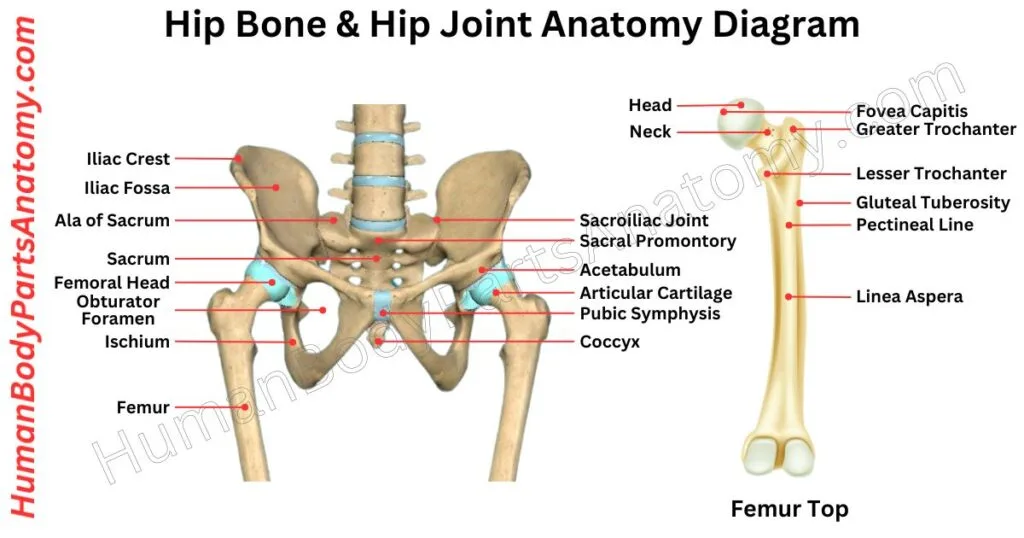
The hip bone, or os coxae, is a large, irregular bone that forms the base of the lower limb.[1] The main functions of the hip bone are to support the body’s weight when standing and provide a base for leg movement.[1][2] It connects to the spine at the sacroiliac joint and the leg at the hip joint.[2][1] Hip bone anatomy comprises three parts that fuse during the teenage years: the ilium, ischium, and pubis.[1][3]
- Ilium: The broad, upper part of the pelvis.
- Ischium: The part of the hip bone you sit on.
- Pubis: The lower, front part of the pelvis.
The acetabulum is a socket on the side of the hip where the head of the thigh bone (femur) fits in, allowing leg movement.[1][2] It is formed by the three parts of the hip bone anatomy.[1] It is separated by cartilage until the ages of 15–17 when they fuse.[3] The hip also contains important nerves and blood vessels for movement and feeling.[1] The femoral nerve is at the front of the thigh,[5] the sciatic nerve runs along the back,[4] and the smaller obturator nerve is deeper.[1] The femoral artery, one of the body’s main arteries, can be felt at the front of the upper thigh.[6]
Detailed Hip Bone Anatomy Diagram
Hip Bones Anatomy
- Ilium
- Ischium
- Pubis
- Acetabulum
Hip Bone Anatomy: Parts & Functions
Ilium
The ilium is a key part of the pelvic bone, comprising two main sections: the body and the wing. Together with the ischium and pubis, the ilium forms the pelvis. Where these bones join, the connections are faint and barely visible.[1]
1. Body of Ilium
The body of the ilium makes up less than 40% of the acetabulum, which is the socket where the thigh bone (femur) fits. It also helps shape part of the acetabular fossa.[1]
Inside, the body lines part of the lesser pelvis and provides an attachment point for some obturator internus muscle fibers.[1]
2. Wing
The wing, also known as the ala, is the wide, flat part that forms the sides of the greater pelvis. It has an outer and inner surface, a crest, and front and back edges.[1]
These features shape the ilium and offer important attachment points for muscles and ligaments, helping with posture and movement.[1]
The ilium also has several important landmarks, including:
- Iliac crest – It is the upper edge of the ilium, the largest bone in the pelvis.[1]
- Anterior superior iliac spine – A prominent bony projection is known as the anterior superior iliac spine at the front of the iliac crest.[1]
- Anterior inferior iliac spine – Just below the anterior superior iliac spine, we can see the anterior inferior iliac spine extends downward.[1]
- Posterior superior iliac spine – On the back side of the ilium, the posterior superior iliac spine marks the rear boundary of the iliac crest.[1]
- Posterior inferior iliac spine – Positioned below the posterior superior iliac spine, adjacent to a roughened surface called the auricular surface, which connects with the sacrum.[1]
- Iliac fossa – It is a smooth, concave region on the inner surface of the upper ilium, where muscles attach.[1]
- Greater sciatic notch – The greater sciatic notch is near the lower rear of the ilium, with a large U-shaped indentation. It allows the sciatic nerve and other structures to pass from the pelvis to the lower limbs.[1][4]
Ischium
The ischium is a key part of the pelvis, playing a major role in both movement and supporting the body weight. It has three main sections:[1]
1. Body
The main part of the ischium features a bump called the ischial spine, where a muscle called the superior gemellus attaches. Below this is a curved area known as the lesser sciatic notch.[1]
Further down is the ischial tuberosity, a rough surface that supports your body when sitting, especially on hard surfaces. It is also where the hamstring muscles and the inferior gemellus muscle begin.[1]
2. Superior Ramus
This section is where muscles like the internal and external obturators attach, which help rotate the thigh.[1]
3. Inferior Ramus
At the front, the inferior ramus connects to the pubic bone. It provides attachment points for muscles such as the adductor magnus and gracilis, essential for leg movement.[1]
The ramus of the ischium features key bony landmarks:
- Ischial tuberosity – A prominent bony structure located at the lower part of the ramus, just below the lesser sciatic notch. It is an attachment point for muscles and bears the body’s weight when sitting.[1]
- Ischial spine – A sharp bony projection positioned between the greater and lesser sciatic notches, playing a role in ligament attachment and serving as a boundary for these notches.[1]
- Lesser sciatic notch – A small curved indentation between the ischial spine and the ischial tuberosity forms a passageway for nerves and vessels.[1]
- Greater sciatic notch – A larger, curved indentation between the posterior inferior iliac and the ischial spine, through which the sciatic nerve exits the pelvis.[1]
Each feature contributes to the pelvis’s structural and functional integrity, supporting movement and protecting neurovascular pathways.[1]
Pubis
In vertebrates, the pubis, or pubic bone, forms the lower front part of the hip on each side. It is the most forward-facing of the three bones that make up the hip, with each pubic bone having three main parts: the body, the superior ramus, and the inferior ramus.[1]
1. Body of the Pubis
The body is the central, flat, and sturdy part of the pubic bone. It connects with the opposite pubic bone at the pubic symphysis, a joint at the body’s center.[1]
The body’s top edge is called the pubic crest, which ends in a small bump known as the pubic tubercle. The body of the pubis has three surfaces: one facing forward (anterior), one facing backward (posterior), and one facing inward (medial).[1]
2. Superior Pubic Ramus
The superior pubic ramus is the upper portion of the pubis. It forms the top boundary of the obturator foramen, a large hole in the pelvis.[1]
The superior pubic ramus extends from the body of the pubis to the other side. It is divided into a flat inner section and a narrower outer section.[1]
3. Inferior Pubic Ramus
The inferior pubic ramus is the lower branch of the pubis. It extends downward and outward, becoming thinner as it goes, and connects with the ischium, another bone in the pelvis, below the obturator foramen.[1]
The obturator foramen lies between the superior and inferior pubic rami, and the two pubic bones meet at the pubic symphysis, allowing them to join at the middle of the pelvis.[1]
Acetabulum
The acetabulum, also known as the cotyloid cavity, is a deep, curved surface on the pelvis that forms part of the hip joint.[2] It is where the head of the femur fits, creating the ball-and-socket structure of the hip.[1][2] Three bones of the hip—ischium, ilium, and pubis—merge to shape the acetabulum.[1]
The ischium forms over two-fifths of the acetabulum, providing support along the lower and side areas. The ilium contributes slightly less than two-fifths to the upper portion. At the same time, the pubis, located near the center, forms the remaining part.[1]
1. Lunate Surface
- It is a crescent-shaped, curved area of the acetabulum.[7]
- It is the primary surface where the head of the femur connects to form the hip joint.[7]
- Provides smooth articulation for the ball-and-socket joint movement.[7]
2. Acetabular Notch
- Located at the lower part of the acetabulum.[7]
- It acts as a gap in the rim of the acetabulum, allowing passage for blood vessels and ligaments.[7]
- It continues with the acetabular fossa, helping support joint stability.[7]
3. Acetabular Fossa
- A circular depression at the bottom of the acetabular cavity.[7]
- It does not directly articulate with the femur but provides space for ligaments and the joint capsule.[7]
- It is important for distributing forces within the hip joint.[7]
FAQ’s
The hip bone, also called the coxal or innominate bone, is a large, irregular bone that forms the pelvis. It connects the spine to the lower limbs, supports body weight, and allows movement such as walking, running, and bending.[1][2]
The hip bone is made up of three fused bones:
Ilium – the broad, upper part.
Ischium – the lower, back part you sit on.[1]
Pubis – the front portion that joins at the pubic symphysis.[1]
The hip bones are located on either side of the pelvis, joining with the sacrum at the back and meeting each other in the front at the pubic symphysis.[1]
Major muscles that attach to the hip bone include the gluteal muscles, the iliopsoas, quadriceps, hamstrings, and adductors.[1][8] These muscles help in walking, running, sitting, and maintaining balance.[1]
Common issues include hip fractures, arthritis, bursitis, hip dysplasia, and labral tears.[9][10] These conditions often cause pain, stiffness, or reduced mobility.[10]
The hip bone is a single bone on each side (left and right), while the pelvic bone refers to the entire pelvic girdle, which includes both hip bones, the sacrum, and the coccyx.[1]
To maintain hip health:
Exercise regularly (walking, stretching, strength training).[11]
Maintain good posture.[1]
Eat a calcium- and vitamin D-rich diet.[12]
Avoid smoking and excessive alcohol.[11]
Seek medical advice if hip pain is severe, lasts more than a few days, limits movement, or follows an injury. Early diagnosis helps prevent long-term problems.[10]
References –
- National Center for Biotechnology Information (NCBI). Anatomy, Abdomen and Pelvis: Bones (Ilium, Ischium, and Pubis). StatPearls Publishing; 2023 (last update July 24). https://www.ncbi.nlm.nih.gov/books/NBK519524/
- National Center for Biotechnology Information (NCBI). Anatomy, Bony Pelvis and Lower Limb: Pelvic Bones. StatPearls Publishing; 2023 (last update July 24). https://www.ncbi.nlm.nih.gov/books/NBK551580/
- Moore KL, et al. (2024). Anatomy, Bony Pelvis and Lower Limb: Hip Joint Development and Ossification. StatPearls Publishing.
https://www.ncbi.nlm.nih.gov/books/NBK526019/ - National Center for Biotechnology Information (NCBI). Anatomy, Sciatic Nerve. StatPearls Publishing; 2023. https://www.ncbi.nlm.nih.gov/books/NBK482431/
- National Center for Biotechnology Information (NCBI). Anatomy, Abdomen and Pelvis: Femoral Region. StatPearls Publishing; 2024 (last update Feb 9). https://www.ncbi.nlm.nih.gov/books/NBK538501/
- National Center for Biotechnology Information (NCBI). Anatomy, Bony Pelvis and Lower Limb: Femoral Artery. StatPearls Publishing; 2023 (last update Nov 29). https://www.ncbi.nlm.nih.gov/books/NBK538262/
- Standring S, et al. (2021). Acetabulum. In: Gray’s Anatomy: The Anatomical Basis of Clinical Practice (42nd ed.). Elsevier.
https://www.elsevier.com/books/grays-anatomy/standring/978-0-7020-7705-0 - National Center for Biotechnology Information (NCBI). Anatomy, Bony Pelvis and Lower Limb: Pelvis Bones. StatPearls Publishing; 2023 (last update Jul 30). https://www.ncbi.nlm.nih.gov/books/NBK545204/
- MedlinePlus (National Library of Medicine). Hip Injuries and Disorders. U.S. National Library of Medicine; 2025 (last reviewed Jun 25). https://medlineplus.gov/hipinjuriesanddisorders.html
- [Mayo Clinic. Hip Dysplasia – Symptoms and Causes. Mayo Foundation for Medical Education and Research; 2024 (last reviewed Mar 5). https://www.mayoclinic.org/diseases-conditions/hip-dysplasia/symptoms-causes/syc-20350209
- [Mayo Clinic. Exercising with Osteoporosis: Stay Active the Safe Way. Mayo Foundation for Medical Education and Research; 2023. https://www.mayoclinic.org/diseases-conditions/osteoporosis/in-depth/osteoporosis/art-20044989
- [National Institutes of Health (NIH). Determinants of Bone Health. In: Bone Health and Osteoporosis: A Report of the Surgeon General. 2023 https://www.ncbi.nlm.nih.gov/books/NBK45503/
Read More-
Human Body-
- Human Anatomy: Guide to Bones, Muscles, Organs, Systems, Functions & Diagram
- Human Skeleton Anatomy: All 206 Bones Explained with Functions & Diagrams
- Human Muscle Anatomy: Validated Guide to Every Major Muscles & Functions
Head, Face & Senses-
- Nose Anatomy: Parts of the Nose, Structure, Nasal Cavity & Sinuses Explained
- Skull Anatomy: Parts of the Skull, Structure, Cranial, Facial Bones & Functions
- Mouth Anatomy: Guide on Parts of Mouth, Lips, Palate, Gums & Oral Cavity
- Eye Anatomy: Parts of the Eye, Cornea, Lens, Retina, Optic Nerve & Diagram
- Ear Anatomy: Parts of the Ear, Outer, Middle & Inner Ear & Structures
Brain & Nervous System-
- Brain Anatomy: Parts of the Brain, Structure, Functions & Regions Explained
- The 4 Lobes of the Brain: Complete Guide with Locations & Functions
Spine & Back-
- Cervical Spine Anatomy: C1–C7 Vertebrae, Muscles & Nerves Explained
- Spine Anatomy: Parts of the Spine, Vertebrae, Curves, Spinal Cord & Diagram
- Neck Muscle Anatomy: Guide with Key Muscles, Groups, Functions & Diagrams
- Rib Cage Anatomy: Ribs, Sternum, Thoracic, Vertebrae & Functions Explained
Organs-
- Stomach Anatomy: Parts of Stomach, Regions, Layers & Digestive Function
- Heart Anatomy: Guide on Parts of Heart, Chambers, Valves & Blood Flow
- Liver Anatomy: Key Parts of Liver, Functions, Lobes, Segments & Diagram
- Kidney Anatomy: Guide on Parts of Kidney, Structure, Functions & Diagram
Upper Limb-
- Forearm Anatomy: Parts of the Forearm, Radius, Ulna, Muscles & Diagram
- Shoulder Anatomy: Parts of the Shoulder, Bones, Joint Structure & Diagram
- Wrist Anatomy: Parts of the Wrist, 8 Carpal Bones, Tendons & Diagram
- Hand Anatomy: Parts of the Hand, Bones, Muscles with Functions & Diagram
- Arm Anatomy: Parts of Arm, Bones, Muscles & Joints with Functions & Diagram
Lower Limb-
- Hip Muscle Anatomy: Guide on Key Muscle Groups, Names, Functions & Diagram
- Femur Anatomy: Parts of Femur, Structure, Functions, Location & Diagram
- Leg Anatomy: Parts of the Leg, Bones, Muscles & Lower Leg with Functions
- Knee Anatomy: Parts of Knee, Bones, Ligaments, Cartilage & Joint Structure
- Thigh Muscle Anatomy: Key Muscle Groups, Names, Functions & Diagram
Medical Disclaimer
All content on HumanBodyPartsAnatomy.com is educational and based on verified, peer-reviewed medical sources. Articles are authored or reviewed by qualified medical or biomedical professionals to ensure accuracy.
This website does not provide medical advice, diagnosis, or treatment. Always consult a licensed healthcare professional for personal medical guidance.
No commercial or promotional interests influence the medical content published on this site.