
📅 Published on August 24, 2024 | 🕒 Last updated on July 15, 2026
Overview of Rib Cage Anatomy
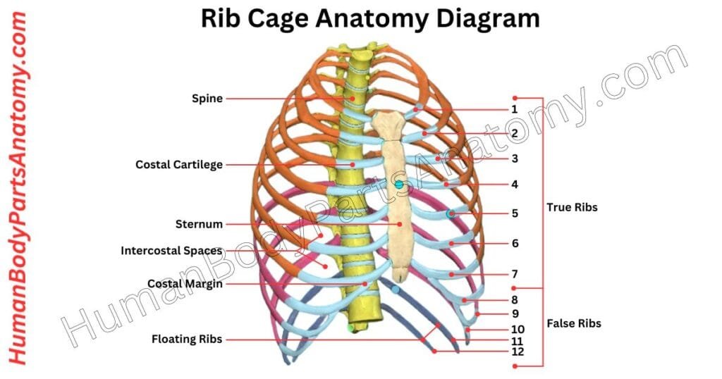
The rib cage is an essential body element that protects the heart and lungs.[1] It creates the chest area known as the thorax, which comprises ribs, the sternum, and a portion of the spine.[5] The rib cage anatomy has 24 ribs, 12 on each side, as well as 12 vertebrae, which are bones that form the spine in the chest area.[1][3] The ribs wrap across the chest, becoming broader as you progress from the top ribs to the lowest ones.[1] While the rib cage is robust, it may expand somewhat, aiding breathing. The tiny joints between the ribs and the spine allow the ribs to move easily when breathing and doing other tasks.[6]
Rib Cage Anatomy Diagram
Anatomy of Rib Cage
- Ribs
- Sternum (Breastbone)
- Costal Cartilages
- Thoracic Vertebrae
- Intercostal Muscles
- Costal Margin
- Costovertebral Joints
- Costocorporeal Joints (Joints of the Head of Ribs):
- Costotransverse Joints:
- Pleura
Rib Cage Anatomy
Ribs
The rib cage is a flat bone collection that protects vital organs such as the heart and lungs. Most humans have 24 ribs grouped into 12 pairs.[1]
Occasionally, someone may have an additional rib, known as a cervical rib, on one or both sides of the neck. Each rib is attached to the spine in the back and numbered according to the thoracic vertebrae it joins, beginning with the first rib (T1).[1]
Most ribs in the front link to the sternum (breastbone) via flexible cartilage. Costovertebral joints are where the ribs connect the vertebrae.[1]
- True Ribs (1-7):
- False Ribs (8-10):
- Floating Ribs (11-12):
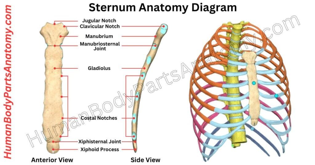
Sternum (Breastbone)
The sternum, or breastbone, is a flat, central bone in your chest that helps form the rib cage. It has three main parts:[2]
Manubrium
It is a top section shaped like a broad, flat rectangle. It has a notch at the top (suprasternal notch) and two side notches for the collarbones to create the sternoclavicular joints.[2]
Body (Gladiolus)
The longest part of the sternum. It has ridges where the cartilages of ribs 3 through 7 attach. This section connects with the manubrium at a prominent ridge called the sternal angle. Also, it connects with the second pair of ribs.[2]
Xiphoid Process
The smallest and lowest section is shaped like a triangle. Its size and shape can differ from person to person.[2]
The sternal angle is the visible bump where the manubrium and body meet. The main job of the sternum is to protect important organs like the heart and lungs.[2]
Costal Cartilages
Costal cartilage is made of hyaline cartilage[8] and helps extend the ribs forward, adding flexibility to the chest wall. It is found only at the front ends of the ribs.[5]
- The first seven pairs of costal cartilage attach to the sternum.[1]
- The next three connect to the rib cartilage above.[1]
- And the last two end in the abdominal wall.[1]
Costal cartilages vary in length, width, and direction. They get longer from the first to the seventh rib, then shorten toward the twelfth. They also get narrower from the first to the last rib.[5]
Most are wider, where they attach to the ribs and taper as they approach the sternum. Except for the first two, they are evenly wide, and the sixth, seventh, and eighth are wider where they meet.[5]
Their direction also varies: the first cartilage slopes slightly downward, the second is horizontal, and the third tilts up a bit. While the others angle, following the rib’s curve briefly before moving up to the sternum or the cartilage of the rib above.[5]
Thoracic Vertebrae
In vertebrates, the thoracic vertebrae form the middle part of the spine. It is between the neck (cervical vertebrae) and the lower back (lumbar vertebrae).[3]
Humans have twelve of these vertebrae, labeled T1 to T12. It starts from the top near the skull and moves down toward the lower back. These vertebrae get bigger as you go down the spine.[3]
They are special because they have surfaces called facets that connect to the ribs. Most of them, except the last two, also have extra facets on their side branches called transverse processes. It helps attach the ribs.[3]
Intercostal Muscles
The intercostal muscles are essential for breathing. It is positioned between your ribs in three layers. These muscles are crucial in altering your chest cavity’s size to allow you to breathe effectively. Each side of your ribcage houses 11 pairs of these muscles, organized into external, internal, innermost layers.[4]
External Intercostal Muscles
It is located on the outer side of your ribcage. These muscles extend from the rear of the ribs toward the front, stopping just before the rib ends. When you inhale, these muscles contract, lifting your ribs and expanding your chest.[4]
Internal Intercostal Muscles
It is found on the inner side of your ribcage. These muscles begin at the front and curve around to the back. They work in tandem with the external intercostals but are oriented to support different movements.[4]
External intercostals are active during inspiration (raise ribs); internal intercostals (particularly the interosseous portion) are primarily active during forced expiration. During normal quiet inspiration external intercostals + diaphragm are principal actors; internal intercostals are mainly for forced expiration.[4]
Costal Margin
The costal margin is also known as the costal arch. It forms the lower boundary of the chest, shaped by the cartilages of the seventh through tenth ribs.[7]
These cartilages connect to the sternum, including its body and xiphoid process. This structure provides an attachment point for the diaphragm, the primary muscle involved in breathing. The costal angle is where the left and right costal margins converge at the sternum.[7]
It creates a protective zone for vital organs in the upper abdomen, such as the liver. This region protects these organs while facilitating respiratory movements.[7]
Costovertebral Joints
The costovertebral joints are two types of joints that connect the ribs to the spine at the back, helping form the chest cage. These are synovial plane joints, which allow limited movement.[6]
- Costocorporeal Joints (Joints of the Head of Ribs): These joints link the heads of the ribs to one or two adjacent vertebrae.[6]
- Costotransverse Joints: These connect the ribs’ necks and tubercles (small rounded projections) to the corresponding thoracic vertebrae’s transverse processes (side projections).[6]
These joints enable small movements, often called ‘pump-handle’ or ‘bucket-handle.’ This limited motion allows the ribs to lift slightly upward and outward during breathing. It increases the chest’s width and helps the lungs expand as you inhale.[6]
FAQ’s
The rib cage is a bony structure made of ribs, the sternum (breastbone), and thoracic vertebrae. It protects vital organs like the heart and lungs, supports breathing, and provides attachment points for muscles.[1]
Most humans have 24 ribs (12 pairs). The first seven pairs are true ribs attached to the sternum, the next three are false ribs, and the last two are floating ribs that don’t attach to the sternum.[1]
Key parts include: [1][2][3][5]
Ribs (12 pairs)
Sternum (manubrium, body, xiphoid process)
Thoracic vertebrae (spine bones)
Costal cartilage (connects ribs to sternum)
During inhalation, muscles like the diaphragm and intercostals expand the rib cage, increasing lung volume and allowing air to flow in. When you exhale, the rib cage returns to a smaller size, pushing air out.[1][4]
True ribs (1–7): Directly attach to the sternum via cartilage[1]
False ribs (8–10): Attach indirectly through cartilage[1]
Floating ribs (11–12): Do not attach to the sternum at all[1]
Common injuries include rib fractures and costochondritis (inflammation of cartilage). Symptoms are sharp chest pain, tenderness, swelling, and pain when breathing or coughing.[9][10]
Yes. Variations like pectus excavatum (sunken chest) or pectus carinatum (protruding chest) can impact breathing, posture, and sometimes self-image. Most cases are mild, but severe forms may require medical evaluation.[11]
Major muscles include the intercostals (between ribs), diaphragm (for breathing), pectoralis major/minor, and serratus anterior, all of which help with breathing and shoulder movements.[4]
References–
- StatPearls – Anatomy, Thorax, Ribs. (2023). National Center for Biotechnology Information (NCBI). https://www.ncbi.nlm.nih.gov/books/NBK538328/ — PMID: 30855912
- StatPearls – Anatomy, Thorax, Sternum. (2023). National Center for Biotechnology Information (NCBI). https://www.ncbi.nlm.nih.gov/books/NBK541141/ — PMID: 31082185
- StatPearls – Anatomy, Back, Thoracic Vertebrae. (2023). National Center for Biotechnology Information (NCBI). https://www.ncbi.nlm.nih.gov/books/NBK459153/ — PMID: 29083651
- StatPearls – Anatomy, Thorax, Muscles. (2023). National Center for Biotechnology Information (NCBI). https://www.ncbi.nlm.nih.gov/books/NBK538321/ — PMID: 30855905
- StatPearls – Anatomy, Thorax, Wall. (2023). National Center for Biotechnology Information (NCBI). https://www.ncbi.nlm.nih.gov/books/NBK535414/ — PMID: 30571035
- Ligaments of the Costovertebral Joints: Biomechanics, Innervations, and Clinical Applications. (2016). Cureus / NCBI PMC. https://pmc.ncbi.nlm.nih.gov/articles/PMC5154401/ — PMCID: PMC5154401, PMID: 27994992, DOI: 10.7759/cureus.874
- Anatomy of the Ribs, Sternum, and Costal Margin. (2024). PubMed / NCBI. https://pubmed.ncbi.nlm.nih.gov/39808712/ — PMID: 39808712
- Hyaline Cartilage – MeSH Descriptor Data. (2006). National Library of Medicine (NLM). https://www.ncbi.nlm.nih.gov/mesh/68051457
- Mayo Clinic. Last Update: February 15, 2023. Broken Ribs – Symptoms and Causes. https://www.mayoclinic.org/diseases-conditions/broken-ribs/symptoms-causes/syc-20350763
- Mayo Clinic. Costochondritis – Symptoms & Causes. https://www.mayoclinic.org/diseases-conditions/costochondritis/symptoms-causes/syc-20371175
- Mayo Clinic. Last Update: March 14, 2025. Pectus Excavatum – Symptoms and Causes. https://www.mayoclinic.org/diseases-conditions/pectus-excavatum/symptoms-causes/syc-20355483
Read More-
Human Body-
- Human Anatomy: Guide to Bones, Muscles, Organs, Systems, Functions & Diagram
- Human Skeleton Anatomy: All 206 Bones Explained with Functions & Diagrams
- Human Muscle Anatomy: Validated Guide to Every Major Muscles & Functions
Head, Face & Senses-
- Nose Anatomy: Parts of the Nose, Structure, Nasal Cavity & Sinuses Explained
- Skull Anatomy: Parts of the Skull, Structure, Cranial, Facial Bones & Functions
- Mouth Anatomy: Guide on Parts of Mouth, Lips, Palate, Gums & Oral Cavity
- Eye Anatomy: Parts of the Eye, Cornea, Lens, Retina, Optic Nerve & Diagram
- Ear Anatomy: Parts of the Ear, Outer, Middle & Inner Ear & Structures
Brain & Nervous System-
- Brain Anatomy: Parts of the Brain, Structure, Functions & Regions Explained
- The 4 Lobes of the Brain: Complete Guide with Locations & Functions
Spine & Back-
- Cervical Spine Anatomy: C1–C7 Vertebrae, Muscles & Nerves Explained
- Spine Anatomy: Parts of the Spine, Vertebrae, Curves, Spinal Cord & Diagram
- Neck Muscle Anatomy: Guide with Key Muscles, Groups, Functions & Diagrams
Organs-
- Pancreas Anatomy: Parts of Pancreas, Structure, Location, Functions & Role
- Thyroid Anatomy: Guide on Key Parts, Location, Structure & Functions
- Stomach Anatomy: Parts of Stomach, Regions, Layers & Digestive Function
- Heart Anatomy: Guide on Parts of Heart, Chambers, Valves & Blood Flow
- Liver Anatomy: Key Parts of Liver, Functions, Lobes, Segments & Diagram
- Kidney Anatomy: Guide on Parts of Kidney, Structure, Functions & Diagram
Upper Limb-
- Forearm Anatomy: Parts of the Forearm, Radius, Ulna, Muscles & Diagram
- Shoulder Anatomy: Parts of the Shoulder, Bones, Joint Structure & Diagram
- Wrist Anatomy: Parts of the Wrist, 8 Carpal Bones, Tendons & Diagram
- Hand Anatomy: Parts of the Hand, Bones, Muscles with Functions & Diagram
- Arm Anatomy: Parts of Arm, Bones, Muscles & Joints with Functions & Diagram
Lower Limb-
- Hip Muscle Anatomy: Guide on Key Muscle Groups, Names, Functions & Diagram
- Hip Bone Anatomy: Parts of Hip Bone, Ilium, Pubis, Functions & Diagram
- Femur Anatomy: Parts of Femur, Structure, Functions, Location & Diagram
- Leg Anatomy: Parts of the Leg, Bones, Muscles & Lower Leg with Functions
- Knee Anatomy: Parts of Knee, Bones, Ligaments, Cartilage & Joint Structure
- Thigh Muscle Anatomy: Key Muscle Groups, Names, Functions & Diagram
Medical Disclaimer
All content on HumanBodyPartsAnatomy.com is educational and based on verified, peer-reviewed medical sources. Articles are authored or reviewed by qualified medical or biomedical professionals to ensure accuracy.
This website does not provide medical advice, diagnosis, or treatment. Always consult a licensed healthcare professional for personal medical guidance.
No commercial or promotional interests influence the medical content published on this site.