📅 Published on March 4, 2024 | 🕒 Last updated on February 3, 2026
Overview of Thigh Anatomy
The thigh is the large part of your leg between your hip and knee. It is the upper leg. Inside, there is a big, strong bone called the femur.[13] It makes a ball-and-socket joint at your hip and a hinge joint at your knee.[13] Soccer players often have defined thigh muscles because they use them a lot. Thigh muscle anatomy is a combination of the 3 compartments.[1] Front Thigh Muscles help bend the hip and straighten the knee. They include the sartorius, quadriceps (rectus femoris, vastus medialis, vastus lateralis, vastus intermedius), and sometimes the articularis genus.[1] Back Thigh Muscles extend the hip and bend the knee. Inner Thigh Muscles bring the thigh toward the body’s center, known as adduction.[1]
In this article, we will examine the anatomy of the thigh, including its different parts and functions, to get detailed information about the thigh.
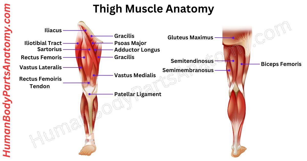
Parts of Thigh Diagram
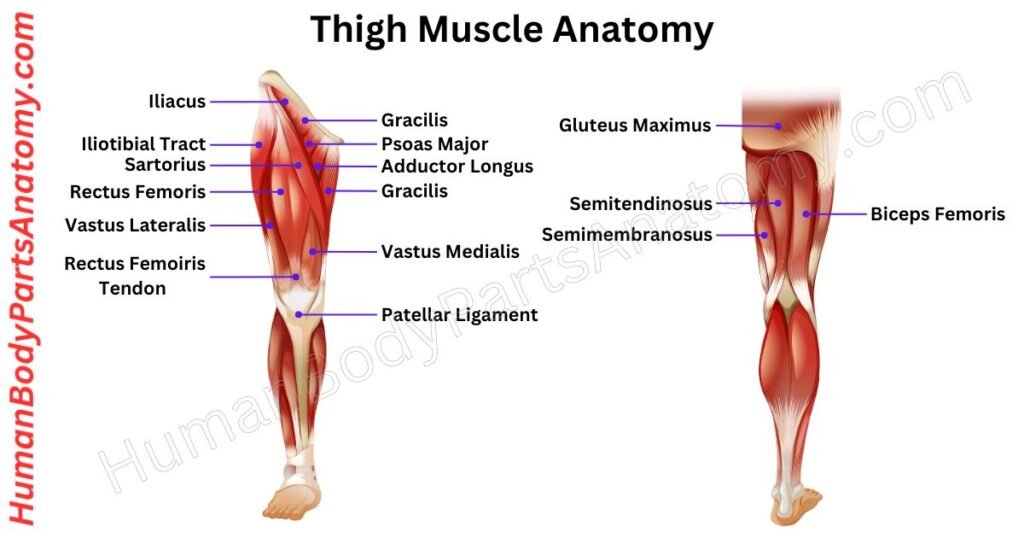
Thigh Muscle Anatomy
Anterior Thigh Muscles
- Sartorius
- Rectus femoris
- Vastus medialis
- Vastus lateralis
- Vastus intermedius
Medial Thigh Muscles
- Gracilis
- Pectineus
- Adductor longus
- Adductor brevis
- Adductor Magnus
- Obturator externus
Anterior Thigh Muscle Anatomy
Sartorius
The sartorius muscle is the body’s longest. It goes from the hip to the knee, like a long strap. It starts at the front upper part of the hip bone and goes diagonally to the inner side of the upper shin.[2]
Its main jobs are bending the knee, lifting the thigh, moving the leg sideways, and turning the thigh outward. It gets signals from the femoral nerve and blood from the femoral artery.[2]
The name “sartorius” comes from the Latin for “tailor” because it relates to the leg position when sewing.[2]
Rectus Femoris
The rectus femoris is a significant thigh muscle in the quadriceps group. Unlike other quadriceps muscles, it is attached to the hip and knee joints. It is known as the “kicking muscle” because it allows the knee to extend forcefully, similar to soccer kicks.[3]
It begins at the front of the hip bone and ends at the kneecap and shin bone. The femoral nerve regulates it.[3]
Squats and lunges, with or without weights, will help strengthen it. These workouts are particularly helpful in targeting the rectus femoris.[3]
Vastus Medicalis
The vastus medialis (VM) is a thigh muscle that helps straighten the knee and keep the kneecap steady. It is part of a group of four muscles called the quadriceps femoris.[4]
The VM has two parts, the upper Vastus Medialis Longus (VML) and the lower Vastus Medialis Obliquus (VMO), each with its job in knee movement.[4]
For blood, the VM gets nourishment from the femoral artery, deep femoral artery, and a branch of the popliteal artery.[4]
Nerve signals come from lower femoral nerve roots and higher lumbar spinal segments, ensuring the muscle works smoothly with the central nervous system.[4]
Vastus Lateralis
The vastus lateralis (VL) is the largest of the four quadriceps muscles in the front part of the thigh. It works with other thigh muscles to help straighten the knee. It is positioned on the thigh’s outer side and connects the thigh bone to the kneecap.[5]
The VL is vital for extending the knee, aiding in movement, building strength, and keeping the hip and knee joints stable. It starts from the hip and thigh bones, attaching to the kneecap through the quadriceps tendon.[5]
The femoral nerve (L2 – L4) controls the VL, and a lack of proper warm-up before activity can lead to tears.[5]
Understanding the basics of the vastus lateralis sheds light on its crucial role in supporting movement, strength, and joint stability, making it essential in musculoskeletal physiology.[5]
Vastus Intermedius
The vastus intermedius is a muscle in your thigh, part of the quadriceps muscle group. It sits beneath the rectus femoris, between the vastus medialis and vastus lateralis muscles. This muscle helps to straighten your knee.[1]
It is shaped like a thick, long piece and attaches to the upper two-thirds of your thigh bone and the tissues around it. It connects to your kneecap through the quadriceps tendon.[1]
The vastus intermedius gets signals from the femoral nerve to work and blood supply from specific arteries in the thigh.[1]
Posterior Thigh Muscle Anatomy
Biceps Femoris
The Biceps Femoris is a muscle at the rear of your thigh that belongs to the hamstring group. It helps you bend your knees and expand your hips. It comprises two parts, the long and short heads, which emerge from the pelvis.[6]
The long head travels around the rear of your leg, traversing both the hip and knee joints before connecting to the fibula’s head. The short head begins on the femur and continues to the fibula head.[6]
Its principal duties are to bend the knee (pull your heel towards your buttocks) and stretch the hip (push your thigh rearward).[6]
This muscle is necessary for running and jumping, and it helps stabilize your knee throughout various actions.[6]
Semimembranosus
The semimembranosus is a hamstring muscle found on the back of the thighs. It is positioned on the inner side, beneath the semitendinosus. Its key actions include bending the knee, straightening the hip, and turning the thigh and knee inward.[6]
It originates from the ischial tuberosity and continues to the shinbone’s inner part. The profound femoris artery supplies blood to the sciatic nerve, which governs movement.[6]
It works with the other hamstrings to stabilize the knee during running and leaping. Strengthening these muscles is essential for avoiding injuries and improving sports performance.[6]
Semitendinosus
The semitendinosus is a muscle in the back of your thigh, along with the biceps femoris and semimembranosus. It helps with bending your knee, straightening your hip, and rotating your thigh and tibia (shin). It also helps to prevent over-bending at the hips.[7]
The semitendinosus starts from the bottom of your pelvis and connects to the inside of your tibia bone (shinbone). The tibial nerve tells it what to do, along with the other muscles in the hamstring.[7]
Athletes often get injured in this muscle, especially when they run fast or make sudden movements.[7]
Medial Thigh Muscle Anatomy
Gracilis
The gracilis muscle is a thin, long muscle found on the inner side of your thigh. It starts from the inner part of the ischiopubic ramus and pubic bones, forming a rounded tendon in the thigh’s lower third. This tendon moves downward.[8]
Its main job is to pull your thigh toward the center of your body and to bend your knee. It also helps keep your knee steady during movement.[8]
Doctors sometimes use the gracilis muscle in surgery to fix torn ligaments by taking it from one place and moving it to another.[8]
Pectineus
The pectineus is a flat muscle in the upper inner part of your front thigh. It is unique because it belongs to the thigh muscles’ front and inner compartments.[9]
Along with other muscles, it helps bring your thigh closer to your body, a movement called adduction. But it also helps with bending your thigh and rotating it outwards and inwards.[9]
This muscle helps you move your thigh and stabilize your pelvis while walking. So, it is not just about moving your leg but also about keeping your body balanced.[9]
Adductor Longus
The adductor longus is a big, fan-shaped muscle in the inner thigh. It is part of a team of muscles that help bring the thigh toward the body’s midline. This muscle gets its commands from a nerve in the lower spine.[10]
It is thought that these muscles developed from both straightening and bending movements. The adductor longus is shaped like a triangle and sits in a specific thigh area.[10]
Starting from the front of the pubic bone, below the pubic ridge, and to the side of the pubic joint, it attaches to the middle part of a bony ridge called the linea aspera. This spot is between where another muscle attaches and where a thigh muscle starts.[10]
Adductor Brevis
The adductor brevis is a flat, triangle-shaped muscle found inside your thigh. It helps with movements like pulling your thigh toward the center of your body. It also plays a role in hip flexion, rotation, and stabilizing your pelvis when standing or walking.[11]
The adductor brevis starts from the front of your pubic bone and part of the side of your pelvis. Then, it widens out as it goes down toward your thigh bone (femur). It attaches to a wide area on the upper part of the inside of your thigh bone.[11]
Although it is one of the shortest muscles in its group, it is not the strongest when pulling your thigh toward your body.[11]
Adductor Magnus
The adductor magnus muscle is a big triangle in your leg. It starts at your hip bone and goes down to the back of your thigh bone.[11]
It is in two compartments in your leg, so it gets two nerves to control it. Even though it is in the back and inside your thigh, we call it a muscle of the inside part.[11]
This muscle is the biggest and strongest in the inside part of your thigh. It works with four other muscles there, adductor longus, adductor brevis, pectineus, and gracilis.[11]
Together, they are called thigh adductors, even though they do more than bring your thigh in.[11]
Besides pulling your thigh in, these muscles help with bending and straightening your thigh, twisting it in and out, and keeping your pelvis steady when you walk.[11]
Obturator externus
The obturator externus muscle is like a flat triangle in the hip area. It helps move your leg in different ways. When your hip is straight, it turns your leg outward.[12]
But when your hip is bent, it moves your leg to the side. This muscle, along with others nearby, keeps your hip joint stable.[12]
This muscle is like a triangle with a wide base and a narrow top. It starts from the side of your pelvis and the bony ring around the hole in it.[12]
Then, its fibers come together into a strong tendon. This tendon runs along the bottom edge of your hip socket, then goes up and out on the back side of the upper leg bone, attaching near the top of the thigh bone.[12]
Femur
Proximal Head
The femur, or thigh bone, has important features at its top end. The rounded part at the top is the femoral head, which fits into the hip socket. The neck of the femur connects the head to the shaft and has different widths along its length.[13]
The angle between the neck and shaft of the femur is important for movement and differs between individuals and genders. The neck also has a twist to it. The top of the neck is almost flat, while the bottom slopes down and out towards the leg.[13]
There is a curved groove on the back of the neck; on the front, it is flat and connects to the rest of the femur. There are two bony bumps at the top of the femur, the greater trochanter on the side and the lesser trochanter towards the back.[13]
These bumps are connected by lines on the front and back of the femur. The front line is called the intertrochanteric line, and the back is the intertrochanteric crest. They help muscles and ligaments attach to the femur.[13]
So, the femur’s top part has important structures like the head, neck, trochanters, and lines that help with movement and stability.[13]
Shaft
The femur, or thigh bone, is shaped like a cylinder but varies in size and shape from person to person. It is wider at the top and narrows towards the middle before expanding again near the bottom. The front is smooth, while the back is rough to help muscles attach.[13]
Three main surfaces and borders are in the middle of the femur. The front is curved and has two rounded edges. Towards the back are two more surfaces, one on each side, divided by a ridge called the “linea aspera.” This ridge starts near the top and runs down the back of the femur.[13]
At the top of the back of the femur, there is a bumpy area called the “gluteal tuberosity” and a smaller ridge called the “pectineal line” towards the inside.[13]
The linea aspera splits into two lines towards the bottom of the femur, with one going towards the inside and one towards the outside. These lines help muscles attach and continue down to the knee area.[13]
The back of the femur near the knee forms a triangle called the “popliteal surface,” which helps make up the bottom of the popliteal fossa, the space behind the knee.[13]
Distal End
The distal femur is the widest part of the thigh bone and connects with the knee and patella. It has two main parts: the medial and lateral condyles. The medial one is smaller but more noticeable, sticking out slightly and causing the knee to bulge inward.[13]
It has a bump called the adductor tubercle, where muscles attach. Below it is the medial epicondyle, where more muscles and ligaments attach.[13]
The lateral condyle is larger and has a groove for the popliteal tendon. Three muscles attach to its back and connect to ligaments and the fibula bone. The space between the condyles is the intercondylar fossa, which has indentations for crucial ligaments.[13]
The patellar surface is on the front of the distal femur, which helps stabilize the kneecap. It is slightly tilted to the side.[13]
- What is Femur Shaft Fracture (Broken Thighbone) & their Types?
- How to do Femur fracture repair?
- All you need to know about Femur X-Ray.
Conclusion
Learning thigh anatomy is essential for various reasons. It helps medical professionals diagnose and treat injuries, ensuring the precision of effective physical therapy and surgery.[1]
Knowledge of thigh anatomy also helps athletes and fitness enthusiasts to improve performance and prevent injuries.[1]
Additionally, a clear understanding of thigh muscle and bone structures reduces the risk of strain in daily activities.[1]
Knowing about thigh anatomy promotes better health, enhances physical capabilities, and contributes to effective medical care.[1]
FAQ’s
The thigh is primarily made up of three muscle groups: the quadriceps (front thigh muscles), hamstrings (back thigh muscles), and adductors (inner thigh muscles). These muscles work together to support walking, running, sitting, and standing.[1]
The main bone in the thigh is the femur, the longest and strongest bone in the human body. It connects the hip joint to the knee joint, supporting body weight and enabling leg movement.[13]
Thigh muscles work across two major joints: the hip joint, which allows movement of the leg in multiple directions, and the knee joint, which enables bending and straightening of the leg.[1]
Thigh muscles are responsible for essential movements like walking, running, jumping, squatting, and maintaining posture. They also provide stability to the hip and knee joints.[1]
Thigh muscle pain is commonly caused by muscle strain, overuse, poor posture, or sudden, intense exercise. In some cases, it can be linked to conditions like arthritis, tendonitis, or nerve compression (sciatica).[10][14]
Some common conditions include quadriceps strain, hamstring tear, groin pull, tendonitis, bursitis, and femur fractures. Athletes are especially prone to hamstring and quadriceps injuries.[10][14]
Strengthening exercises like squats, lunges, leg presses, cycling, and resistance band workouts are highly effective for building thigh muscle strength and improving stability.[3]
You should consult a doctor if thigh pain is severe, persistent, accompanied by swelling, numbness, weakness, or difficulty walking. These could be signs of a serious injury or medical condition.[10][14]
References-
- Launico M, Sinkler M, Nallamothu S. Anatomy, Bony Pelvis and Lower Limb: Femoral Muscles. StatPearls. 2025. https://www.ncbi.nlm.nih.gov/books/NBK500008/ — PMID: 29763184
- Walters B, Varacallo M. Anatomy, Bony Pelvis and Lower Limb: Thigh Sartorius Muscle. StatPearls. 2025. https://www.ncbi.nlm.nih.gov/books/NBK532889/ — PMID: 30335342
- Murdock C, Mudreac A, Agyeman K. Anatomy, Abdomen and Pelvis: Rectus Femoris Muscle. StatPearls. 2025. https://www.ncbi.nlm.nih.gov/books/NBK539897/ — PMID: 30725901
- Bordoni B, Varacallo M. Anatomy, Bony Pelvis and Lower Limb: Thigh Quadriceps Muscle. StatPearls. 2025. https://www.ncbi.nlm.nih.gov/books/NBK513334/ — PMID: 30020699
- Biondi N, Varacallo M. Anatomy, Bony Pelvis and Lower Limb: Vastus Lateralis Muscle. StatPearls. 2025. https://www.ncbi.nlm.nih.gov/books/NBK532309/ — PMID: 30335341
- Vaughn J, Cohen-Levy W. Anatomy, Bony Pelvis and Lower Limb: Posterior Thigh Muscles. StatPearls. 2025. https://www.ncbi.nlm.nih.gov/books/NBK542215/ — PMID: 31082095
- Mathew K, Pillarisetty LS. Anatomy, Bony Pelvis and Lower Limb: Thigh Semitendinosus Muscle. StatPearls. 2025. https://www.ncbi.nlm.nih.gov/books/NBK539862/ — PMID: 30725900
- Khan I, Bordoni B, Varacallo M. Anatomy, Bony Pelvis and Lower Limb: Thigh Gracilis Muscle. StatPearls. 2025. https://www.ncbi.nlm.nih.gov/books/NBK538229/ — PMID: 30860724
- Khan A, Arain A. Anatomy, Bony Pelvis and Lower Limb: Anterior Thigh Muscles. StatPearls. 2025. https://www.ncbi.nlm.nih.gov/books/NBK538425/ — PMID: 30860723
- StatPearls Publishing. Anatomy, Bony Pelvis and Lower Limb: Tensor Fasciae Latae Muscle. 2023. https://www.ncbi.nlm.nih.gov/books/NBK534775/
- Jeno S, Launico M, Schindler G. Anatomy, Bony Pelvis and Lower Limb: Thigh Adductor Magnus Muscle. StatPearls. 2025. https://www.ncbi.nlm.nih.gov/books/NBK534842/ — PMID: 30725700
- Gudena R, et al. The anatomy and function of the obturator externus. Hip Int. 2015;25(5):424–427. doi:10.5301/hipint.5000249. https://pubmed.ncbi.nlm.nih.gov/25952918/ — PMID: 25952918
- Chang A, Breeland G, Black A, Hubbard J. Anatomy, Bony Pelvis and Lower Limb: Femur. StatPearls. 2025. https://www.ncbi.nlm.nih.gov/books/NBK532982/ — PMID: 30335349
- Kiel J, Kaiser K. Adductor Strain. StatPearls. 2025. https://www.ncbi.nlm.nih.gov/books/NBK493166/ — PMID: 29630218
Read More-
Lower Limb
- Hip Bone Anatomy – Complete Guide with Parts, Names, Functions & Diagram
- Complete Guide on Leg Anatomy with Parts, Functions & Diagram
- Knee Anatomy: Complete Guide to Parts, Names, Functions & Diagram
- Femur Anatomy: Complete Guide with Parts, Names, Functions & Diagram
- Hip Muscle Anatomy – Complete Guide with Parts, Names, Functions & Diagram
Upper Limb
- Complete Guide to Finger Anatomy with Parts, Names, Functions & Diagram
- Complete Guide to Forearm Anatomy: Parts, Names, Functions & Diagram
- Comprehensive Guide to Arm Anatomy: Parts, Names & Diagram
- Comprehensive Guide to Hand Anatomy: Parts, Functions & Diagram
- Ultimate Guide to Bicep Anatomy: Parts, Names, Functions & Diagram
- Shoulder Anatomy: Ultimate Guide to Parts, Names, Functions & Diagram
- Wrist Anatomy: Ultimate Guide to Parts, Names, Functions & Diagram
- Complete Guide to Nail Anatomy with all Parts, Names & Diagrams
External Sources-
- Wikipedia
- KenHub
- Optometrists
- Cleveland Clinic
- American Academy of Ophthalmology
Official websites of the United States government.
Medical Disclaimer
All content on HumanBodyPartsAnatomy.com is educational and based on verified, peer-reviewed medical sources. Articles are authored or reviewed by qualified medical or biomedical professionals to ensure accuracy.
This website does not provide medical advice, diagnosis, or treatment. Always consult a licensed healthcare professional for personal medical guidance.
No commercial or promotional interests influence the medical content published on this site.
whoah this weblog is magnificent i really like studying your articles.
Keep up the good work! You already know, many persons are looking around for this info, you could aid them greatly.