📅 Published on June 25, 2025 | 🕒 Last updated on February 25, 2026
Overview of Basal Ganglia Anatomy
The basal ganglia, also known as the basal nuclei, are a group of deep brain structures located in the lower part of the forebrain, near the midbrain.[1][4] These structures are present in all vertebrates and play an important role in how the brain controls movement and behavior.[1][3] In humans, the basal ganglia form essential connections with the cerebral cortex, thalamus, and brainstem, helping to regulate both physical actions and mental functions.[1][2] The basal ganglia help the brain manage voluntary movements, such as walking, writing, or reaching for an object.[1][2] They are also involved in learning by repetition, forming habits, and responding to environmental cues.[2][4] Beyond movement, they influence thinking, emotions, and eye coordination.[1][3] The basal ganglia anatomy is made up of five main pairs of nuclei-
- Caudate nucleus – supports learning, memory, and processing feedback from actions. It helps us adjust behavior based on outcomes.[1][2]
- Putamen – works closely with the caudate to control movement and build habits through repeated actions.[1][2]
- Globus pallidus – comes in two parts: internal and external. It fine-tunes muscle movements and keeps motion smooth and accurate.[1][2]
- Subthalamic nucleus – acts as a relay station that adjusts signals coming from other basal ganglia parts.
- Substantia nigra – produces dopamine, a chemical crucial for movement control. Its proper function is vital for preventing motor disorders.[1][2]
These five pairs of nuclei form complex circuits, allowing them to send and receive signals that control how and when muscles move. They help filter out unwanted actions and make movements more fluid and intentional.[1][2]
In the article, we’ll take a closer look at each part, exploring its structure, role, and how all parts work together to support body movements and mental tasks.
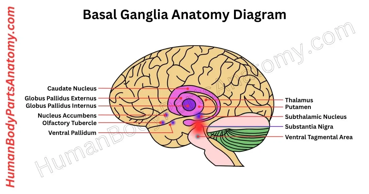
Anatomy of Basal Ganglia Diagram
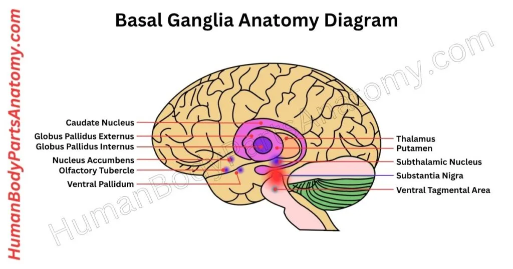
Anatomy Basal Ganglia
- Striatum
- Caudate Nucleus
- Putamen
- Nucleus Accumbens & Olfactory Tubercle
- Globus Pallidus
- Subthalamic Nucleus
- Substantia Nigra
Basal Ganglia Anatomy
1. Striatum
The striatum is a key structure located deep within the forebrain, situated near the insular cortex. It plays a central role in motor control and several cognitive functions.[1][2]
The striatum divides into two main parts: the dorsal striatum and the ventral striatum. Since the ventral part mainly processes emotions and rewards as part of the limbic system, we’ll focus on the dorsal striatum.[1][2]
The dorsal striatum, often referred to as the striatum in the neuroscience literature, is a major component of the basal ganglia anatomy. It consists of two structures: the caudate nucleus and the putamen.[1][2]
These are separated by the internal capsule, which is a bundle of nerve fibers that gives the striatum its striped (or striated) appearance.[1][2] Together with the globus pallidus, these structures form the corpus striatum.[1][3]
Functionally, the striatum serves as the main input center of the basal ganglia. It receives excitatory signals from various regions of the cerebral cortex via glutamatergic neurons.[1][2]
Different areas of the cortex send signals to matching regions within the striatum in a topographically organized manner.[1][2]
Medium spiny neurons (MSNs), which make up around 80–95% of the striatum’s cells, dominate the region. These neurons possess many small branches or spines and act as inhibitory GABAergic neurons.[2][3]
They send outputs through two major pathways—the direct and indirect pathways—which influence movement by connecting to the globus pallidus and substantia nigra.[1][2]
Caudate Nucleus
The caudate nucleus is a long, curved structure in the brain shaped like the letter “C.” It sits just in front of the thalamus, next to the lateral ventricles, and lies on the inner side of the internal capsule.[1][2]
It has three parts: the head, body, and tail. The head helps form the outer wall of the lateral ventricle. The tail curves around, creating the top part of the ventricle’s lower horn, passes over the underside of the thalamus, enters the temporal lobe, and ends near the amygdala.[1][2]
At its front end, the caudate blends with the putamen, and its lower edge is close to the nucleus accumbens.[1][2]
Functionally, the caudate nucleus is involved in motor control.[1][2] It helps the brain understand the body’s position in space and adjust movement accordingly.[1][4]
It allows for smoother, more accurate actions and supports posture control and fine-tunes the speed and precision of voluntary movements.[1][2]
Putamen
The putamen is a rounded structure located at the base of the forebrain. It is one of the main components of the basal ganglia. On an axial brain section, it appears as the outermost basal ganglia anatomy.[1][2]
It sits to the side of the globus pallidus and just inside the external capsule, wrapping around the globus pallidus like a shell.[1][2]
Toward the front and back, it extends in line with the caudate nucleus, although the two are divided by the internal capsule.[1][2]
A thin layer of white matter known as the medial medullary lamina separates the putamen from the globus pallidus.[1][2]
The putamen plays a key role in controlling voluntary movements and contributes to motor learning. It interacts closely with the neurotransmitter dopamine, which helps modulate movement and reinforce motor skills through practice and experience.[1][2]
Nucleus Accumbens & Olfactory Tubercle
The nucleus accumbens and olfactory tubercle form two paired brain structures at the base of the forebrain. Both belong to the ventral striatum, which plays a key role in processing rewards in the brain.[1][2]
They also receive major input from the ventral tegmental area (VTA)—a region that drives motivation and pleasure.[1][2]
The nucleus accumbens sits in the front part of the brain, where the caudate nucleus and putamen come together. Just below it lies the olfactory tubercle, positioned between the optic chiasm and the olfactory tract.[1][2]
Unlike brain areas that control movement, these structures are central to the brain’s reward system. When you experience something enjoyable—like eating, listening to music, or using addictive substances—dopamine-producing neurons in the VTA become active.[1][2]
These neurons send signals to the nucleus accumbens and olfactory tubercle, causing dopamine levels to rise.[1][2]
This surge of dopamine reinforces the behavior, encouraging you to seek out the rewarding experience again. Because of this, these regions are often called the “limbic-motor interface”, linking emotional states with action.[1][2]
Globus Pallidus
The globus pallidus consists of a pair of deep brain structures located on both sides of the brain, just inside the putamen. It plays a key role in controlling movement. [1][2]
GABA-producing neurons within the globus pallidus actively send out high, irregular signals to inhibit other brain regions.[1][2]
A thin vertical band of white matter, called the medial medullary lamina, separates the structure into two parts: the external segment (GPe) and the internal segment (GPi).[1][2]
The internal capsule is a major nerve fiber bundle that borders the globus pallidus on its upper and inner sides, separating it from the caudate nucleus.[1][2]
Below, it rests near the subthalamic nucleus and zona incerta, which act as boundaries between it and the thalamus.[1][2]
Toward the front, the globus pallidus lies close to the substantia innominata and the hypothalamus. Further back, it approaches the optic tract, which carries visual information.[1][2]
Due to their tight connection and bean-like combined shape, the putamen and globus pallidus together are called the lentiform nucleus.[1][2]
2. Subthalamic Nucleus
The subthalamic nucleus (STN) or Luys body is a small, lens-shaped structure located in the subthalamus (a region of the brain situated between the thalamus and the midbrain).[1][2]
Although it is not anatomically classified within the basal ganglia. Its strong functional connections make it an important part of the basal ganglia circuitry.[1][2]
It is positioned just below the thalamus and next to the red nucleus. The STN lies near several key brain regions. It borders the substantia nigra at the front and the internal capsule toward the middle.[1][2]
Surrounding the STN are a set of fiber pathways known as Forel’s fields and pallidothalamic fibers. These fibers curve around the STN and help define its boundaries, separating it from the zona incerta beneath and the thalamus above.[1][2]
The neurons in the subthalamic nucleus are excitatory and release glutamate. This chemical messenger stimulates activity in other parts of the brain.[1][2]
The STN receives organized signals from the frontal cortex, which allows it to be divided into three functional regions:
- The dorsal part- Involved in movement control, receives signals from the motor cortex.[2][3]
- The ventrolateral part is linked to decision-making and eye movements and connects to the prefrontal cortex and the frontal eye fields.[2][3]
- The ventromedial part is related to emotion and motivation and receives input from the anterior cingulate cortex.[2][3]
Researchers are still studying the exact role of the STN, but they believe it plays a key role in regulating movement. It likely acts through a brain circuit called the hyperdirect pathway, which controls and adjusts actions before the body carries them out.[1][2]
Because it fires rhythmically, scientists often call the STN the “pacemaker” of the basal ganglia, as it helps coordinate the overall function of this critical motor system.[2][3]
3. Substantia Nigra
The substantia nigra is a small, darkly pigmented structure located in the front part of the midbrain, between the cerebral peduncle and the tegmentum.[1][2]
Although it is part of the midbrain anatomically, its function links it closely to the basal ganglia, which are involved in controlling movement.[1][2]
The substantia nigra is divided into two distinct regions:
- Pars compacta (SNc) – This is the upper (dorsal) part, containing tightly packed neurons filled with melanin, giving it a dark appearance. These neurons release dopamine, a chemical essential for regulating movement. The SNc sends dopamine signals to a region called the striatum, which plays a key role in the basal ganglia motor circuit. This pathway is called the nigrostriatal pathway, and it involves both D1 and D2 dopamine receptors.[1][2]
- Pars reticulata (SNr) – It is located below the pars compacta (ventrally). This region is larger but contains fewer nerve cells. Instead of sending dopamine, it mostly handles signals coming into the basal ganglia. It processes these signals and relays them to the thalamus, a part of the brain that helps coordinate movement.[1][2]
Next to the substantia nigra, on its inner side, is the ventral tegmental area (VTA). Though smaller and less densely packed, the VTA also contains dopamine-producing cells and is considered an extension of the pars compacta in terms of function.[1][2]
Overall, the substantia nigra acts as a critical hub in the brain’s motor control network. The pars compacta sends dopamine to help initiate movement. In contrast, the pars reticulata helps transmit processed movement signals back to other brain areas.[1][2]
FAQ’s
The basal ganglia are a group of subcortical (deep‐brain) nuclei found at the base of the forebrain and top of the midbrain. They include structures such as the caudate nucleus, putamen, globus pallidus, subthalamic nucleus and substantia nigra.[1][4]
Their key role is to regulate voluntary movement by refining and modulating signals from the cortex before they are sent to muscles. They also play roles in procedural learning, habit formation, motivation and reward.[1][2]
Key components include the striatum (caudate nucleus and putamen), globus pallidus, subthalamic nucleus, and substantia nigra. Each part contributes differently to motor control and cognitive functions.[1][2]
They process signals from the cerebral cortex and send feedback through the thalamus to adjust body movements. The direct pathway promotes movement, while the indirect pathway helps suppress unwanted actions.[1][2]
Damage or dysfunction can cause movement disorders such as Parkinson’s disease, Huntington’s disease, or dystonia, leading to tremors, stiffness, or involuntary movements.[1][4]
Because they help refine and modulate movement commands and integrate feedback, the basal ganglia are key in procedural learning: as you practise a motor skill, you rely less on conscious control and more on these deeper circuits.[1][2]
Yes. Beyond movement, the basal ganglia are involved in cognitive functions (like decision-making), emotional processing, habit formation and reward signalling.[1][4]
References–
- National Institutes of Health (NIH) / National Center for Biotechnology Information (NCBI) Year of last review: 2023 Full source title: Neuroanatomy, Basal Ganglia Direct URL: https://www.ncbi.nlm.nih.gov/books/NBK537141
- Cold Spring Harbor Laboratory Press Year of publication: 2012 Full source title: Functional Neuroanatomy of the Basal Ganglia Direct URL: https://pmc.ncbi.nlm.nih.gov/articles/PMC3543080 PMID: 23071379; DOI: 10.1101/cshperspect.a009621
- Frontiers Media S.A. Year of publication: 2023 Full source title: Basal Ganglia for Beginners: The Basic Concepts You Need to Know and Their Role in Movement Control Direct URL: https://pmc.ncbi.nlm.nih.gov/articles/PMC10435282 PMID: 37593541; DOI: 10.3389/fnsys.2023.1211218
- Cleveland Clinic Year of last review: 2022 Full source title: Basal Ganglia: What It Is, Function & Anatomy Direct URL: https://my.clevelandclinic.org/health/body/23962-basal-ganglia
Read More-
Human Head
- Skull Anatomy: Complete Guide with Parts, Names, Functions & Diagram
- Ultimate Guide to Eye Anatomy: Parts, Structure, Functions & Diagram
- Tongue Anatomy: Complete Guide with Parts, Names, Functions & Diagram
- Mouth Anatomy: Complete Guide with Parts, Names, Functions & Diagram
- Complete Guide to Tooth Anatomy: Learn Parts, Names & Diagram
- Ultimate Guide to Ear Anatomy: Parts, Structure, Functions & Diagram
- Nose Anatomy: Complete Guide with Parts, Names, Functions & Diagram
Brain
- Basal Ganglia Anatomy: Complete Guide with Names, Functions & Diagram
- Lobes of the Brain: Complete Guide with Names, Functions & Diagram
Organs
- Kidney Anatomy: Complete Guide with Parts, Names, Functions & Diagram
- Liver Anatomy: Complete Guide with Parts, Names, Functions & Diagram
- Heart Anatomy: Complete Guide with Parts, Names, Functions & Diagram
External Sources-
- Wikipedia
- KenHub
- Optometrists
- Cleveland Clinic
- American Academy of Ophthalmology
Medical Disclaimer
All content on HumanBodyPartsAnatomy.com is educational and based on verified, peer-reviewed medical sources. Articles are authored or reviewed by qualified medical or biomedical professionals to ensure accuracy.
This website does not provide medical advice, diagnosis, or treatment. Always consult a licensed healthcare professional for personal medical guidance.
No commercial or promotional interests influence the medical content published on this site.