📅 Published on May 16, 2024 | 🕒 Last updated on November 12, 2025
Overview of Leg Anatomy
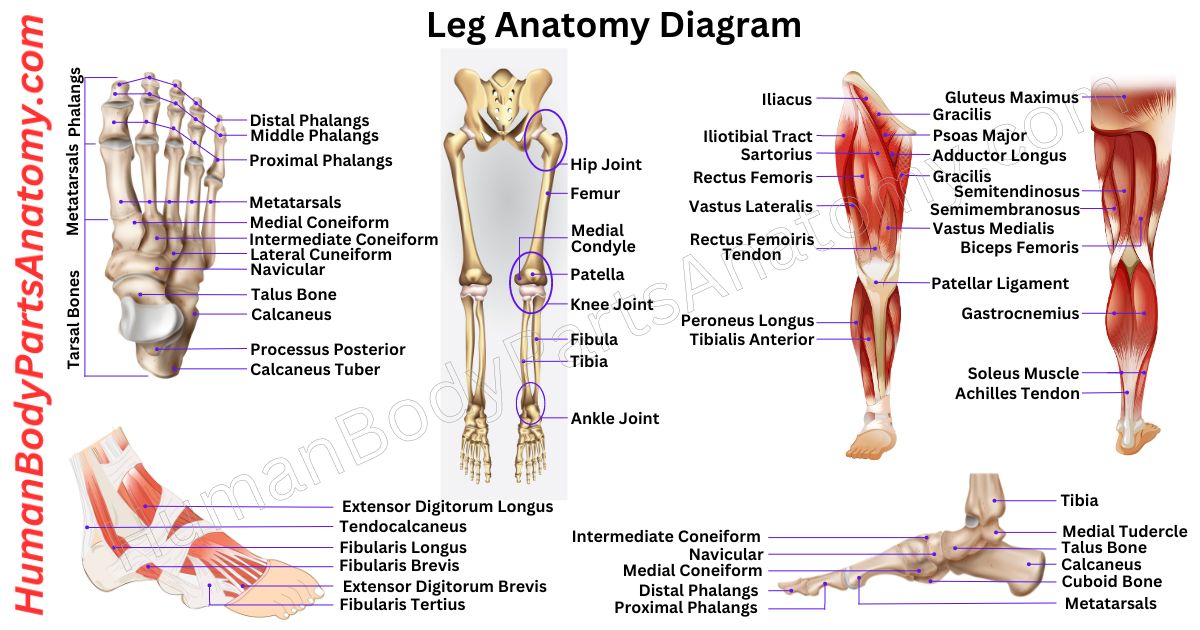
The legs are a part of the lower limbs of the human body, located between the knee and ankle. They play a vital role in supporting body weight and maintaining balance.[1] It also enables movement such as standing, walking, running, climbing, and even complex activities like dancing or jumping.[1] The parts of leg anatomy comprise several key components, including bones, muscles, tendons, ligaments, joints, and nerves. They work together to provide strength, flexibility, and coordination.[1][11] The largest bone in the body, the femur (thigh bone), connects the hip to the knee.[2] Below the knee, the tibia (shin bone) and the thinner fibula form the lower leg.[4][5] The thigh lies between the hip and the knee, while the calf comprises the back portion of the lower leg.[1] The shin refers to the front part of the lower leg where the tibia is located.[1][4]
Human legs are unique in evolution because they are specially adapted for bipedal locomotion (walking on two feet). This adaptation gives us efficiency in movement compared to most animals.[1][2][19] Anatomical differences exist between male and female legs—for example, in hip angle, knee alignment, and bone length—but the overall structure and function remain the same.[17][2]
In this article, we will explore leg anatomy in detail, covering the major bones, muscles, tendons, ligaments, joints, and nerves, to understand how our legs work and why they are so essential for daily activities and overall mobility.
Leg Anatomy Diagram
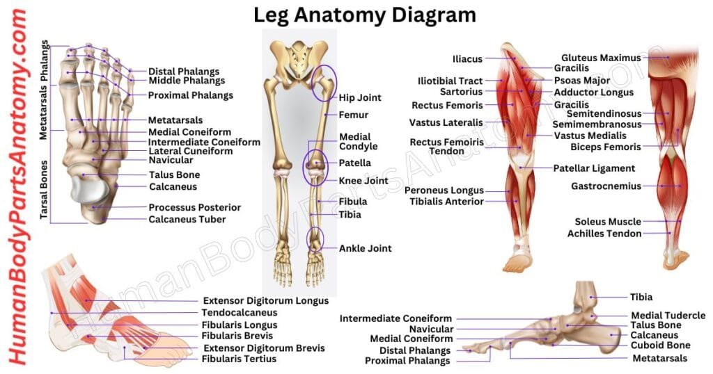
Parts of the Leg
Leg Muscles
- Thigh
- Gastrocnemius
- Soleus
- Tibialis Anterior
- Peroneus Longus
- Peroneus Brevis
Tendons
- Achilles Tendon
- Tibialis Posterior Tendon
Ligaments
- Anterior Cruciate Ligament (ACL)
- Posterior Cruciate Ligament (PCL)
- Medial Collateral Ligament (MCL)
- Lateral Collateral Ligament (LCL)
Leg Bone Anatomy
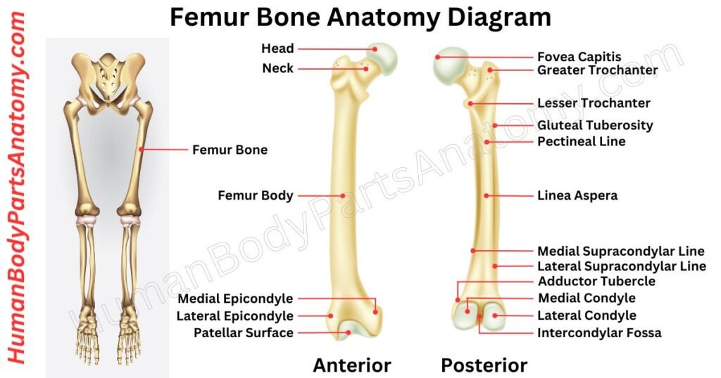
Femur
The femur or thigh bone is the longest, heaviest, and strongest bone in the human body. It plays a crucial role in supporting body weight, maintaining posture, and enabling smooth movements like walking, running, and jumping.[2][1]
The femur is divided into three main parts:
- Proximal end (upper part): It connects with the hip bone at the hip joint.[2][15]
- Shaft (middle portion): It is the long, cylindrical body of the bone.[2][15]
- Distal end (lower part): It connects with the tibia and patella (knee cap) to form the knee joint.[2][15]
The femur also acts as a weight-bearing bone, transmitting forces from the hip to the knee and giving the body strength and stability.[2] It serves as an anchor point for several muscles, ligaments, and tendons that control leg movement and balance.[15]
Inside the femoral shaft, there are two types of bone marrow:
- Red bone marrow – It is responsible for producing red blood cells, white blood cells, and platelets (essential for oxygen transport and immunity).[2]
- Yellow bone marrow – It mainly stores fat and serves as an energy reserve.[2]
Because of its strength and vital functions, the femur is often studied in orthopedics, anatomy, and sports medicine to understand mobility, fractures, and overall skeletal health.
Read More – Femur Anatomy: Complete Guide with Parts, Names, Functions & Diagram
- What is a Femur Shaft Fracture (Broken Thighbone) & its Types?
- How to do Femur fracture repair?
- All you need to know about the Femur X-Ray.
Patella
The patella or kneecap is a triangular and flat bone in a central position at the front of the knee joint. It helps to connect the femur and tibia in the knee area.[3][11][16]
The main role of the patella comes during the process of knee extension.[3][11][16] Also, it has another critical function, which is to facilitate the easy movement of the knee during both flexion and extension.[3][11][16]
Additionally, it acts as a protective shield for the anterior surface of the knee joint, safeguarding it from potential harm.[16]
- Patella Fractures: Approach to Treatment.
- Patella fractures treated with suture tension band fixation.
Tibia
The tibia, commonly known as the shin bone, is one of the two major bones in the lower leg. It serves as the main weight-bearing bone of the body. It is larger, stronger, and more vital for support compared to the fibula.
The tibia connects with the femur at the knee joint and with the fibula and talus at the ankle joint. It is positioned on the inner side of the leg and runs parallel to the fibula, stretching from just below the knee down to the ankle.[4]
From a functional perspective, the tibia is responsible for supporting body weight, maintaining balance, and ensuring stability during standing, walking, and running.[4]
It acts as a crucial link between the thigh and the foot. It allows smooth movement while protecting the internal structures of the leg.[4]
- Cross-Union Surgery for Congenital Pseudarthrosis of the Tibia.
- Knee Pain After Intramedullary Nailing in the Tibia.
- Tibia (Shinbone) Shaft Fractures.
Fibula
The fibula is a long, thin bone in the lower leg, located on the outer side next to the larger tibia. Unlike the tibia, it does not carry much body weight, but it is essential for leg stability, balance, and ankle movement.[5]
- Upper End (Head & Neck): At the top, the fibula has a rounded head just behind the tibia’s head, followed by a narrow neck.[5]
- Shaft: The middle section of the fibula is called the shaft. It is triangular in shape with three surfaces—lateral (outer), medial (facing the tibia), and posterior (back).
- Lower End (Lateral Malleolus): At the ankle, the fibula widens to form the lateral malleolus, the bony bump on the outer side of the ankle. This part connects with the talus bone, helping form the ankle joint and preventing excessive side-to-side movement.
The fibula is tightly linked to the tibia by the interosseous membrane.[5] It is a strong sheet of connective tissue that runs between the two bones.
This connection provides stability and ensures proper leg function during walking, running, and standing.[5]
- Nutrient foramina of human fibula: morphometric analysis and clinical relevance.
- Innovative approach to intramedullary nailing of the fibula: a technical note.
Leg Muscle Anatomy
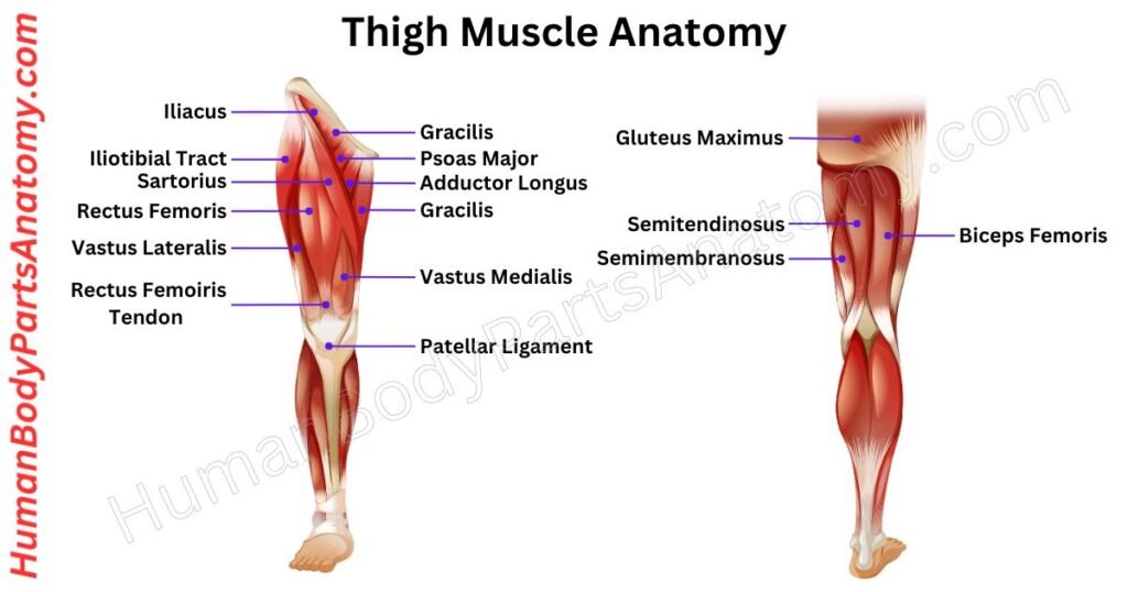
Thigh
The thigh is the upper part of the leg, located between the hip and the knee. Its main bone is the femur, the longest and strongest bone in the human body. The femur supports body weight and helps with movement.
The thigh is divided into three main compartments, and each has its own muscles, nerves, and blood vessels that work together for movement and stability:
- Anterior Compartment – It contains the quadriceps muscles, which straighten the knee. They are important for walking, running, climbing, and kicking.[7][15]
- Medial Compartment – It is known as the adductor group. These muscles pull the legs inward toward the body’s midline. They help with balance, side-to-side motion, and controlled leg positioning.[7][15]
- Posterior Compartment – It includes the hamstring muscles, which bend the knee and extend the hip. These muscles are essential for sitting, standing, jumping, and sprinting.[7][15]
All these compartments are separated by a strong connective tissue called fascia, which gives structure, protection, and support to the thigh muscle.
Read More – Complete Guide to Thigh Muscle Anatomy: Learn Parts, Names & Diagram
Gastrocnemius
The gastrocnemius muscle, commonly called the calf muscle, is one of the largest and strongest muscles in the lower leg. It has two parts (the medial and lateral heads) that start just above the knee and join together at the back of the leg, attaching to the Achilles tendon at the heel.[6][10]
This muscle crosses three important joints:
- Knee Joint
- Ankle joint
- Subtalar joint (in the foot)
Because it spans multiple joints, the gastrocnemius plays a key role in leg movement and stability. Its main functions are:
- Plantar flexion – pointing the toes downward, such as when standing on tiptoe, walking, or pushing off the ground.[6][10]
- Knee flexion – helping bend the knee during movement.[6][10]
These actions make the gastrocnemius essential not only for daily activities like walking, climbing stairs, and standing, but also for athletic movements such as running, sprinting, and jumping.
From a muscle fiber perspective, the gastrocnemius is rich in fast-twitch fibers, which are designed for quick and powerful movements. This makes the muscle highly effective for bursts of speed and strength, though less suited for long-term endurance compared to muscles with more slow-twitch fibers.[6]
For sports, exercise, and rehabilitation, keeping the gastrocnemius strong and flexible is crucial. Proper strengthening and stretching improve ankle stability, balance, mobility, and overall athletic performance.
Soleus
The soleus muscle is important in the lower leg, located deep in the calf, and works closely with the gastrocnemius muscle. Together, these two muscles form the triceps surae.[10]
The soleus has a complex structure. It has many muscle fibers that attach to different places on the leg bones. Most of these fibers start below the knee and go down to the heel bone, while some start at various spots on the back of the leg bones.[10]
Some of them attach to a flat structure called the anterior aponeurosis.[10] In contrast, others connect to another similar structure called the posterior aponeurosis. This muscle helps us stand and walk.[10]
Tibialis anterior
The tibialis anterior muscle is the largest in the front part of the lower leg. It starts from the upper section of the shin bone (tibia) and extends downward toward the foot.
This muscle performs two main movements:
- Dorsiflexion – lifting the foot upward toward the shin.
- Inversion – turning the sole inward.
Although the tibialis anterior is a muscle, it works closely with important foot and ankle bones such as the calcaneus (heel bone), talus (ankle bone), navicular, cuneiform bones, and the first three metatarsals.[8][11] Together, they make smooth and controlled foot movement possible.
The tibialis anterior is vital for walking, running, climbing stairs, and balance. It lifts the foot during movement, supports the natural arch, and helps reduce strain on the lower leg and foot.[8][12]
A strong tibialis anterior is crucial for maintaining healthy gait mechanics, foot stability, and preventing injuries. Weakness or injury in this muscle may cause issues such as foot drop, shin pain, or walking instability.[8][12]
Peroneus Longus
The peroneus longus is one of the two main muscles on the outer side of the lower leg, along with the peroneus brevis. It is supplied with blood by the fibular artery and branches of the tibial artery. Its movements are controlled by the superficial fibular (peroneal) nerve, which comes from the spinal nerves L5–S2.[3][5]
This muscle begins at the upper and outer surface of the fibula (the long bone of the leg) and extends downward toward the foot. It attaches at the base of the first metatarsal and the medial cuneiform bone near the big toe.[3][5]
Key Functions of the Peroneus Longus
- Plantarflexion – pointing the foot downward.
- Eversion – turning the sole outward.
- Arch support – helping stabilize the transverse arch of the foot.
This muscle works together with the peroneus brevis & ensures smooth foot movements, balance, and stability during walking, running, and side-to-side activities.[3][5]
The peroneus longus can be affected by:
- Strains or tears – often from sudden twisting motions.
- Tendonitis – inflammation caused by overuse or repetitive stress.
- Ankle sprains – especially in sports like basketball, football, or running.
Symptoms may include pain in the outer ankle, swelling, weakness, or difficulty moving the foot.
Peroneus Brevis
The peroneus brevis is an important muscle on the outer side of the lower leg. It is located just beneath the peroneus longus.
Its main role is to help turn the foot outward (eversion) and point the toes downward (plantar flexion). These movements are essential for walking, running, and keeping proper balance.[9][5]
This muscle gets its nerve supply from the superficial peroneal nerve and its blood supply from the peroneal artery.
It begins from the lower part of the fibula (the thin bone on the outside of the leg) and attaches to the base of the fifth metatarsal bone on the outer edge of the foot.[9][5]
Unlike the longer and slimmer peroneus longus, the peroneus brevis is shorter and thicker, which makes it more stable. Its tendon runs slightly above the outer ankle bone (lateral malleolus), curves around the ankle, and firmly attaches to the fifth metatarsal.
This strong attachment helps stabilize the ankle and protects it from common injuries such as ankle sprains.[9][5]
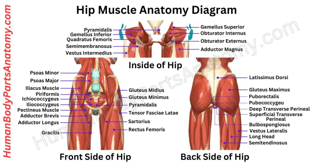
Read More – Hip Muscle Anatomy – Complete Guide with Parts, Names, Functions & Diagram
Leg Anatomy: Joint
Hip Joint
The hip joint is also known as a ball-and-socket joint. It has a “ball” at the top of the thigh bone (femur) that fits into a “socket” in the hip bone (acetabulum).[13]
This design allows the hip joint to move in many directions. It can bend and straighten (flexion and extension), rotate (internal and external rotation), and move sideways (abduction and adduction).
One of the primary functions of the hip joint is to support the body’s weight when standing, walking, or running. It also helps transfer force from your upper body to your legs, allowing you to move.[13]
The hip joint is strong and stable, partly because of the deep socket that holds the femur in place. There is also a ring of tough cartilage called the acetabular labrum around the edge of the socket, which helps keep the joint secure.[13]
This cartilage has a few important roles:
- It helps spread the pressure when you put weight on your hips.
- It creates a suction that helps keep the joint stable, like a vacuum seal.
- It also helps control the fluid flow that lubricates the joint, which keeps everything moving smoothly.
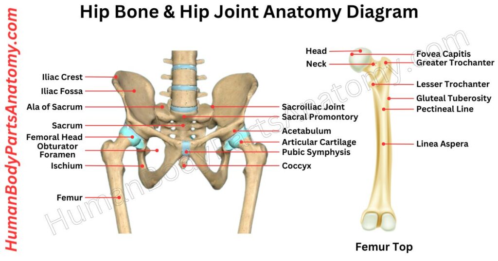
Read More – Hip Bone Anatomy – Complete Guide with Parts, Names, Functions & Diagram
- What is a Hip Fracture? Symptoms and Signs of Hip Fractures.
- What is Hip Replacement Surgery & why is it needed?
- How to do a Hip joint replacement?
- What are Hip Injuries and Disorders?
- How to take care of your new hip joint?
- What are the Hip Dislocation treatments?
Knee Joint
The knee is the largest joint in our body. It mainly operates as a hinge for bending and straightening the leg. Its structure involves two key articulations: the tibiofemoral and patellofemoral joints, which create a compound synovial joint together.[18][15]
This joint is essential for efficient walking, running, and jumping. The bones involved in knee articulation are complex.
The femur has a slight inward slant, while the tibia is almost vertical. The patella, the largest sesamoid bone in the body, sits at the front of the knee. It serves as the endpoint for the quadriceps tendon and shields the front surface of the femur.[18][15]
The articulating surfaces at the knee include the lateral and medial condyles of the femur and tibia, and the front-to-back connection between the patella and femur.[18]
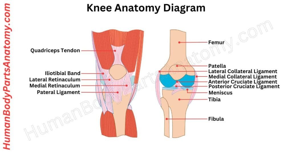
Read More – Knee Anatomy: Complete Guide to Parts, Names, Functions & Diagram
- How to do a Knee MRI scan?
- How to do a Knee CT Scan?
- What is Knee Pain & how to diagnose it?
- What are the Knee Injuries and Disorders?
- How to do a Knee Replacement?
- How to do a Partial Knee replacement?
- How does Intensive weight loss help knee arthritis?
- How To Do Knee Arthrocentesis?
Ankle Joint
The ankle joint is formed by joining three bones: the talus, tibia, and fibula. It is a socket where the talus bone sits, surrounded by the tibia and fibula. The bony prominence on the lower fibula, the malleoli, helps form the edges of this joint.[5][11]
During walking, the ankle adjusts to different surfaces. It can move in four main ways: pushing your toes down (plantarflexion), pulling them up (dorsiflexion), tilting the sole of your foot inwards (inversion), or tilting it outwards (eversion).[5][11]
- What is Ankle Pain? Causes & Care.
- How to do an Ankle Replacement?
- What are the Ankle Injuries and Disorders?
- What is an Ankle Fracture & how to diagnose it?
Subtalar Joint
The subtalar joint is also known as the talocalcaneal joint. It is where the talus bone and the calcaneus bone meet in the foot.
There are three points where they connect, two in the front and one in the back. These connection points are called facets, with one at the back, one in the middle, and one in the front.[11]
At the front and middle connections, the shape of the talus fits into the shape of the calcaneus, with the talus being convex and the calcaneus being concave. In the back connection, the talus is concave, and the calcaneus is convex.[11]
The middle connection has a structure called the sustentaculum tali as its floor, and the front connection fits snugly against the head of the talus.
Sometimes, the middle and front connections are combined into one. The back connection is the largest and is separated from the others by a tarsal canal structure.
This joint allows the foot to move from side to side (inversion and eversion). However, it does not have much to do with moving the foot up or down (dorsiflexion or plantarflexion).[11]
FAQ’s
The human leg consists of bones (femur, tibia, fibula, patella), muscles (quadriceps, hamstrings, calf muscles, tibialis anterior, peroneal muscles, etc.), joints (hip, knee, ankle, subtalar), ligaments, tendons, blood vessels, and nerves.[1][4][5][7][11]
Each lower limb’s main long bones include the femur, tibia, fibula, and patella, plus numerous tarsal, metatarsal, and phalangeal bones in the foot. The major weight-bearing leg bones are the femur, tibia, fibula, and patella, referenced above.[1][4][5]
Major leg muscles include the quadriceps and hamstrings in the thigh, and the calf muscles (gastrocnemius and soleus), tibialis anterior in the anterior compartment, and the peroneal (fibularis) muscles in the lateral compartment; additional deeper muscles control toe and foot movements.[7][6][8][9]
The tibia is commonly called the shin bone because it forms the front portion of the lower leg. It bears most of the body’s weight and connects the knee to the ankle.[4]
The tibia is larger, weight-bearing, and forms the shin. The fibula is thinner, lies beside the tibia, and provides stability to the ankle but does not bear much weight.[4][5]
The sciatic nerve is the largest nerve supplying the leg. It branches into the tibial and common peroneal nerves, which control movement and sensation in the thigh, calf, and foot.[14][20]
Leg muscles (quadriceps, hamstrings, calves, gluteals, and intrinsic foot muscles) stabilize joints and coordinate movements that maintain posture and enable safe, balanced gait and stance.[6][11][20]
References-
- Berry K, et al. (2024). Anatomy, Bony Pelvis and Lower Limb: Leg Bones. StatPearls. NCBI Bookshelf. https://www.ncbi.nlm.nih.gov/books/NBK537024/
- Berry K, et al. (2023). Anatomy, Bony Pelvis and Lower Limb: Femur. StatPearls. NCBI Bookshelf. https://www.ncbi.nlm.nih.gov/books/NBK532982/
- Lezak B, Varacallo M. (2023). Anatomy, Bony Pelvis and Lower Limb: Calf Peroneus Longus Muscle. StatPearls. NCBI Bookshelf. https://www.ncbi.nlm.nih.gov/books/NBK546650/
- Johnson EC, et al. (2023). Anatomy, Bony Pelvis and Lower Limb: Tibia. StatPearls. NCBI Bookshelf. https://www.ncbi.nlm.nih.gov/books/NBK526053/
- Gupton M, et al. (2023). Anatomy, Bony Pelvis and Lower Limb: Fibula. StatPearls. NCBI Bookshelf. https://www.ncbi.nlm.nih.gov/books/NBK470591/
- Bordoni B, et al. (2023). Anatomy, Bony Pelvis and Lower Limb: Gastrocnemius Muscle. StatPearls. NCBI Bookshelf. https://www.ncbi.nlm.nih.gov/books/NBK532946/
- Berry K, et al. (2022). Anatomy, Bony Pelvis and Lower Limb: Thigh Muscles. StatPearls. NCBI Bookshelf. https://www.ncbi.nlm.nih.gov/books/NBK482445/
- Juneja P, et al. (2023). Anatomy, Bony Pelvis and Lower Limb: Tibialis Anterior Muscle. StatPearls. NCBI Bookshelf. https://www.ncbi.nlm.nih.gov/books/NBK513304/
- Khawaji MT, et al. (2023). Anatomy, Bony Pelvis and Lower Limb: Foot Peroneus Brevis Muscle. StatPearls. NCBI Bookshelf. https://www.ncbi.nlm.nih.gov/books/NBK535427/
- Binstead JT, et al. (2023). Anatomy, Bony Pelvis and Lower Limb: Calf. StatPearls. NCBI Bookshelf. https://www.ncbi.nlm.nih.gov/books/NBK459362/
- Manganaro D, et al. (2023). Anatomy, Bony Pelvis and Lower Limb, Foot Joints. StatPearls. NCBI Bookshelf. https://www.ncbi.nlm.nih.gov/books/NBK536941/
- Juneja P, Hubbard J. (2023). Anatomy, Bony Pelvis and Lower Limb: Tibialis Anterior Muscles. StatPearls. NCBI Bookshelf. https://www.ncbi.nlm.nih.gov/books/NBK513304/
- Gold M, et al. (2023). Anatomy, Bony Pelvis and Lower Limb, Hip Joint. StatPearls. NCBI Bookshelf. https://www.ncbi.nlm.nih.gov/books/NBK470555/
- Machi M, et al. (2023). Anatomy, Sciatic Nerve. StatPearls. NCBI Bookshelf. https://www.ncbi.nlm.nih.gov/books/NBK482431/
- Cleveland Clinic Medical Education Team. (2022). Femur (Thighbone): Anatomy, Function & Common Conditions. Cleveland Clinic. https://my.clevelandclinic.org/health/body/22503-femur
- Berry K, Varacallo M. (2023). Anatomy, Bony Pelvis and Lower Limb, Knee, Patella. StatPearls. NCBI Bookshelf. https://www.ncbi.nlm.nih.gov/books/NBK519534/
- Tetsworth K, et al. (2017). Gender-related differences in lower limb alignment, range of joint motion, and the incidence of sports injuries in Japanese university athletes. Knee Surg Sports Traumatol Arthrosc. PMC. https://pmc.ncbi.nlm.nih.gov/articles/PMC5300795/
- Berry K, et al. (2023). Anatomy, Bony Pelvis and Lower Limb, Knee. StatPearls. NCBI Bookshelf. https://www.ncbi.nlm.nih.gov/books/NBK500017/
- Lovejoy CO, Suwa M, Spurlock L, et al. (2009). Fossils, feet, and the evolution of human bipedal locomotion. J Anat. PMC. https://pmc.ncbi.nlm.nih.gov/articles/PMC1571304/
- Varacallo M, et al. (2023). Anatomy, Bony Pelvis and Lower Limb: Nerves. StatPearls. NCBI Bookshelf. https://www.ncbi.nlm.nih.gov/books/NBK532304/
Read More-
Lower Limb
- Hip Bone Anatomy – Complete Guide with Parts, Names, Functions & Diagram
- Complete Guide on Leg Anatomy with Parts, Functions & Diagram
- Complete Guide to Thigh Muscle Anatomy: Learn Parts, Names & Diagram
- Knee Anatomy: Complete Guide to Parts, Names, Functions & Diagram
- Femur Anatomy: Complete Guide with Parts, Names, Functions & Diagram
- Hip Muscle Anatomy – Complete Guide with Parts, Names, Functions & Diagram
Upper Limb
- Complete Guide to Finger Anatomy with Parts, Names, Functions & Diagram
- Complete Guide to Forearm Anatomy: Parts, Names, Functions & Diagram
- Comprehensive Guide to Arm Anatomy: Parts, Names & Diagram
- Comprehensive Guide to Hand Anatomy: Parts, Functions & Diagram
- Ultimate Guide to Bicep Anatomy: Parts, Names, Functions & Diagram
- Shoulder Anatomy: Ultimate Guide to Parts, Names, Functions & Diagram
- Wrist Anatomy: Ultimate Guide to Parts, Names, Functions & Diagram
- Complete Guide to Nail Anatomy with all Parts, Names & Diagrams
- Spine Anatomy: Complete Guide with Parts, Names, Functions & Diagram
Official websites of the United States government.
- How to do a Leg MRI scan?
- Anatomy, Bony Pelvis and Lower Limb: Leg Bones
- Leg skeletal anatomy
- What is Restless Legs Syndrome?
- How To Measure Compartment Pressure in the Lower Leg
Medical Disclaimer
All content on HumanBodyPartsAnatomy.com is educational and based on verified, peer-reviewed medical sources. Articles are authored or reviewed by qualified medical or biomedical professionals to ensure accuracy.
This website does not provide medical advice, diagnosis, or treatment. Always consult a licensed healthcare professional for personal medical guidance.
No commercial or promotional interests influence the medical content published on this site.