
📅 Published on July 5, 2025 | 🕒 Last updated on February 25, 2026
Overview of Cerebellum Anatomy
The cerebellum is located at the back of your head, just beneath the larger cerebral hemispheres.[1] It is protected above by a tough membrane called the tentorium cerebelli.[1] While the cerebellum doesn’t start muscle movements, it plays a key role in refining them. It helps you walk smoothly, stay balanced, and adjust your posture.[1][2] If the cerebellum gets injured, you might have trouble walking steadily, staying upright, or learning new movements, like playing a musical instrument.[2] Structurally, the cerebellum is small in size but contains nearly 80% of all the brain’s neurons, making it incredibly dense and active.[1] In cerebellum anatomy, there are two rounded halves called hemispheres, connected in the middle by a narrow part known as the vermis.[1]
Each hemisphere is split into three main lobes:[1]
- The anterior lobe (at the front)
- The posterior lobe (at the back)
- The flocculonodular lobe (near the brainstem)
These lobes are divided into three main grooves or fissures:[1]
- Primary fissure
- Posterolateral fissure
- Horizontal fissure
In the article, we will see the cerebellum anatomy with different parts and their functions in detail.
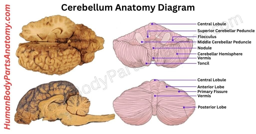
Cerebellum Anatomy Diagram
Anatomy of the Cerebellum
- Cerebellar Hemisphere
- Relations
- Superior
- Anterior
- Posterior and Lateral
- Surfaces
- Lobes
- Anterior Lobe
- Posterior Lobe
- Flocculonodular Lobe
- Fissures
- Primary Fissure
- Posterolateral Fissure
- Horizontal Fissure
- Zones
- Vermal Zone
- Paravermal (Intermediate) Zone
- Lateral Zones
- Cerebellar Cortex
- Molecular layer
- Purkinje cell layer
- Granule layer
- Deep Nuclei
- Fastigial Nucleus
- Globose Nucleus
- Emboliform Nucleus
- Dentate Nucleus
- Cerebellar Peduncles
- Superior
- Middle
- Inferior
- White Matter (Arbor Vitae)
Cerebellum Anatomy
Cerebellar Hemisphere
The cerebellum is a key part of the brain responsible for movement control and coordination. It has three main sections: a central vermis and two side hemispheres.[1]
These parts are connected and share a similar structure. The vermis is a narrow, ridge-like strip in the middle, while the hemispheres are larger and sit on each side.[1]
The cerebellum helps the body perform smooth and accurate movements. It manages tasks like:[1][2]
- Keeping balance and an upright posture.[1][2]
- Guiding voluntary movements such as walking or reaching.[1][2]
- Controlling muscle tension and tone.[1][2]
- Helping the brain learn and fine-tune motor skills.[1][2]
Each hemisphere has a specific function:
- The intermediate zone (also called the spinocerebellum) adjusts and smooths out movements of the arms and legs.[1][2]
- The outer zone (known as the cerebrocerebellum or pontocerebellum) is involved in planning precise and complex actions, like playing an instrument or typing. This part is the most developed and evolved region of the cerebellum.[1][2]
Altogether, the cerebellum allows the brain and body to work in sync, making movement automatic, efficient, and well-coordinated.
Relations in Cerebellum Anatomy
1. Superior
The cerebellum is located at the back of the brain, just below a layer of tissue called the tentorium cerebelli.[1][2]
Above it, there’s a large vein called the great cerebral vein of Galen, which drains into the straight sinus—a channel that carries blood away from the brain. Also above the cerebellum is part of the brain’s occipital lobe, called the lingual gyrus.[1][2]
2. Anterior
The brainstem sits in front of the cerebellum and connects the brain to the spinal cord. Behind the brainstem, the fourth ventricle forms a fluid-filled space, partly covered by thin membranes called the medullary velum.[1]
The aqueduct of Sylvius ends near this area and carries brain fluid to it. Just above, four bumps called the corpora quadrigemina rise, surrounded by a fluid-filled space known as a cistern.[1]
Below and in front of the cerebellum are the medulla oblongata, the foramen of Magendie (an opening that allows brain fluid to flow), and the foramen magnum—the large hole at the base of the skull.[1]
These areas help move cerebrospinal fluid (CSF) around the brain and spinal cord, including through side openings called the foramina of Luschka.[1]
3. Posterior and Lateral
Behind and to the sides of the cerebellum is the occipital bone, covered by a tough protective layer called dura mater. Below the cerebellum are curved blood channels called the sigmoid sinuses.[1]
At the very back, you might also find the occipital sinus and the confluence of sinuses, where several blood channels join.[1]
Surfaces in Cerebellum Anatomy
When you look at the cerebellum from above, you can see a raised middle part called the superior vermis. It starts at the front notch of the cerebellum, curves around at the back notch, and then continues underneath as the inferior vermis.[1]
On both sides of the vermis are the cerebellar hemispheres, which help control movement and balance.[1]
Lobes in Cerebellum Anatomy
The cerebellum is a key part of the brain responsible for coordination and balance. It is organized into three main lobes—each with a unique location and function:
- Anterior Lobe – It is found above the cerebellar peduncles. This lobe includes the front two-thirds of the vermis (the central ridge) and the front one-third of each hemisphere. It stops at the primary fissure and mainly helps control the movements of the trunk and limbs.[1]
- Posterior Lobe – It starts at the primary fissure and extends toward the back. It is the largest cerebellar lobe, plays a major role inadjusting and refining voluntary movements, and supports motor planning for precise actions.[1]
- Flocculonodular Lobe – The flocculonodular lobe sits below the posterolateral fissure and above the inferior medullary velum and cerebellar peduncles. This small lobe maintains balance and guides eye movements.[1][2]
The surface of the cerebellum divides further into smaller sections—nine segments along the vermis and five paired sections on each side.[1]
These divisions help organize the cerebellum’s complex structure and support its fine-tuned control of body movements.[1]
Fissures
The surface of the cerebellum has several grooves that give it a layered look. These grooves help divide the cerebellum into different parts called lobes and lobules.[1]
- One major groove is the horizontal fissure, which wraps around the sides and back of the cerebellum. Just below it is a deeper groove called the posterolateral fissure, found beneath a small part called the flocculonodular lobe.[1]
- Another important groove is the postlunate fissure, which curves across the upper back part of the cerebellum, called the tentorial surface. This fissure is located just behind the primary fissure, which is a key landmark separating the anterior lobe from the posterior lobe. Both of these fissures also continue onto the underside of the cerebellum.[1]
- Lastly, the retrotonsillar fissure is a shallow groove located just behind the rounded cerebellar tonsil on each side.[1]
These grooves help shape the cerebellum and serve as important landmarks for understanding its structure and function.[1]
Zones of Cerebellum Anatomy
Suppose the cerebellum is sliced along the horizontal fissure and laid flat with its outer surface facing up. In that case, it can be functionally divided into four main zones:
1. Vermal Zone
It is located along the midline structure called the vermis. The vermal zone plays a key role in maintaining balance and posture.[1][2]
It receives sensory information about muscle tone from the hips, trunk, shoulders, and neck through the spinocerebellar tracts. When body alignment shifts, thefastigial nucleus sends corrective signals to help restore proper posture.[1][2]
2. Paravermal (Intermediate) Zone
It is positioned just beside the vermis. This zone is crucial for coordinating skilled, voluntary movements. It processes input from the hands, feet, and other distal muscles via the spinocerebellar tracts. The interposed nuclei (globose and emboliform) manage their motor output to fine-tune these movements.[1][2]
3. Lateral Zones
These are the largest regions found on either side of the paravermal zone, forming most of the cerebellar hemispheres. They help plan and adjust complex motor activities across the entire body.[1][2]
They receive signals from the cerebral cortex through the corticopontocerebellar tracts, and the dentate nucleus handles motor planning based on this input.[1][2]
Cerebellar Cortex (outer layer of cerebellum)
The cerebellum’s grey matter, known as the cortex, is organized into three distinct layers. The outermost is the molecular layer, which contains few cells but many branching nerve fibers.[1]
Beneath it lies the Purkinje cell layer, made up of large, specialized neurons that play a key role in sending signals from the cerebellum. The deepest layer is the granular layer, packed with small neurons that receive input from other brain regions.[1]
Together, these layers hold various nerve cells, support cells (glia), and fibers that work in harmony to control and fine-tune body movements.[1]
1. Molecular Layer
The molecular layer is the topmost part of the cerebellar cortex, located just under the pia mater. This layer has few nerve cell bodies but is filled with thin nerve fibers.[1]
The long, branching extensions of Purkinje cells stretch into this layer and connect with parallel fibers, which come from granule cells. Two types of small inhibitory neurons—stellate cells (found near the surface) and basket cells (found deeper)—are present here.[1]
Both types help control Purkinje cell activity by reducing their signals. However, the parallel fibers of granule cells first activate them.[1]
2. Purkinje Cell Layer
Right below the molecular layer is a thin zone that contains a single row of Purkinje cells—large, uniquely shaped neurons. These are the only cells that send signals out of the cerebellar cortex.[1]
Their messages go to deeper brain centers like the cerebellar and vestibular nuclei and are always inhibitory. Purkinje cells receive excitatory input from two main sources:
- Climbing fibers from the inferior olivary nucleus.[1]
- Parallel fibers from granule cells.[1]
This setup allows them to play a key role in adjusting body movement and balance.
3. Granular Layer
The deepest layer, just above the white matter, is the granular layer. It is tightly packed with tiny granule cells and larger Golgi cells. Granule cells receive excitatory signals from mossy fibers, which bring sensory and motor information from the spinal cord and brainstem.[1]
Golgi cells, on the other hand, help regulate granule cell activity by sending inhibitory feedback. This feedback loop helps the cerebellum refine its output for smooth, coordinated movement.[1]
Deep Nuclei (inside the cerebellum)
1. Dentate nucleus
The dentate nucleus is the largest of the cerebellar nuclei. It is found in the outer part of the cerebellar hemispheres. It looks like a folded or crumpled pouch, with its open end facing toward the front and middle of the brain.[1]
Unlike other cerebellar nuclei, the dentate nucleus wraps around bundles of nerve fibers. These fibers form important pathways called the dentatorubrothalamic tract and the dentatoolivary tract.[1]
The main job of the dentate nucleus is to help control voluntary movements. It plays an important role in making sure movements are well-timed, properly planned, and smoothly carried out.[1]
2. Fastigial nucleus
The fastigial nuclei are the most centrally located of the cerebellar nuclei. They are closely linked to the vermis, the midline region of the cerebellum. It is positioned just behind the roof of the fourth ventricle. These nuclei play a key role in maintaining posture and balance.[1][2]
These nuclei receive information from the spinal cord and the inner ear (which helps with balance). After processing this information, they send signals to other parts of the brain, like the vestibular nuclei and the thalamus.[1][2]
From there, messages go to the motor area of the brain (called the precentral gyrus), which controls movement.[1][2]
In the end, the brain sends signals to the trunk and nearby limb muscles to help keep the body stable and balanced.[1][2]
3. Emboliform & Globose
The emboliform nucleus is one of the key deep nuclei inside the cerebellum. It sits next to the dentate nucleus on its inner side and partially overlaps the dentate opening, known as the hilum.[1]
If you move from the outer edge of the cerebellum inward, you will find the deep nuclei arranged in the following order: dentate, emboliform, globose, and fastigial. A special microscope stain called Weigert’s elastic stain reveals these structures in detail.[1]
In many lower animals, the emboliform and globose nuclei do not appear separately; instead, they join together and form what is called the interposed nucleus.[1]
The emboliform primarily supports the spinocerebellar system, which adjusts and smooths out limb movements.[1]
It sends signals through the superior cerebellar peduncle to the red nucleus in the midbrain and to parts of the thalamus. From there, these signals travel to regions of the cerebral cortex that plan and control limb movement.[1]
The globose nucleus lies between the emboliform and fastigial nuclei. Like the emboliform, it is also part of the interposed nuclei and works in motor control.
It sends movement-related signals down the spinal cord, helping to manage the accuracy and coordination of arm and leg actions.[1]
Cerebellar Peduncles (Connect to Brainstem)
Superior
The superior cerebellar peduncle works as the main communication route between the cerebellum and other parts of the brain. It carries messages from the deep cerebellar nuclei to the thalamus, helping coordinate movement.[1]
Some fibers from the fastigial nucleus also take a separate path to reach the vestibular nuclei, playing a role in maintaining balance and posture.[1]
At the same time, this peduncle brings signals into the cerebellum, such as body position information from the ventral spinocerebellar tract, touch signals from the trigeminal nucleus, and regulating messages from the locus coeruleus.[1]
Middle
The middle cerebellar peduncle carries incoming signals from the pons to the cerebellum, specifically targeting the neocerebellum. These signals, called pontocerebellar fibers, reach areas on both sides of the paravermal zone, which lies next to the central vermal region.[1]
Inferior
The inferior cerebellar peduncle links the spinal cord and brainstem to the cerebellum through two key pathways.[1]
- The first is the restiform body, which carries only sensory signals to the cerebellum. These include input from the dorsal spinocerebellar, olivocerebellar, and cuneocerebellar tracts, all of which help the brain understand body position and movement.[1]
- The second pathway is the juxtarestiform body, which carries both incoming and outgoing signals. It brings balance-related input from the vestibular system. It sends signals back through cerebellovestibular fibers, helping the body stay upright and coordinated.[1]
White Matter (Arbor Vitae)
The arbor vitae is the white matter located at the core of the cerebellum. It is named for its tree-like shape that resembles branching leaves and is found in both cerebellar hemispheres.
It serves as a vital pathway for carrying information to and from the cerebellum, helping manage body movement and coordination.
Within this white matter lie four important groups of nerve cells known as the deep cerebellar nuclei: the dentate, emboliform, globose, and fastigial nuclei.
These act as the cerebellum’s main output hubs, processing signals and sending them to other brain regions. Their role is essential in fine-tuning motor activity, ensuring smooth movement, balance, and control of posture.
FAQ’s
The cerebellum is a distinct structure at the back of the brain, tucked underneath the occipital lobes and just above the brainstem. It sits in the posterior cranial fossa and wraps around the brainstem in a half-circle shape.[1][3]
The cerebellum is divided into:
Two lateral hemispheres and a midline vermis.
Anatomical lobes: the anterior lobe, posterior lobe, and flocculonodular lobe.
Functional zones: cerebro-cerebellum (planning & motor learning), spinocerebellum (error correction & posture), and vestibulocerebellum (balance & eye movement).[1][2]
Although the cerebellum makes up only about 10% of total brain volume, it contains more than 50% of the brain’s neurons. This high neuron density allows it to process massive amounts of sensory and motor information quickly and accurately.
During early brain development:
The cerebellum arises from the hindbrain region (specifically the metencephalon).
It develops below the occipital lobes and temporally behind the brain-stem region as the skull and cranial vault evolve.[1]
Cerebellar damage can lead to ataxia (loss of coordination), tremors, unsteady walking, slurred speech, dizziness, and difficulty judging distance or speed. Severe cases may cause problems with eye movements or fine motor skills.[2]
Conditions include cerebellar stroke, tumors, multiple sclerosis, hereditary ataxias, infections, and chronic alcohol-related degeneration. Nutritional deficiencies (especially vitamin B12) and toxins can also impair cerebellar function.[2][3]
The cerebellum helps control balance, coordination, posture, and smooth muscle movement. It fine-tunes motor actions so movements are accurate and well-timed, and it supports motor learning (learning physical skills). Damage to the cerebellum can cause poor coordination, unsteady walking, and tremors.[1][2]
References-
- Jimsheleishvili S, Dididze M. (2023). Neuroanatomy, Cerebellum. StatPearls Publishing (NCBI Bookshelf). https://www.ncbi.nlm.nih.gov/books/NBK538167/ — PMID: 30969714
- Roostaei T, Sadaghiani S. (2023). Neuroanatomy, Cerebellar Dysfunction. StatPearls Publishing (NCBI Bookshelf). https://www.ncbi.nlm.nih.gov/books/NBK545251/ — PMID: 30422530
- Cleveland Clinic. (2022). Cerebellum: What It Is, Function & Anatomy. Cleveland Clinic Health Library. https://my.clevelandclinic.org/health/body/23418-cerebellum — Clinical Educational Source
Read More-
Lower Limb
- Hip Bone Anatomy – Complete Guide with Parts, Names, Functions & Diagram
- Complete Guide on Leg Anatomy with Parts, Functions & Diagram
- Complete Guide to Thigh Muscle Anatomy: Learn Parts, Names & Diagram
- Knee Anatomy: Complete Guide to Parts, Names, Functions & Diagram
- Femur Anatomy: Complete Guide with Parts, Names, Functions & Diagram
- Hip Muscle Anatomy – Complete Guide with Parts, Names, Functions & Diagram
Upper Limb
- Complete Guide to Finger Anatomy with Parts, Names, Functions & Diagram
- Complete Guide to Forearm Anatomy: Parts, Names, Functions & Diagram
- Comprehensive Guide to Arm Anatomy: Parts, Names & Diagram
- Comprehensive Guide to Hand Anatomy: Parts, Functions & Diagram
- Ultimate Guide to Bicep Anatomy: Parts, Names, Functions & Diagram
- Shoulder Anatomy: Ultimate Guide to Parts, Names, Functions & Diagram
- Wrist Anatomy: Ultimate Guide to Parts, Names, Functions & Diagram
- Complete Guide to Nail Anatomy with all Parts, Names & Diagrams
- Spine Anatomy: Complete Guide with Parts, Names, Functions & Diagram
Human Head
- Skull Anatomy: Complete Guide with Parts, Names, Functions & Diagram
- Ultimate Guide to Eye Anatomy: Parts, Structure, Functions & Diagram
- Tongue Anatomy: Complete Guide with Parts, Names, Functions & Diagram
- Mouth Anatomy: Complete Guide with Parts, Names, Functions & Diagram
- Complete Guide to Tooth Anatomy: Learn Parts, Names & Diagram
- Ultimate Guide to Ear Anatomy: Parts, Structure, Functions & Diagram
- Nose Anatomy: Complete Guide with Parts, Names, Functions & Diagram
Brain
- Basal Ganglia Anatomy: Complete Guide with Names, Functions & Diagram
- Lobes of the Brain: Complete Guide with Names, Functions & Diagram
- Parts of the Cerebrum Anatomy: Complete Guide with Names, Functions & Diagram
- Midbrain Anatomy: Complete Guide with Parts, Names, Functions & Diagram
- The Cerebellum Anatomy: Complete Guide with Names, Functions & Diagram
Organs
- Kidney Anatomy: Complete Guide with Parts, Names, Functions & Diagram
- Liver Anatomy: Complete Guide with Parts, Names, Functions & Diagram
- Heart Anatomy: Complete Guide with Parts, Names, Functions & Diagram
External Sources-
- Wikipedia
- KenHub
- Optometrists
- Cleveland Clinic
- American Academy of Ophthalmology
Medical Disclaimer
All content on HumanBodyPartsAnatomy.com is educational and based on verified, peer-reviewed medical sources. Articles are authored or reviewed by qualified medical or biomedical professionals to ensure accuracy.
This website does not provide medical advice, diagnosis, or treatment. Always consult a licensed healthcare professional for personal medical guidance.
No commercial or promotional interests influence the medical content published on this site.