📅 Published on April 10, 2026 | 🕒 Last updated on May 20, 2026
Overview of Human Anatomy and Physiology
The human body has 206 bones, about 650 muscles, 78 to 80 organs, and a wide network of blood vessels. All of these parts work together, with each cell playing its own role to keep us alive. Two main fields help us understand how our bodies function: physiology, which looks at how the body works inside, and anatomy, which studies its structure. Anatomy examines everything from the smallest cells to tissues, organs, and entire systems. By learning about human anatomy, we better understand how our bodies are built and how all the parts work together to keep us alive.
Human Anatomy Diagram
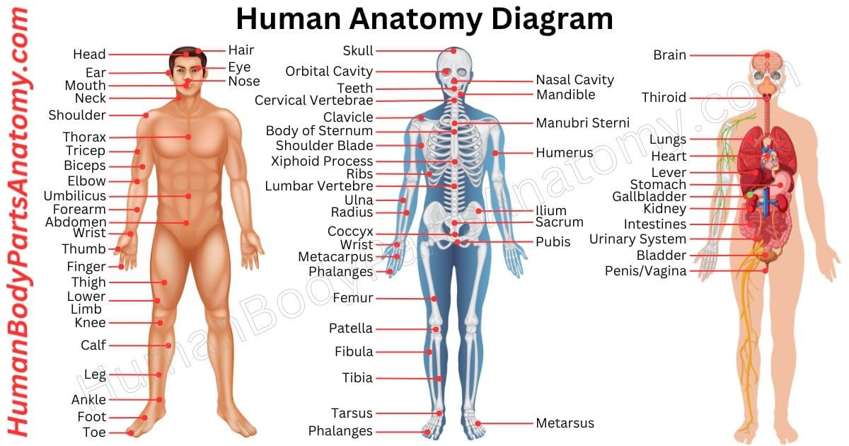
Human Body Parts Name
Skeletal System
- Axial Skeleton
- Skull
- Cranial Bones
- Frontal bone
- Parietal bones (2)
- Temporal bones (2)
- Occipital bone
- Sphenoid bone
- Ethmoid bone
- Facial Bones
- Nasal bones (2)
- Maxilla bones (2)
- Zygomatic bones (2)
- Lacrimal bones (2)
- Palatine bones (2)
- Inferior nasal conchae (2)
- Vomer bone
- Mandible
- Cranial Bones
- Hyoid Bone
- Auditory Ossicles
- Malleus (hammer)
- Incus (anvil)
- Stapes (stirrup)
- Vertebral Column (Spine)
- Cervical Vertebrae (7)
- Thoracic Vertebrae (12)
- Lumbar Vertebrae (5)
- Sacrum (5 fused vertebrae)
- Coccyx (3-5 fused vertebrae)
- Ribs
- True Ribs (1-7)
- False Ribs (8-12)
- Vertebrochondral Ribs (8-10)
- Floating Ribs (11-12)
- Skull
- Sternum (Breastbone)
- Manubrium
- Body (gladiolus)
- Xiphoid process
- Thoracic cage
- Thoracic cavity
- Superior thoracic aperture (thoracic inlet)
- Inferior thoracic aperture
- Intercostal space
- Infrasternal angle
- Appendicular Skeleton
- Pectoral Girdle (Shoulder Girdle)
- Clavicle (Collarbone)
- Scapula (Shoulder Blade)
- Upper Limb (Arm)
- Humerus
- Radius
- Ulna
- Carpal Bones
- Metacarpal Bones
- Phalanges (Fingers Bones)
- Pelvic Girdle (Hip Girdle)
- Ilium
- Ischium
- Pubis
- Acetabulum
- Lower Limb (Leg)
- Femur
- Patella (Kneecap)
- Tibia
- Fibula
- Tarsal Bones
- Metatarsal Bones
- Phalanges (Toe Bones)
- Pectoral Girdle (Shoulder Girdle)
- Joints
- Head and Neck Joints
- Temporomandibular Joint (TMJ)
- Atlanto-occipital Joint
- Spinal Joints
- Intervertebral Joints
- Facet Joints (Zygapophyseal Joints)
- Atlantoaxial Joint
- Shoulder Joints
- Glenohumeral Joint
- Acromioclavicular Joint
- Sternoclavicular Joint
- Elbow Joint
- Humeroulnar Joint
- Humeroradial Joint
- Proximal Radioulnar Joint
- Wrist and Hand Joints:
- Radiocarpal Joint
- Intercarpal Joints
- Carpometacarpal Joints
- Metacarpophalangeal Joints (MCP Joints)
- Interphalangeal Joints (IP Joints)
- Hip Joint (Coxal Joint)
- Acetabulofemoral Joint
- Knee Joint
- Tibiofemoral Joint
- Patellofemoral Joint
- Ankle and Foot Joints
- Talocrural Joint (Ankle Joint)
- Subtalar Joint
- Midtarsal Joint (Chopart’s Joint)
- Tarsometatarsal Joints
- Metatarsophalangeal Joints (MTP Joints)
- Interphalangeal Joints (IP Joints)
- Head and Neck Joints
- Cartilage
- Ligaments
- Tendons
- Bone Marrow
- Periosteum
- Sesamoid Bones
Female Reproductive System
- Ovary
- Ligament of ovary
- Suspensory ligament of ovary
- Fallopian tube
- Uterus
- Cervix of uterus
- Round ligament of uterus
- Pubocervical ligament
- Cardinal ligament
- Uterosacral ligament
- Va*ina
- Hymen
- Epoophoron
- Paroophoron
- Vulva
- Mons pubis
- Labia
- Vestibule of the vagina
- Bulb of the vestibule
- Cliteris
- Glans
- Clitoral hood
- Urinary meatus
- Female urethra
- Bartholin’s gland
- Skene’s gland
Male Reproductive System
- Testicle
- Tunica vaginalis
- Tunica albuginea
- Seminiferous tubules
- Straight tubules
- Rete testis
- Epididymis
- Paradidymis
- Spermatic cord
- Cremaster
- Vas deferens
- Seminal vesicle
- Seminal gland
- Ejaculatory duct
- Prostate
- Bulbourethral gland
- Penis
- Glans
- Foreskin
- Body of the penis
- Corpus cavernosum penis
- Corpus spongiosum penis
- Helicine arteries
- Fascia of the penis
- Suspensory ligament of the penis
- Urinary meatus
- Male urethra
- Scrotum
- Dartos fascia
- Perineum
- Perineal body
- Subcutaneous perineal pouch
- Superficial perineal pouch
- Deep perineal pouch
- Ischio-anal fossa
Sense Organs
Integumentary System
- Skin
- Hair
- Nail
- Breast
- Subcutaneous tissue
Human Muscle Anatomy
- Upper Body Muscles
- Thorax Muscles
- Pectoralis major
- Pectoralis minor
- Subclavius
- Serratus anterior
- Levatores costarum
- External intercostal muscle
- Internal intercostal muscle
- Innermost intercostal muscle
- Subcostales
- Transversus thoracic
- Pectoral fascia
- Clavipectoral fascia
- Thoracic fascia
- Endothoracic fascia
- Thoracic diaphragm
- Shoulder Muscles (Deltoid Muscles)
- Anterior Deltoid
- Medial Deltoid
- Posterior Deltoid
- Upper Arm Muscles (Arm Muscles)
- Biceps Brachii
- Long Head
- Short Head
- Brachialis
- Brachioradialis
- Biceps Brachii
- Back Muscles
- Trapezius
- Latissimus dorsi
- Rhomboid major
- Rhomboid minor
- Levator scapulae
- Serratus posterior inferior
- Serratus posterior superior
- Anterior cervical intertransversarii
- Lateral posterior cervical intertransversarii
- Intertransversarii laterales lumborum
- Erector spinae
- Erector spinae aponeurosis
- Iliocostalis
- Longissimus
- Spinalis
- Spinotransversales
- Splenius
- Transversospinales
- Multifidus
- Semispinalis
- Rotatores
- Interspinales
- Intertransversarii
- Thoracolumbar fascia
- Neck Muscles:
- Platysma
- Longus colli
- Longus capitis
- Scalenus anterior
- Scalenus medius
- Scalenus posterior
- Sternocleidomastoid
- Suboccipital muscles
- Suprahyoid muscles
- Infrahyoid muscles
- Rotator Cuff Muscles:
- Supraspinatus
- Infraspinatus
- Teres Minor
- Subscapularis
- Abdominal Muscles (Upper Abdomen)
- Rectus abdominis
- Pyramidalis
- External oblique
- Inguinal ligament
- Superficial inguinal ring
- Internal oblique
- Cremaster
- Transversus abdominis
- Inguinal falx
- Deep inguinal ring
- Linea alba
- Linea semilunaris
- Inguinal canal
- Quadratus lumborum
- Abdominal fascia
- Pelvic fascia
- Pelvic diaphragm
- Levator ani
- Ischiococcygeus
- External anal sphincter
- Triceps Brachii
- Serratus Anterior
- Thorax Muscles
- Lower Body Muscles
- Hip Muscles:
- Gluteus Maximus
- Gluteus Medius
- Gluteus Minimus
- Thigh Muscles (Quadriceps)
- Rectus Femoris
- Vastus Lateralis
- Vastus Medialis
- Vastus Intermedius
- Thigh Muscles (Hamstrings)
- Biceps Femoris
- Semimembranosus
- Semitendinosus
- Adductors (Inner Thigh Muscles):
- Adductor Magnus
- Adductor Longus
- Adductor Brevis
- Gracilis
- Hip Flexors:
- Iliopsoas
- Tensor Fasciae Latae (TFL)
- Calf Muscles:
- Gastrocnemius
- Soleus
- Tibialis Posterior
- Shin Muscles (Anterior leg)
- Tibialis Anterior
- Hip Rotators (Deep Muscles):
- Piriformis
- Gemellus Superior and Inferior
- Obturator Internus and Externus
- Hip Muscles:
Alimentary System
Respiratory System
- Nose
- Larynx
- Trachea
- Bronchi
- Lungs
Urinary System
- Kidney
- Nephrons
- Renal arteries
- Renal veins
- Renal pelvis
- Ureter
- Urinary bladder
- Female urethra
- Male urethra
Human Nervous System
- Central nervous system
- Meninges
- Spinal cord
- Brain
- Peripheral nervous system
- Cranial nerves
- Spinal nerves
- Autonomic division (Autonomic nervous system)
Cardiovascular System
Human Bone Anatomy
In human anatomy, the skeleton is the internal framework of the body. It is responsible for both structure and function. At birth, it is composed of approximately 270 bones. However, by adulthood, this number reduces to roughly 206 due to bone fusions. This skeletal system accounts for around 14% of the average person’s body weight, which ranges from 10 to 11 kg. Bone mass reaches its peak between the ages of 25 and 30.
Skull
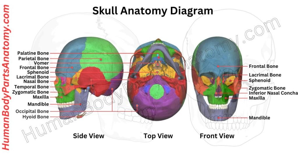
The skull is a bony structure that covers and protects the brain. It comprises three main types of bones: cranial bones, facial bones, and ear ossicles.
In humans, the skull is divided into the neurocranium (the braincase) and the viscerocranium (the facial skeleton), which includes the mandible. This structure is an example of cephalization, where the brain and sensory organs are concentrated at the head.
The skull is located at the front of the skeleton, a result of cephalization. It houses the brain along with key sensory organs such as the eyes, ears, nose, and mouth.
The human skull is made up of 22 bones, or 29 if you include the inner ear bones and the hyoid bone. These bones are mainly connected by ossified joints known as sutures.
The skull has several crucial functions: it protects the brain, maintains the proper distance between the eyes for stereoscopic vision, and positions the ears to help with sound localization.
In certain animals, like horned ungulates (hoofed mammals), the skull also serves a defensive role by supporting the horns on the frontal bone.
Read More – Skull Anatomy: Complete Guide with Parts, Names, Functions & Diagram
Vertebral Column or Spine
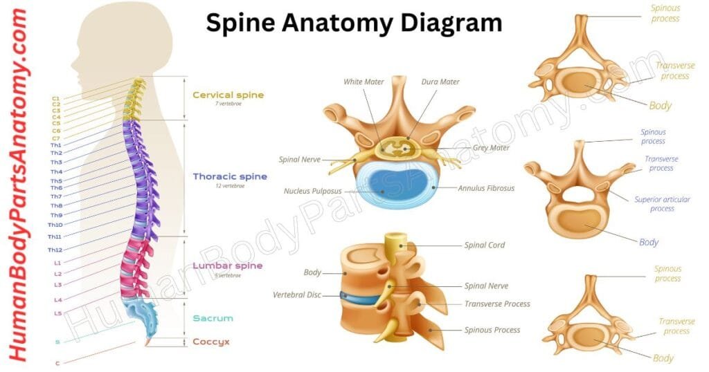
The vertebral column, or the spine, is an essential human body part of the axial skeleton. It safeguards the spinal cord and nerves while maintaining an upright posture.
This complex skeletal framework bears most of the body’s weight to maintain a vertical pose. Its different feature lies in a flexible rod found in all chordates, into a segmented array of bones referred to as vertebrae.
These vertebrae are interposed with intervertebral discs, which enhance the spine’s durability and flexibility.
Each vertebra is named according to its position within the spinal column.
The spinal canal is enclosed within the vertebral column, a protective cavity that envelops and shields the spinal cord.
Read More – Spine Anatomy: Complete Guide with Parts, Names, Functions & Diagram
- What is the Spine?
- What are the causes of back pain? Know about its diagnosis and treatments?
- When do you need spine surgery?
- What are the exercises for a healthy spine?
- 10 exercises for a strong spine after 60.
Hip Bone
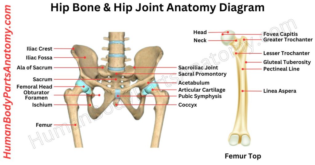
The hip is also known as the coxa in medical terms. It is a key area in vertebrate anatomy found on the outer side of the pelvis. It is located to the side and front of the buttocks, below the bony ridge of the iliac crest, and beside the obturator foramen.
This area includes muscles, tendons, and soft tissues that cover the prominent greater trochanter of the femur.
In adults, the hip bone forms from the fusion of three pelvic bones (the ilium, ischium, and pubis). It creates the sturdy inner and upper walls of the hip region.
Read More – Hip Bone Anatomy: Complete Guide with Parts, Names, Functions & Diagram
- What is Hip Injuries and Disorders?
- What is Hip Fracture? Symptoms and Signs of Hip Fractures.
- How to take care of your new hip joint?
Femur
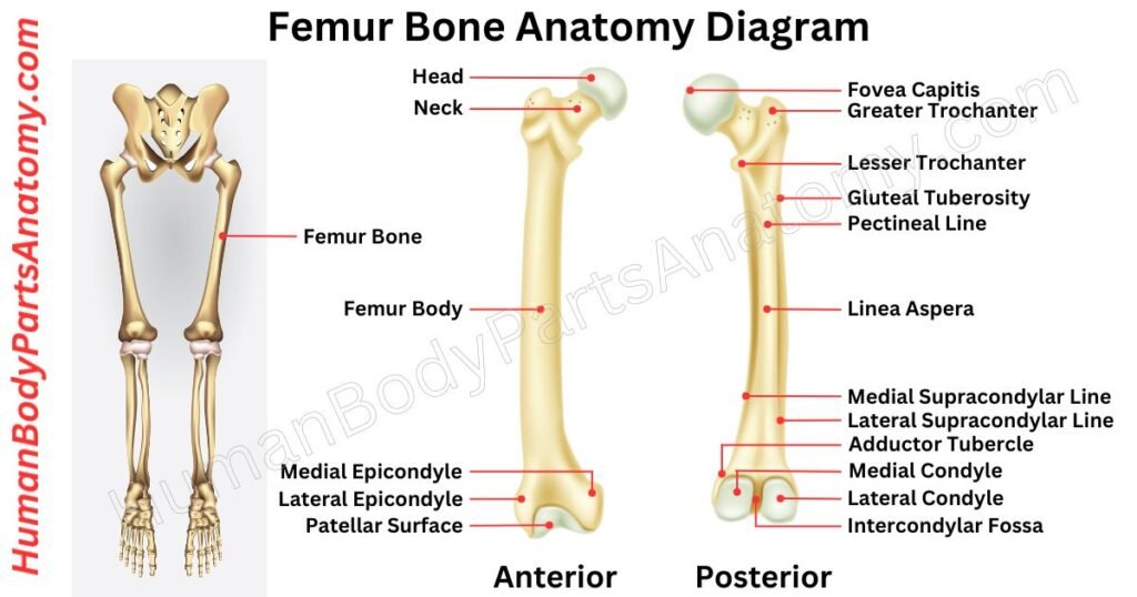
The femur, scientifically called the thigh bone, is essential within the human skeletal system. It is in the lower limb and bone between the hip joint and knee joints. This bone shapes the hip joint as its proximal end and forms an articulation point with the pelvic socket.
Moreover, the femur‘s distal end engages with the tibia and patella to form a knee joint structure. Beyond this, the femur bears the human body’s weight during stationary and dynamic activities.
Additionally, the femur is an essential anchor point for muscles, tendons, and ligaments that help move the hip joint and knee joints.
Read More – Femur Anatomy: Complete Guide with Parts, Names, Functions & Diagram
- Severe Upper Leg Pain Symptoms, Causes & Common Questions
- 6 Reasons You Might Have Thigh Pain — Plus, What to Do
- What are the best upper leg exercises?
- How is a femur shaft fracture treated?
Rib Cage
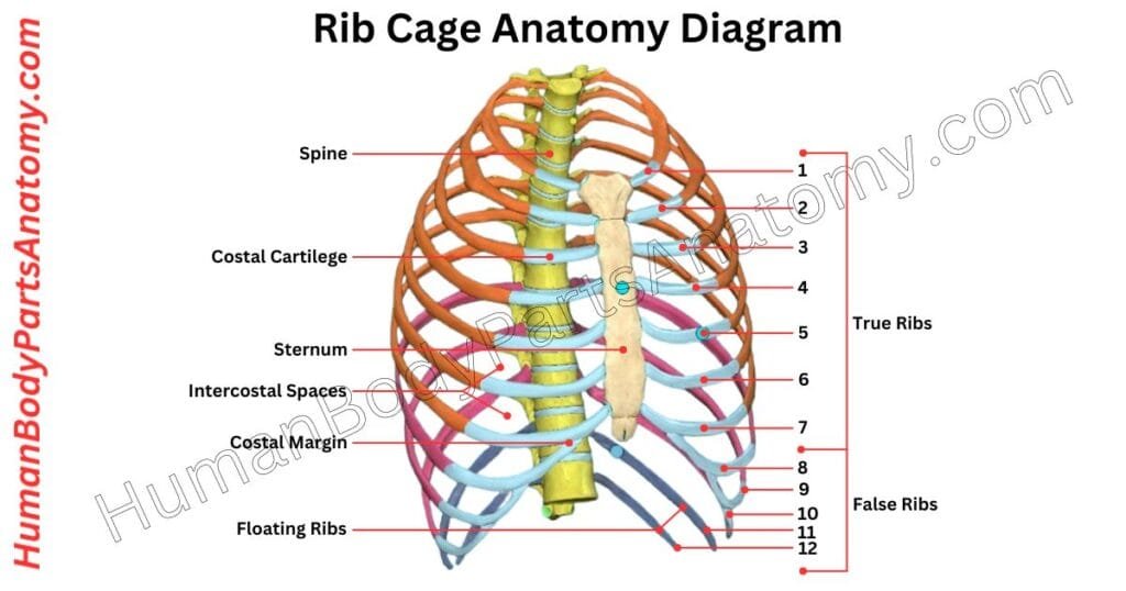
The rib cage, or thoracic cage, is an important component of the skeleton in most vertebrates. It comprises the ribs, the vertebral column, and the sternum.
This structure safeguards vital organs such as the heart, lungs, and major blood vessels. It also supports the shoulder girdle, contributing to the central framework of the axial skeleton.
In humans, the thoracic cage consists of 12 ribs connected to the sternum via costal cartilage. The sternum itself has three parts: the manubrium, the body, and the xiphoid process.
The cage also includes 12 thoracic vertebrae that interact with the ribs. This setup provides attachment points for muscles in the neck, upper limbs, abdomen, and back. Along with the skin and other tissues, it forms the chest wall.
Read More – Rib Cage Anatomy: Complete Guide with Parts, Names, Functions & Diagram
Sternum
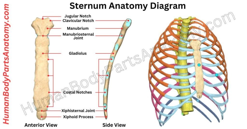
The sternum, or breastbone, is a flat, vertical bone situated in the center of your chest. It forms a key part of the rib cage. It consists of three distinct sections:
- Manubrium: The uppermost section, shaped like a broad, quadrilateral. It has a notch at the top, known as the suprasternal notch, and two side notches for the collarbones (clavicles). It creates the sternoclavicular joints.
- Gladiolus (Body): This is the longest section of the sternum. It has ridges where the cartilages of ribs 3 through 7 attach. The body joins the manubrium at a prominent bump called the sternal angle. It also connects with the second pair of ribs.
- Xiphoid Process: The smallest and lowest section of the sternum, which has a triangular shape. Its size and shape can vary among individuals.
The sternal angle, or angle of Louis, is the noticeable bump where the manubrium and body connect. The primary function of the sternum is to shield vital organs such as the heart and lungs.
Human Muscle Anatomy
In human anatomy, muscle tissues are made up of specialized cells that can contract and allow movement. This movement includes not just the motion of body parts and limbs but also the flow of blood, food, and other substances within the body.
Muscles are essential for moving the skeleton and making the heartbeat. They are found in the walls of organs like the intestines, uterus, and stomach.
Numerous muscles exist in our bodies, each serving various functions. Let’s examine the major muscles, understanding their different parts and how they contribute to movement and strength.
Biceps
The biceps brachii is a large muscle in the anterior upper arm that extends from the shoulder to the elbow. It has two unique heads, the long and short heads, which emerge from the scapula. These heads join together to produce a muscular system that joins to the upper section of the forearm.
Function—The biceps brachii is responsible for forearm flexion and supination. It helps with various activities and daily tasks. Curling the forearm at the elbow joint is referred to as forearm flexion.
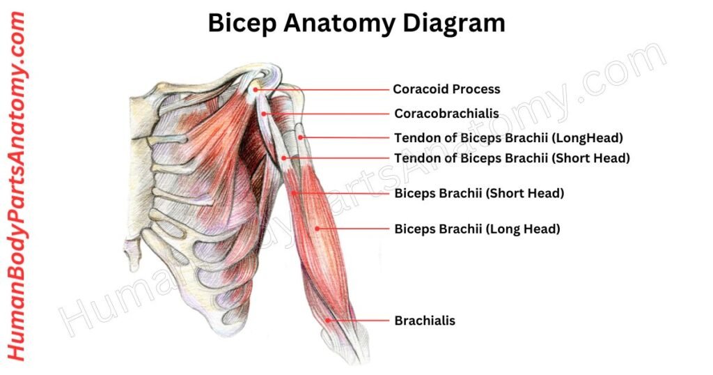
Read More – Ultimate Guide to Bicep Anatomy: Parts, Names, Functions & Diagram
Triceps
The triceps brachii is an extensor muscle in various vertebrates at the back of the upper limb. These muscles originate from the humerus and scapula, which comprise three distinct parts: the medial, lateral, and long heads.
Function—The triceps brachii muscle extends the forearm at the elbow joint. Its long head helps extend and adduct the arm at the shoulder joint.
Forearm
The forearm is the part of your arm between the elbow and wrist. It is made up of two bones: the outer radius and the inner ulna.
It has 20 muscles grouped into front (flexor) and back (extensor) compartments, which control elbow, wrist, and hand movements.
There are two types of muscles: front flexors and back extensors. Fascia organizes and supports these muscles around the ulna and radius.
Two structures, the intermuscular septum and interosseous membrane, create compartments and offer extra support.
The septum starts from the front of the radius, connecting with the forearm fascia, while the membrane forms between the radius and ulna.
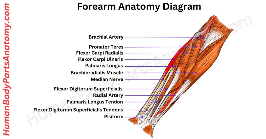
Read More – Complete Guide to Forearm Anatomy: Parts, Names, Functions & Diagram
Hip Muscles
The muscles around the hip joint are crucial for its movement in human anatomy. Typically, anatomists identify 17 primary muscles involved in hip motion; also, more muscles are included.
These muscles are categorized into four groups based on their location around the hip joint: the gluteal group, the lateral rotator group, the adductor group, and the iliopsoas group.
Hip movements are achieved through the coordinated action of multiple muscles. Most muscles contribute to more than one type of movement. These movements are described using specific anatomical terms.
- Flexion: Brings the thigh closer to the abdomen.
- Lateral Rotation: Outward leg turns, like in the lotus yoga position.
- Medial Rotation: Inward turning of the leg, opposite to lateral rotation.
- Abduction: Moving the thigh away from the body’s midline, like spreading the thighs apart.
- Adduction: Bringing the thigh back towards the midline, closing the thighs together.
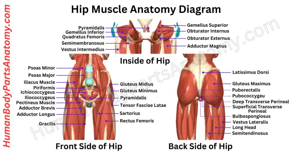
Read More – Hip Muscle Anatomy: Complete Guide with Parts, Names, Functions & Diagram
- What is Hip Fracture? Symptoms and Signs of Hip Fractures.
- What Is Hip Replacement Surgery & why it is needed?
- How to do Hip joint replacement?
- What are the Hip Dislocation treatments?
Thigh
The thigh is a significant part of human anatomy in the lower limb. It is between the hip and houses the pelvis and the knee joint. The femur is the prominent bone within the thigh and has exceptional strength, density, and robustness.
Functionally, the femur is a ball and socket joint at the hip and a modified hinge joint at the knee. Remarkably, the thigh region houses various main muscles in the human body.
These muscles enable various body movements, including bending, flexing, and rotational.
Additionally, they bear most of the body’s total weight. Furthermore, these muscles help maintain the structural integrity of the hips and legs.
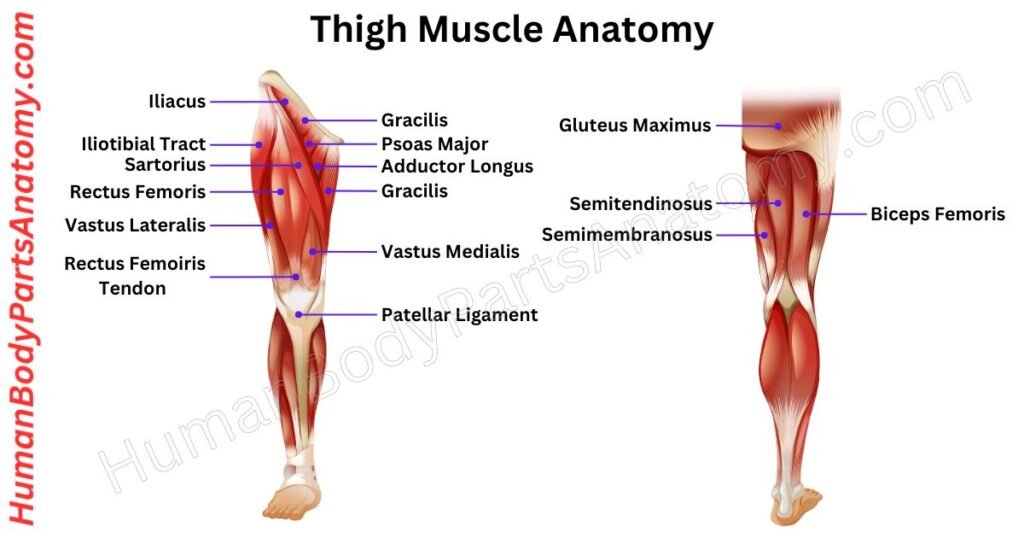
Read More – Complete Guide to Thigh Muscle Anatomy: Learn Parts, Names & Diagram
Human Body Parts – Joints
Wrist Joint
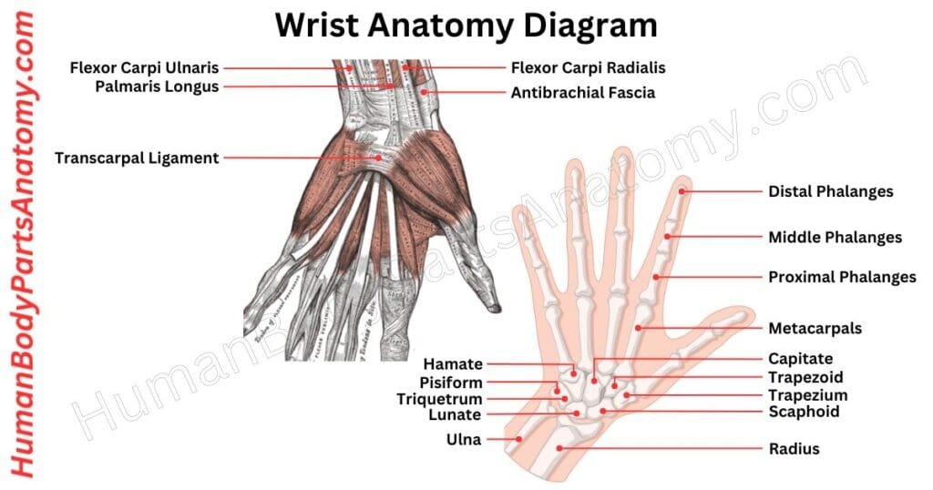
In human anatomy, the wrist is scientifically termed the carpus or carpal bones. It is a crucial part of the hand‘s structure, consisting of eight distinct bones that create the foundational framework for the upper part of the hand.
The wrist joint is scientifically known as the radiocarpal joint. It acts as the vital connection between the radius and the carpal bones. It includes both the carpus and the lower portions of the forearm bones.
The metacarpus is formed by the proximal sections of the five metacarpal bones. A network of interconnected joints exists among these anatomical components, making hand movement possible.
Read More – Wrist Anatomy: Ultimate Guide to Parts, Names, Functions & Diagram
Hip Joint
The hip joint connects your thigh bone (femur) to your hip bone (pelvis). It is a crucial body part, second in size only to your knee joint.
This ball-and-socket joint consists of the rounded head of the femur fitting snugly into a cup-like cavity in the pelvis, known as the acetabulum. This structure allows for extensive movement and helps your legs support your body weight.
It is located between your torso and lower legs. The hip joint serves several vital functions:
- Balances and supports your upper body.
- Facilitates the movement of your upper leg.
- Bears and distributes your body weight.
The ball-and-socket configuration of the hip joint permits your upper leg to move in three primary ways:
- Flexion (bending).
- Extension (straightening).
- Rotation (twisting).
This universal joint is essential for everyday activities, enabling a wide range of motions and providing stability and support.
Read More – Hip Bone Anatomy: Complete Guide with Parts, Names, Functions & Diagram
Knee Joint
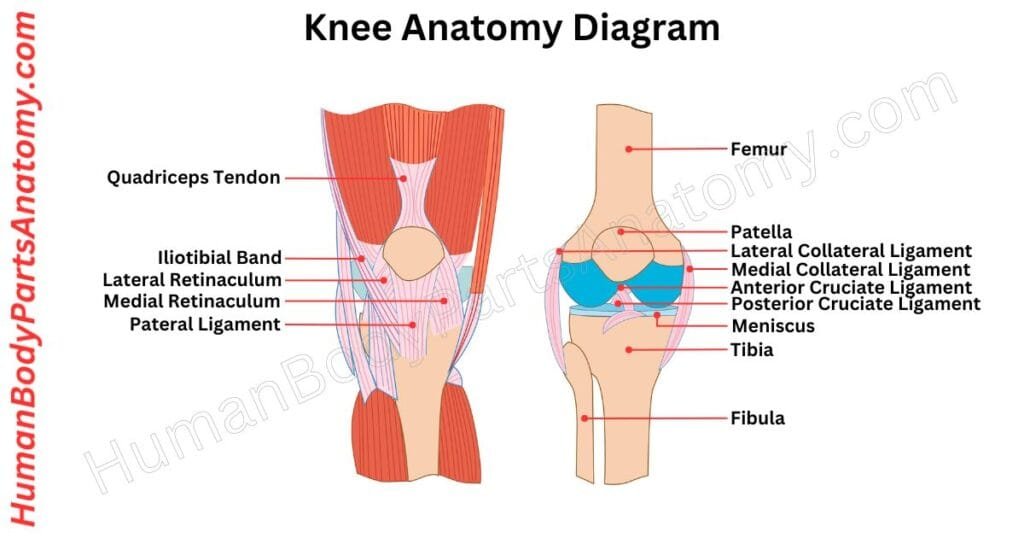
The knee joint, or a synovial joint, is an essential link between the femur, tibia, and patella bones. It is the body’s largest joint, mainly allowing leg bending and straightening. It contains two primary components: the tibiofemoral and patellofemoral articulations.
The tibiofemoral joint forms a connection between the tibia and the femur, while the patellofemoral joint forms with the patella with the femur.
Your knees are vital in supporting your body weight and allowing leg movement. This joint helps in activities like walking, running, and jumping.
Read More – Knee Anatomy: Complete Guide to Parts, Names, Functions & Diagram
- How to do Knee MRI scan?
- How to do Knee CT Scan?
- What is Knee Pain & how to diagnose it?
- What are the Knee Injuries and Disorders?
Ankle Joint
Your ankle is a hinge joint connecting your lower leg and foot. It is a hinge-like joint formed by the talus, tibia, and fibula bones.
The bony bump on the lower fibula (lateral malleolus) forms the outer boundary on one side, and the bony bump on the lower tibia (medial malleolus) creates the inner boundary. Together, they make up the ankle mortise.
The talus bone acts like a connector, linking with the calcaneus below and the navicular in front. The top part of the talus has a smooth surface, allowing comfortable up-and-down movement of your foot.
It snugly fits between the bony bumps, making the ankle most stable when you lift your toes towards your shin (dorsiflexion).
Strong ligaments act like rugged rubber bands on either side of the ankle to provide stability.
Shoulder Joint
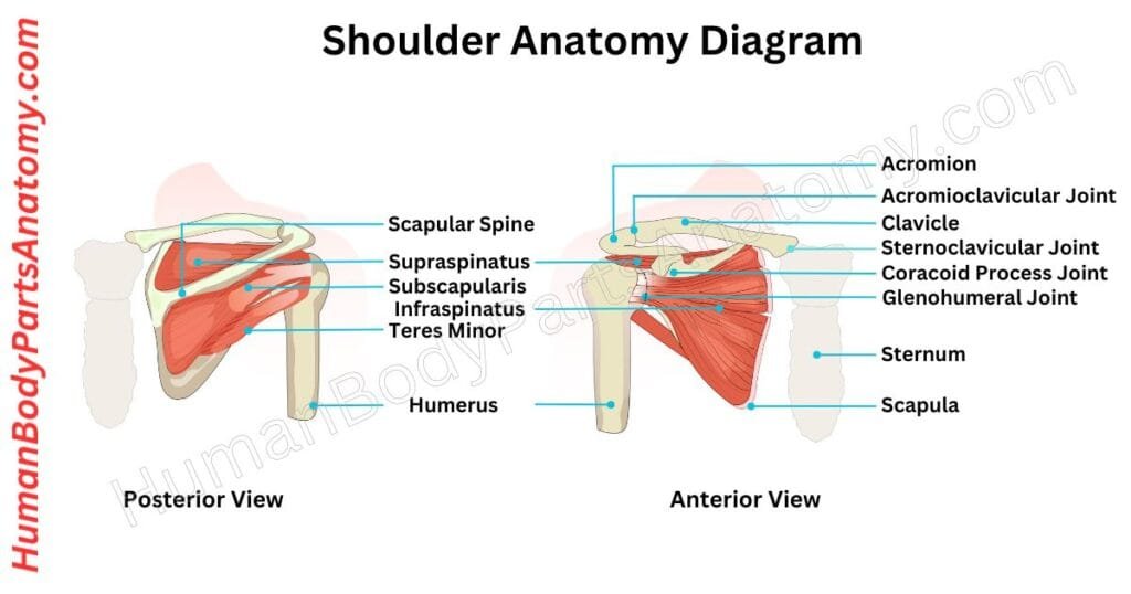
The human shoulder anatomy has three bones: the collarbone, shoulder blade, and upper arm bone. These bones are connected by joints, with the main one being the shoulder joint or glenohumeral joint.
Other joints, like the acromioclavicular joint, are also part of the shoulder. The shoulder joint allows circular rotation and lifting of the arm away from the body.
It is like a ball in a socket formed by the shoulder blade. A soft tissue envelope called the joint capsule surrounds the shoulder joint, lined with a smooth synovial membrane.
A group of four muscles maintains the shoulder’s stability, called the rotator cuff. These muscles attach to the shoulder blade and the upper arm bone. They are the supraspinatus, subscapularis, infraspinatus, and teres minor.
Read More –
- Shoulder Anatomy: Ultimate Guide to Parts, Names, Functions & Diagram
- What is a Shoulder?
- What are the causes of shoulder pain? Know about its diagnosis and treatments?
- Do you need shoulder surgery?
- 5 ways to help shoulder pain?
Human Anatomy – Alimentary System
Mouth
The mouth is necessary for digestion. It is a complex structure with different parts that work together to make the digestion system more efficient.
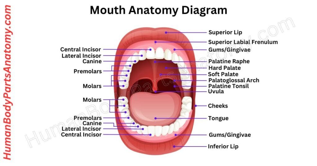
The lips create two regions: the vestibule and the oral cavity. The tongue occupies the central cavity and is surrounded by teeth, cheeks, and the isthmus of the fauces at the back.
The hard palate forms the front roof, and the soft palate makes up the rear, with the uvula hanging down.
The inner lining is called the oral mucosa. It is made of stratified squamous epithelium.
Salivary glands provide fluid to keep the mouth moist. Nerves and blood vessels form a network essential for the mouth’s diverse functions in human life.
Read More – Mouth Anatomy: Complete Guide with Parts, Names, Functions & Diagram
Teeth
Teeth are essential for chewing food and helping with digestion. Although they may look like bones, they’re ectodermal organs similar to hair and skin.
In adults, the 32 permanent teeth work together to cut, tear, mix, and grind food into smaller pieces. The tongue and oropharynx shape the food into a ball for easy swallowing.
Teeth have four main layers. The outer layer, called Enamel, is the hardest substance in the body and protects against cavity-causing bacteria.
Below the Enamel is dentin, a less intense layer. If Enamel wears away, it exposes dentin, increasing the risk of cavities.
The tooth root is covered by cementum, which, along with periodontal tissues, anchors the tooth in the jaw. The innermost layer, tooth pulp, houses nerves, blood vessels, and connective tissues, contributing to overall tooth health.
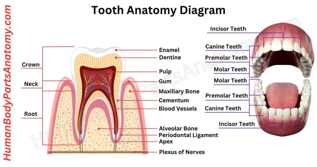
Read More – Complete Guide to Tooth Anatomy: Learn Parts, Names & Diagram
Lips
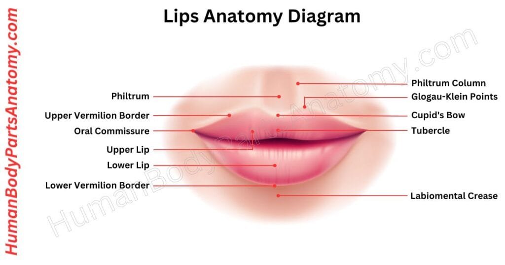
The lips are an essential part of the human face, pivotal in expressing emotions, talking, feeling, chewing, and romantic moments. Soft structures connected to the jaws are visible in many animals, including humans.
The upper and lower lips are scientifically called labium superius oris and labium inferius oris. Both lips have inner mucosal membranes, a colored vermilion layer, and outer skin.
In animals, including humans, lips are soft and flexible, helping with tasks like eating (such as sucking and swallowing) and forming sounds for speech.
Stomach
The stomach is a J-shaped crucial component of the digestive system. It makes enzymes and acids that chemically decompose food.
This process helps digestion before the food passes into the small intestine via the gastrointestinal (GI) tract. This tube extends from the mouth to the anus, through which food travels and waste exits.
The primary function of the stomach is to temporarily store food, mixing and breaking it down through muscular contractions and producing specialized cells and enzymes necessary for digestion.
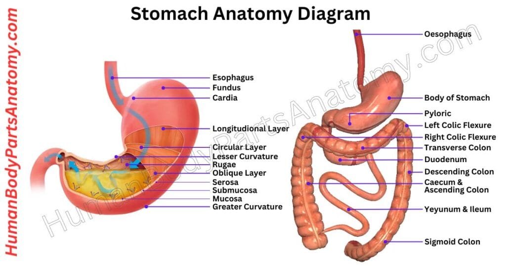
Read More – Stomach Anatomy: Complete Guide with Parts, Names, Functions & Diagram
Intestine
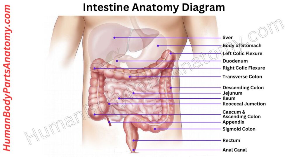
The intestine is also known as the bowel. It is a long, coil-shaped muscular tube that runs from the stomach to the anus. Its primary function is digestion, but it also helps produce hormones that regulate physiological activities and help in immunological protection.
The small intestine is directly connected to the stomach. It is 10 to 16 feet long and has three sections: the duodenum, jejunum, and ileum. Its inner lining is folded like an accordion, considerably increasing its surface area.
Enzymes present in the small intestine convert food into sugars, amino acids, and fatty acids. The nutrients are later taken into the circulation and distributed throughout the body.
The large intestine is present in the lower right abdomen and spans about 3 to 5 feet. It includes the cecum, appendix, colon, and rectum, terminating at the anus.
The main function of the large intestine is to absorb water and salts from digested food and convert them into solid waste (stool). Muscular contractions along the intestine propel waste toward the anus for elimination.
Liver
The liver is a critical organ found only in vertebrate animals that helps maintain the body healthy. It performs multiple critical functions, like removing toxins from the blood and producing proteins and other compounds required for digestion and development.
In humans, the liver is positioned in the upper right abdomen, just below the diaphragm, and protected by the lower ribs.
One of the liver‘s primary functions is to assist in controlling the body’s carbohydrate utilization, which includes storing and releasing energy like glucose and glycogen. It also promotes the breakdown of old red blood cells and the production of hormones.
In addition, the liver produces bile, which aids in the digestion of fats. Bile is stored in the gallbladder, a tiny pouch behind the liver, and discharged into the small intestine when needed to help digestion.
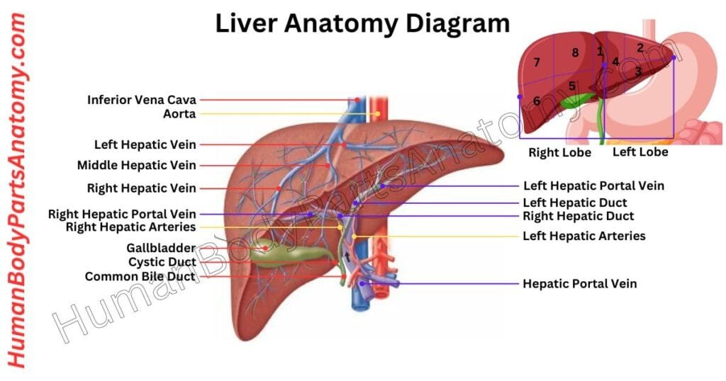
Read More – Liver Anatomy: Complete Guide with Parts, Names, Functions & Diagram
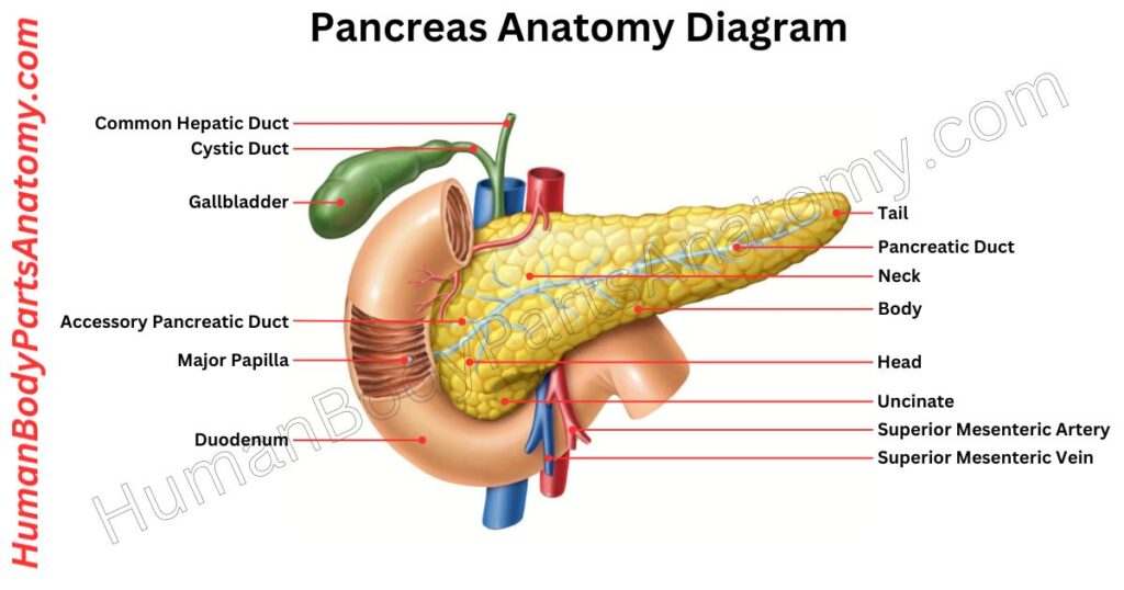
Pancreas
The pancreas is a big gland found deep within the belly. It works in both your digestive and endocrine systems. This dual-role organ functions as a factory with two independent manufacturing lines:
- Enzymes for Digestion: It creates enzymes that help break down the food you ingest.
- Hormones for Blood Sugar Regulation: It secretes hormones that control blood sugar levels in your body.
Beyond these primary functions, the pancreas supports other vital organs, including the heart, liver, and kidneys. Each day, it secretes about 1 to 4 liters of enzyme-rich juice, with the exact amount depending on your food intake.
Human Anatomy – Respiratory System
Nose
The nose is an essential part of our face. Its primary function is to let air inside our body. The nose filters, warms, and adds moisture to the air during breathing. It has bones and cartilage, which give it a unique shape.
Inside the nose, there are shell-like bones called nasal conchae. The tiny hairs in our nostrils act as filters that stop large particles from entering our lungs.
If something irritates the inside of our nose, like dust or allergens, our body makes us sneeze to get rid of them.
The nose is also essential for our sense of smell. It gives each person a unique look, which adds beauty to our face. Common issues like a stuffy nose or nosebleeds can affect how well our nose works and how we feel.
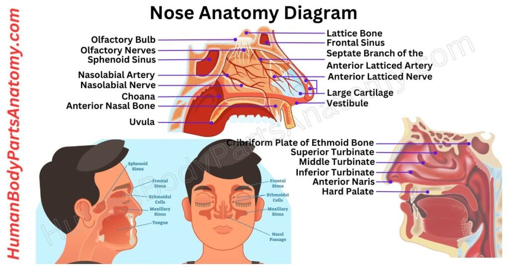
Read More –Nose Anatomy: Complete Guide with Parts, Names, Functions & Diagram
Human Anatomy – Sense Organs
Eye
Our eyes are incredible organs that respond to light and allow us to see and understand the world around us. The human brain can’t sense the environment directly.
Our eyes collect crucial information about what’s happening and help us to see things and keep our body balanced.
Most people have two eyes that work together to give us a broad view—about 200 degrees side-to-side and 135 degrees up and down. When our eyes cooperate well, we can perceive depth and see things in 3D and colors.
It’s important to note the difference between sight and vision. Sight is what our eyes do, capturing images and light. Vision is the whole process—from the eyes sending signals to the brain interpreting those signals into meaningful images.
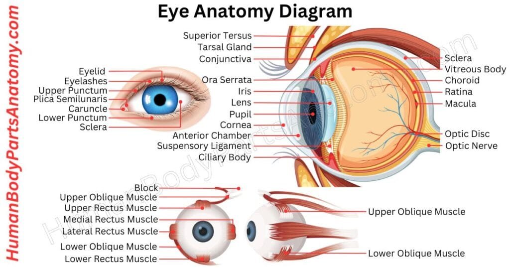
Read More – Ultimate Guide to Eye Anatomy: Parts, Structure, Functions & Diagram
Ear
Your ears help us hear and stay balanced. When sound enters your ear, it makes your eardrum vibrate. This vibration passes through tiny bones in your middle ear, making the sound louder. Then, in your inner ear, small hair cells turn the vibrations into electrical signals and send them to your brain.
Your inner ear also has fluid-filled canals that help you stay balanced. These canals have hair-like sensors. When you move, the fluid shifts and sends signals to your brain.
Your brain uses these signals to help your muscles keep you steady. So, your ears do much more than hear—they help you stay on your feet!
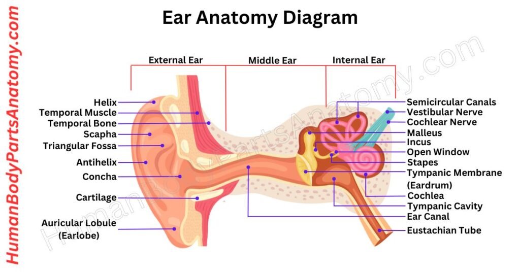
Read More – Ultimate Guide to Ear Anatomy: Parts, Structure, Functions & Diagram
Tongue
The tongue is a muscle in your mouth that helps you eat, talk, and taste food. It is covered in tiny bumps called taste buds, which let you taste sweet, sour, salty, and bitter. The tongue is always wet because of saliva, which also helps you taste and chew.
When you eat, the tongue helps move food around so you can chew it properly. It also enables you to swallow by pushing food down your throat.
In humans, the tongue plays a big role in talking, helping to form words and sounds. In other animals, it helps make different noises or vocalizations.
The tongue has two main parts: the front part, which is in the mouth, and the back part, which is closer to the throat. A line down the middle of the tongue separates it into left and right halves.
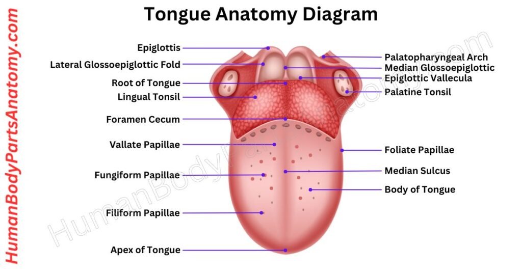
Read More – Tongue Anatomy: Complete Guide with Parts, Names, Functions & Diagram
Human Body Parts – Integument
Nails
Nails, found on our fingers and toes, are rigid plates made of a protein called alpha-keratin. This protein is also in other animals’ claws, hooves, and horns.
Nails are attached to the nail bed and can be used for scratching. The visible part is the “nail plate,” made of hard keratin and about half a millimeter thick.
Nails have lateral folds on each side and a proximal nail fold at the base. The cuticle, a thin layer of skin, protects and enhances sensory experiences.
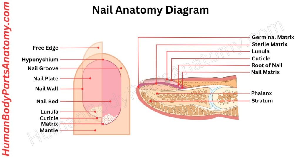
Read More – Complete Guide to Nail Anatomy with all Parts, Names & Diagrams
Hair
Hair is a protein-based filament that emerges from follicles embedded in the dermis layer of the skin. It is a distinctive feature of mammals.
Except for areas of smooth, hairless skin, the human body is largely covered with follicles that produce two types of hair: thick terminal hair and fine vellus hair.
While much attention is given to hair growth, types, and care, hair also serves as a significant biomaterial, primarily composed of alpha-keratin protein.
Many mammals have hair that serves various purposes. Hair helps animals stay warm and can help them blend into their surroundings. For some, it also sends signals to other animals, like warnings or attracting a mate.
In some cases, hair can even help defend the animal or, though rarely, be used for attack. Hair can also act like a sensor, enhancing the sense of touch.
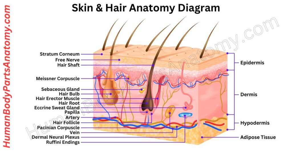
Skin
Skin is the soft outer layer that covers and protects the bodies of humans and many animals. It has three main jobs: protecting, controlling, and sensing.
First, the skin acts as a shield, keeping out harmful things like germs and preventing the body from losing too much water. It also helps keep us warm or cool by controlling our body temperature.
Additionally, the skin lets us feel sensations like touch. When exposed to sunlight, skin helps make vitamin D, which is important for our health.
If the skin gets hurt, it can heal itself by forming scar tissue, which might look different from the surrounding skin.
The thickness of the skin changes depending on where it is on the body. For example, the skin around the eyes is very thin, only about 0.5 mm thick, making it more prone to wrinkles.
On the other hand, the skin on the palms of our hands and the soles of our feet is much thicker, up to 4 mm. Hormones like estrogen can help skin wounds heal faster.
Human Anatomy – Nervous System
Brain
The brain is a vital organ that acts as the control center of the nervous system in all vertebrates and most invertebrates. It sits inside the skull, where it is cushioned and protected by cerebrospinal fluid.
As one of the most complex and essential organs, the brain works closely with the spinal cord to form the central nervous system.
This system manages nearly all body functions by processing information from the senses and sending out appropriate responses.
At birth, a baby’s brain weighs around 350 to 400 grams—only about 25% of the adult brain’s weight, which averages around 1.4 to 1.45 kilograms.
Despite making up just 2% of total body weight, the brain plays a huge role in overall function and development.
The most rapid brain growth happens during the first three years of life, and by age five, it reaches about 90% of its adult size.
On average, the adult brain measures roughly 167 mm in length, 140 mm in width, and 93 mm in height.
While the brain keeps changing throughout life, the most dramatic structural changes occur in early childhood. After the age of four, brain growth continues but at a slower and more gradual pace.
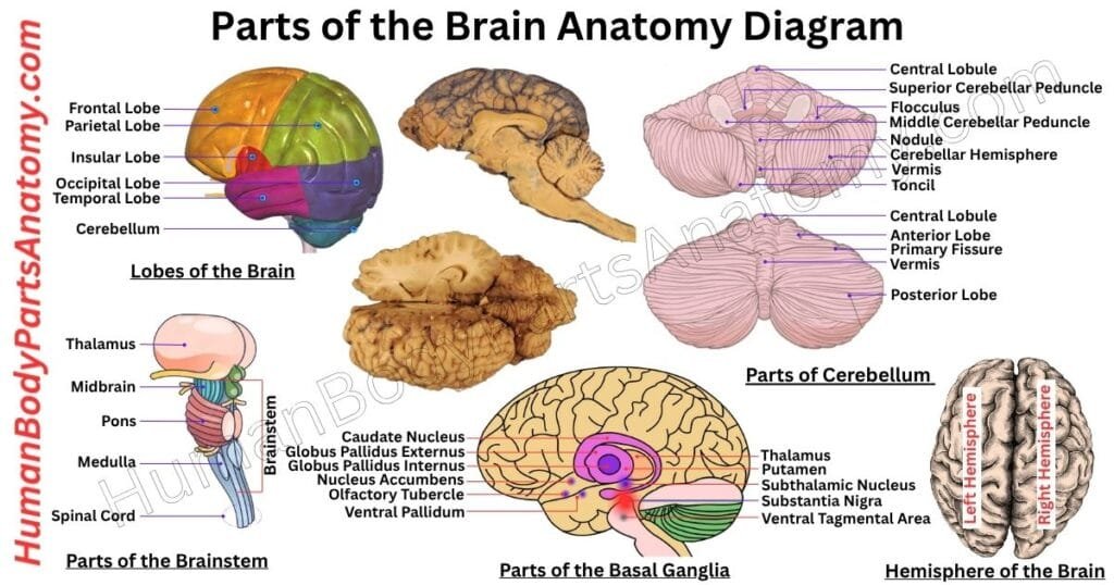
Read More – Parts of the Brain Anatomy: Complete Guide with Names, Functions & Diagram
- Parts of the Cerebrum Anatomy: Complete Guide with Names, Functions & Diagram
- Basal Ganglia Anatomy: Complete Guide with Names, Functions & Diagram
- The Cerebellum Anatomy: Complete Guide with Names, Functions & Diagram
- Brainstem Anatomy: Complete Guide with Names, Functions & Diagram
Lobes of the Brain
The cerebrum is the largest portion of the brain and sits at the top of the skull. Its outer layer is folded into grooves called sulci and raised ridges called gyri.
These folds let the brain fit more surface area inside the skull without getting bigger. Just beneath the folds, there is a thin layer—about 2 to 4 millimeters thick—of gray matter.
This gray matter contains neuron cell bodies and is where information is processed. Below it lies the white matter, made up of long fiber tracts that carry signals in and out of the gray layer.
The cerebrum is split into two halves, known as the left and right hemispheres. A bundle of fibers called the corpus callosum connects these halves and lets them send messages back and forth.
Each hemisphere mainly controls the opposite side of the body: the left side of the brain handles movement and sensation on the right side of the body, and vice versa.
Finally, each hemisphere is divided into four regions, or lobes, which specialize in tasks like processing touch, planning movements, handling vision, and managing speech. These lobes are Frontal Lobe, Parietal Lobe, Temporal Lobe, and Occipital Lobe.

Read More – Lobes of the Brain: Complete Guide with Names, Functions & Diagram
Human Anatomy – Cardiovascular System
Heart
The heart is a vital organ of muscles that pumps blood throughout the body and delivers oxygen and nutrients to every human body part. While doing this, it removes waste like carbon dioxide from the body.
In humans, the heart is located in the chest’s central space between the lungs and leaning left. It is around the size of a closed fist and weighs around 10 ounces in adults. However, it varies with factors like body size and gender.
Humans, birds, and mammals have four heart chambers – right atria, upper left, lower left, and right ventricles. The right side is the right heart, and the left is the left heart.
The heart is separated by the muscular wall called the septum. Blood is pumped from the right side of the heart through the pulmonary arteries for oxygen, and this blood goes to the lungs.
Special valves on the right side of the heart prevent blood from backflowing into the heart. After the lungs receive oxygen, the left side gets the blood through the pulmonary veins.
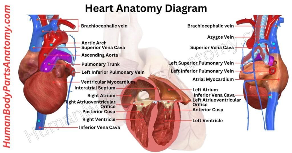
Read More – Heart Anatomy: Complete Guide with Parts, Names, Functions & Diagram
Arteries
Arteries carry oxygen-rich blood from the heart to all our organs. They work closely with veins and the heart, like tubes that transport blood from the heart to all parts of the body.
This blood, with oxygen and nutrients, is essential for adequately functioning the different organs. Arteries can change based on signals from the nervous system and outside factors like pressure and temperature.
Nerves in the arteries help them respond to these signals. Hormones like catecholamines can narrow or widen arteries, influencing blood pressure and flow. So, arteries are dynamic vessels that ensure our body gets the oxygen and nutrients it needs.
Human Anatomy – Urinary System
Ureter
The ureters are two muscular tubes that carry urine from the kidneys to the bladder for storage before it is excreted from the body.
After blood is filtered in the kidneys, the resulting liquid, called filtrate, goes through several stages of reabsorption in the kidneys tubules.
Eventually, the liquid becomes urine and passes into the collecting ducts. From there, urine moves into the calyces and then the renal pelvis, which is the starting point of the ureters.
The ureters get their blood supply directly and indirectly from the abdominal aorta. While there are no nerve ganglia on the ureters, they do receive signals from both the sympathetic and parasympathetic nervous systems.
In adults, the ureters are usually 20 to 30 centimeters long and 3 to 4 millimeters wide. They are lined with urothelial cells, a type of transitional epithelium, and have an extra layer of smooth muscle in the lower third to help move urine through peristalsis (wave-like muscle contractions).
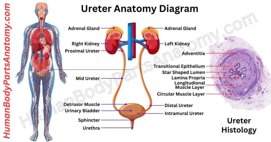
Kidney
The kidneys are two bean-shaped organs in your urinary system that filter your blood. Every day, they process about 200 quarts of fluid, which is enough to fill a large bathtub.
They remove waste products, excreted as urine, amounting to about two quarts per day. The remaining 198 quarts of fluid are reabsorbed and reused by your body.
In addition to waste removal, the kidneys maintain fluid balance and regulate electrolytes, including essential minerals like sodium and potassium.
They play a crucial role in filtering out toxins and waste from your blood, such as urea, creatinine, and acids, processing about half a cup of blood every minute.
Each kidney houses over a million filtering units called nephrons. Nephrons consist of :
- Glomeruli: These are clusters of tiny blood vessels that initiate the blood filtration process, a step known as glomerular filtration. They filter substances, which are then passed to the renal tubules.
- Renal Tubules: These small tubes reabsorb water, nutrients, and essential minerals, including sodium and potassium. They also remove waste and excess acids, sending these to the kidney’s collecting chambers. The waste is eventually excreted as urine.
This streamlined process ensures that your body efficiently removes waste and maintains a balanced internal environment.
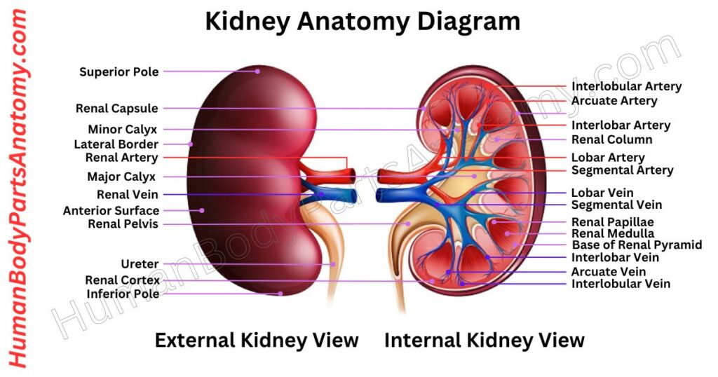
Read More – Kidney Anatomy: Complete Guide with Parts, Names, Functions & Diagram
Human Body Parts
Leg
The leg is part of your body between your knee and foot. It is made up of two bones: the tibia and the fibula. These bones give support and balance to your body, and they work with muscles to help you move around.
The tibia connects with the femur at your knee, and at the bottom, it joins with the fibula to form the ankle joint with the talus bone. This ankle joint is special because it helps your foot move smoothly while also keeping it stable.
When your ankle joint works properly, it lets your foot move. It makes the human body easier to walk and move around comfortably.
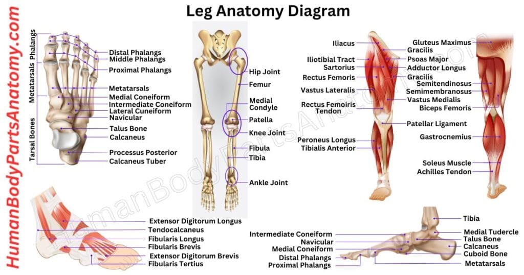
Read More – Complete Guide on Leg Anatomy with Parts, Functions & Diagram
Foot
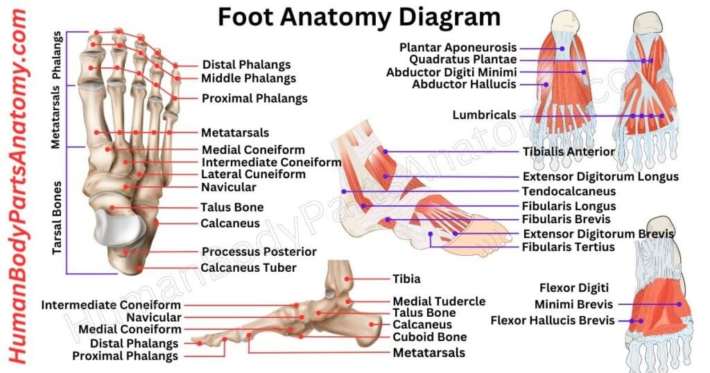
The foot is a complicated part of the human anatomy, consisting of many bones, joints, muscles, and tendons. It helps us walk and stand up straight. The foot includes everything below the ankle joint.
The ankle joint is where the shinbone (tibia), the thinner bone next to it (fibula), and a bone called the talus meet.
There are 26 bones in the foot, divided into three groups: the hindfoot, midfoot, and forefoot. These bones have cartilage covering their surfaces, where they meet each other to form joints.
The joints are surrounded by capsules and ligaments, which keep them stable. Twenty-nine muscles move the foot and ankle bones, which are connected to the bones by tendons.
Arm
The upper extremity, or arm, is a crucial part of the human anatomy. It has three main sections: the upper arm, forearm, and hand. It starts from the shoulder to the fingers and includes 30 bones, nerves, blood vessels, and muscles.
Starting at the shoulder joint, often called a ball-and-saucer joint. It allows for a wide range of movement, though it’s less stable than the hip joint.
Next is the elbow joint, a hinge joint that facilitates arm bending and straightening. This joint also gives the forearm the unique abilities of pronation and supination.
The wrist joint is ellipsoidal or condyloid, providing a good range of motion. The carpal bones have intercarpal joints, which allow some movement. The interphalangeal joints in the fingers act as basic hinge joints.
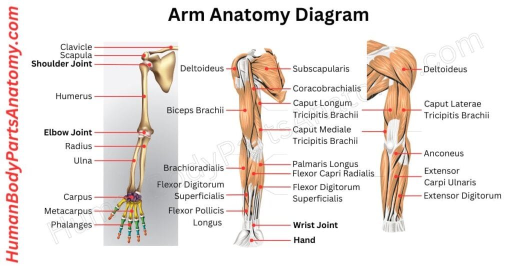
Read More – Comprehensive Guide to Arm Anatomy: Parts, Names & Diagram
Hand
A hand is a helpful part at the end of our arm. Humans and some animals like monkeys and koalas have hands. Even raccoons are said to have hands but don’t have thumbs like we do.
A human hand usually has five parts called fingers. We count the thumb as one of them. There are 27 bones in a hand, not depending on a particular bone. There are 14 finger bones connecting to the wrist bones.
Each hand has five long metacarpal bones and eight small carpal bones. Thus, a hand comprises fingers, thumbs, and bones that help it move and work.
Also, it contains various muscles, tendons, and ligaments, which help to do multiple operations like gripping and holding something in hand.
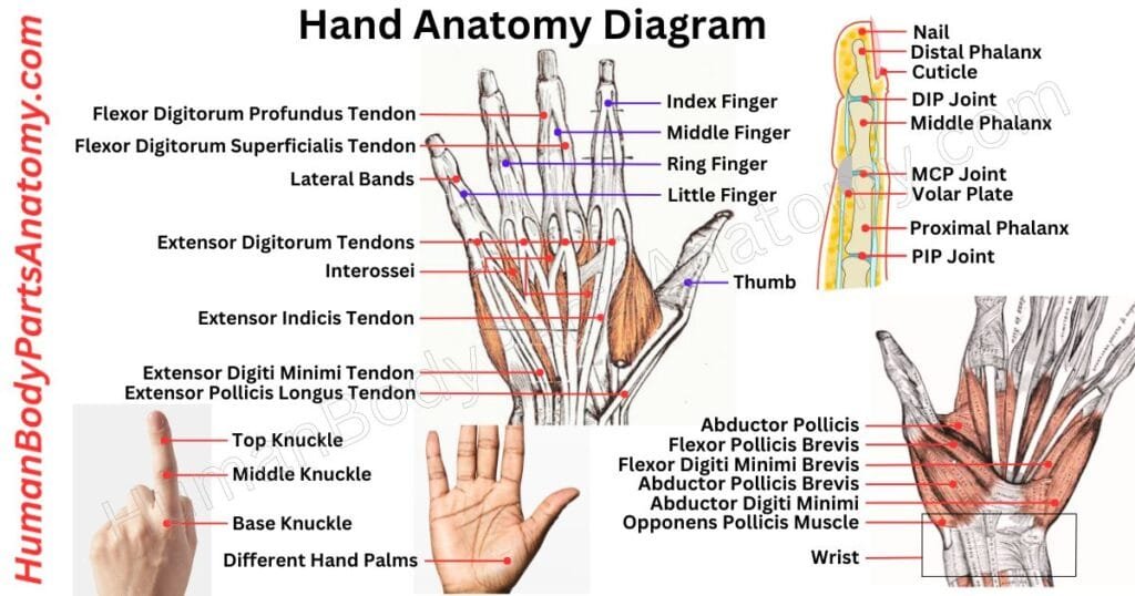
Read More – Comprehensive Guide to Hand Anatomy: Parts, Functions & Diagram
Finger
Fingers are essential parts of our hands and similar limbs in many animals. Most animals with limbs, like humans and primates, have five fingers, while shorter ones are called toes.
Fingers are flexible and opposable in humans. They help us feel things and make precise movements, and they are vital for skills like grabbing and moving objects.
The thumb is the first digit, followed by the index finger, the middle finger, the ring finger, and the little finger, also known as the pinkie.
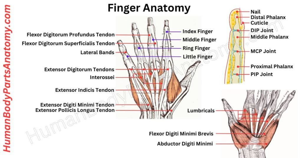
Read More – Complete Guide to Finger Anatomy with Parts, Names, Functions & Diagram
Thumb
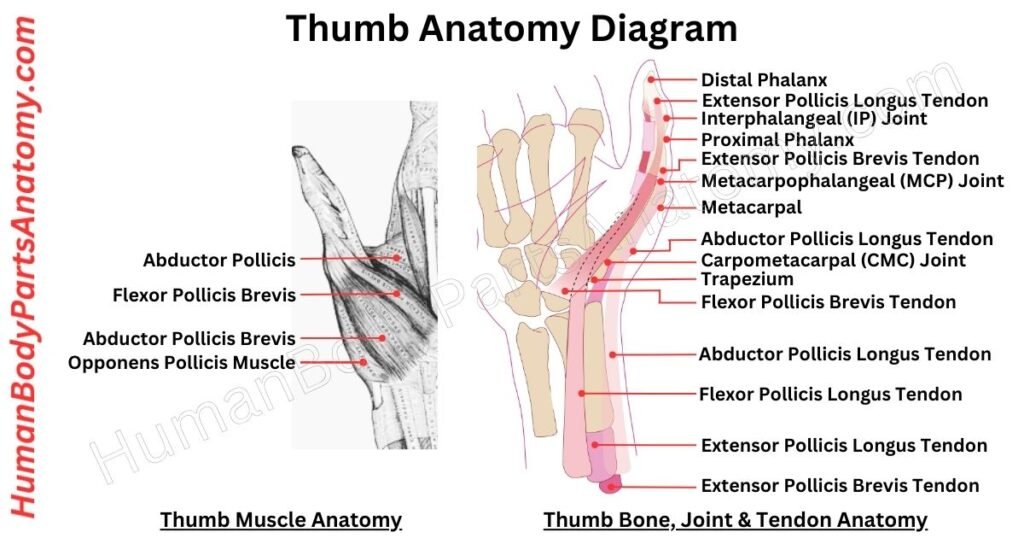
The thumb is a particular part of the hand with impressive flexibility. It can bend at the knuckle and touch the tips of other fingers. It enables various essential movements for holding and grasping objects.
The thumb consists of the metacarpal bone connected to the trapezium in the wrist. This bone is linked to the proximal phalanx, which then connects to the distal phalanx, forming the tip of the thumb.
Unlike the other fingers, the thumb lacks an intermediate phalanx bone. Oxygenated blood is mainly supplied to the thumb through the Princeps pollicis artery.
The thumb muscles, labeled ‘pollicis,’ include the extensor, flexor, opponents, and abductor muscles, with additional distinctions like longus and brevis.
One crucial muscle, the first dorsal interosseus, plays a significant role in thumb movement.
FAQ’s
The human body has around 600 to 650 muscles. These muscles help with movement, posture, breathing, and essential internal functions like digestion and blood circulation.
Muscles are divided into three types: skeletal (movement), smooth (internal organs), and cardiac (heart). The exact number may vary slightly based on classification, but most anatomy sources agree on over 600 muscles in the human body.
The human body has about 86 billion neurons (nerve cells). These neurons form the central nervous system (brain and spinal cord) and the peripheral nervous system, which includes 12 pairs of cranial nerves and 31 pairs of spinal nerves. Together, they transmit signals that control movement, sensation, thinking, and vital body functions.
The human body has about 78 organs, based on modern anatomy. These organs work together to carry out essential functions like breathing, digestion, circulation, and thinking.
The exact number may vary slightly depending on how an organ is defined, but 78 organs is the most widely accepted and commonly referenced figure.
The human body has 11 major body systems, each performing essential functions to keep the body healthy and alive. These systems include the circulatory, respiratory, digestive, nervous, endocrine, muscular, skeletal, integumentary, urinary, reproductive, and lymphatic systems. Understanding these systems is crucial for studying human anatomy, healthcare, and biology.
The human body has over 78 organs, but the most vital ones include the brain, heart, lungs, liver, and kidneys. Together with bones, muscles, and joints, they maintain life functions such as movement, circulation, and digestion.
An adult human has 206 bones, while a newborn has about 270 bones that gradually fuse as the body grows. Bones provide structure, protect organs, and store essential minerals like calcium.
Joint cracking often happens when gas bubbles in the synovial fluid burst or when tendons move slightly out of place. It’s usually harmless, but persistent pain or swelling may indicate arthritis or joint problems.
Muscle pain, or myalgia, can result from overuse, strain, dehydration, poor posture, or medical conditions like fibromyalgia. Most mild cases improve with rest, hydration, and stretching, but chronic pain should be checked by a doctor.
Commonly injured parts include the knee, ankle, lower back, shoulder, and wrist. These areas are highly mobile and bear significant stress during daily activities and sports.
Muscles generate movement by contracting.
Tendons connect muscles to bones, helping transfer force.
Ligaments connect bones to other bones, stabilizing joints.
It works by contracting and relaxing muscles to enable movement, support joints, aid breathing and digestion, pump blood through the heart, and produce heat to regulate body temperature.