
📅 Published on October 16, 2024 | 🕒 Last updated on July 4, 2026
Overview of Skull Anatomy
The skull is a complex, protective structure made of bone.[1] It is essential for safeguarding the brain and supporting various sensory functions.[1] The skull anatomy is divided into two main sections: the cranium, which encloses and shields the brain, and the facial bones, including the mandible or jawbone, that shape the face.[2] In scientific terms, the part that encases the brain is known as the neurocranium.[1] At the same time, the viscerocranium forms the facial skeleton, including the jaw.[1]
Beyond its role in protecting the brain, the skull supports vital sensory organs like the eyes, ears, nose, and mouth.[2] It ensures that the eyes are positioned correctly for optimal vision and secures the placement of the ears to assist in hearing and determining the direction of sound. Additionally, the skull aids breathing and eating by housing the nasal cavity and jaw.[2]
The skull’s intricate design ensures protection and functionality, playing a central role in survival by enabling sensory perception and environmental interaction.[1]
Skull Anatomy Labeled Diagram
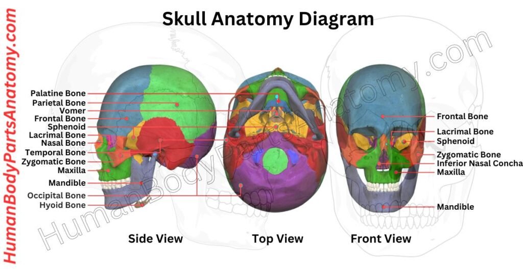
Parts of Skull
Cranial Bones (8 bones)
- Frontal Bone
- Parietal Bones (2)
- Temporal Bones (2)
- Occipital Bone
- Sphenoid Bone
- Ethmoid Bone
Facial Bones (14 bones)
- Nasal Bones (2)
- Maxillae (2) & Mandible
- Zygomatic Bones (2)
- Lacrimal Bones (2)
- Palatine Bones (2)
- Inferior Nasal Conchae (2)
- Vomer
Additional Structures
- Sutures
- Coronal Suture
- Sagittal Suture
- Lambdoid Suture
- Squamous Suture
- Foramina
- Foramen Magnum
- Optic Foramen
Human Skull Anatomy: Parts & Functions
Cranial Bones (8 bones)
The cranium, or neurocranium, consists of eight bones that cover and safeguard the brain. It is divided into two main parts: the cranial roof and the cranial base.[2]
The cranial roof, known as the calvarium, includes the frontal, occipital, and parietal bones.[2] The cranial base comprises all eight cranial bones. These protect the brain and support structures of the face.[2]
1. Frontal Bone
The frontal bone has two main parts: the squamous part and the orbital part. The squamous part is the large, flat area that makes up most of the forehead.[3]
The orbital part is smaller and lies horizontally, forming the top of the eye sockets and helping shape the nasal cavities.[3]
Sometimes, there is a third part called the nasal part, which connects the brow ridges to the nasal bones below and the bones on the sides of the nose (lacrimal and maxilla).[3]
The key function of the frontal bone is to shape the forehead and protect the brain’s frontal lobe. It also helps form the eye sockets and the front of the brain cavity.[3]
2. Parietal Bones (2)
The parietal bones are paired structures on each side of the skull. It forms the upper and side portions of the head. They rest above the brain’s parietal lobes and are shielded by a fibrous tissue called the epicranial aponeurosis.[2]
These bones are part of the neurocranium—the part of the skull that houses and protects the brain—alongside other key bones like the frontal, ethmoid, sphenoid, temporal, and occipital bones. The parietal bones mainly shape the top of the skull, with a smaller section contributing to the skull base.[2]
Their primary function is to safeguard the brain from injury. Slightly curved and rectangular, each parietal bone has two surfaces, four borders, and four corners.[2]
These borders link with other skull bones at junctions called cranial sutures. The inner surfaces of the parietal bones feature grooves and depressions that accommodate blood vessels and other essential structures.[2]
3. Temporal Bones (2)
The temporal bones are a pair of bones on either side of the skull. It forms parts of both the base and the sides of the head. They have a complex shape because they anchor various muscles and connect with neighboring bones.[4]
These bones are also crucial passageways for nerves and blood vessels moving in and out of the skull. Hidden within the temporal bones are the essential structures that control hearing and balance, including portions of the middle and inner ear.[4]
From the back of the skull, the temporal bones are visible on the sides, with the mastoid process—a noticeable, rounded bump—standing out.[4]
The temporal bone comprises several distinct sections:[4]
- The broad,
- Flat squamous part
- The pyramid-shaped petrous part
- The tympanic part, which surrounds the ear canal
- The slender styloid process is a pointed projection beneath the ear.
4. Occipital Bone
The occipital bone is located at the back and base of the skull. It is a crucial structure that protects the brain’s occipital lobes.[5]
The bone is curved trapezoid and has a key feature called the foramen magnum. This large oval opening allows the spinal cord to connect with the brain.[5]
The occipital bone is divided into three distinct sections based on its features.
- The front section, the basilar part or basioccipital, is a thick, rectangular segment extending forward.[5]
- On either side of the foramen magnum lie the lateral parts, known as the exoccipitals, which support the skull’s connection to the spine.[5]
- The back section, or squamous part, forms the largest and most curved area, supporting the skull’s rear.[5]
This design plays a vital role in brain protection and the connection between the brain and spinal cord. It makes it essential for structural integrity and neurological function.[5]
5. Sphenoid Bone
The sphenoid bone is one of the most intricate bones in the human body. Because of its unique shape, it is sometimes called the “wasp bone.”[6]
It forms a large part of the base of the skull, especially in the middle, and helps create the floor of the middle section of the skull’s interior.[6]
This bone is closely connected to important soft tissues like the cranial nerves and parts of the brain. Its main role is to provide openings and pathways (foramina and canals) for nerves and blood vessels to pass in and out of the skull.[6]
6. Ethmoid Bone
The ethmoid bone is one of the eight bones that make up the skull. It is a small, light bone located between the eyes, at the top of the nasal cavity. Even though it is delicate, it plays an important role in breathing, smell, vision, and protecting the brain.[7]
This bone separates the nasal cavity below from the cranial cavity above, where the brain sits. It also helps form the inner walls of the eye sockets, part of the base of the skull, and key parts of the nasal septum and nasal walls.[7]
The ethmoid bone is closely connected to the sense of smell. The olfactory nerve (cranial nerve I) passes through many tiny holes in the ethmoid bone to carry smell signals from the nose to the brain. Because of this, damage to the ethmoid bone—such as from injury or infection—can reduce or eliminate the sense of smell.[7]
The ethmoid bone has three main parts, each with a specific function:
- The cribriform plate forms the roof of the nasal cavity. It has many small holes that allow smell nerve fibers to pass through, giving it a sieve-like appearance. A vertical projection called the crista galli rises from the cribriform plate. It provides an attachment point for the falx cerebri, a membrane that separates the left and right sides of the brain.[7]
- The perpendicular plate extends downward from the cribriform plate and forms the upper two-thirds of the nasal septum. This septum divides the nasal cavity into left and right passages, helping guide airflow during breathing.[7]
- On each side of the perpendicular plate are the ethmoidal labyrinths, which contain multiple ethmoid air cells (sinuses). These air-filled spaces make the skull lighter, help warm and moisten inhaled air, and improve voice resonance.[7]
Each ethmoidal labyrinth includes two thin bony plates:
- Orbital plate (lamina papyracea): forms the inner wall of the eye socket.[7]
- Medial plate: forms part of the upper side wall of the nasal cavity and gives rise to the superior and middle nasal conchae, which help filter, warm, and humidify air.[7]
Facial Bones (14 bones)
The cranium has two main parts: the neurocranium and the viscerocranium. The neurocranium protects the brain, while the viscerocranium, also called the facial skeleton, shapes our face.[2]
The facial skeleton has 14 bones.
- Six of them come in pairs, with one on each side of the face. These paired bones are the inferior nasal conchae, nasal bones, maxillae (upper jaw), palatine bones, lacrimal bones, and zygomatic bones (cheekbones).[2]
- There are also two single bones: the mandible (lower jaw) and the vomer.[2]
Besides giving shape to the face, the viscerocranium protects important organs like the eyes and mouth. It has spots for muscles to attach and tiny holes, called foramina, that let nerves and blood vessels pass through.[2]
These shape the face and provide cavities for the sense organs (eyes, nose, and mouth).[2]
1. Nasal Bones (2)
The nasal bones are two small, oblong bones located in the upper central area of the face. These bones join at the midline through the internasal suture and form the bridge of the upper third of the nose.[8]
It is small, and its size and shape can vary significantly between individuals, contributing to the diverse range of nose shapes seen in humans.[8]
This variation extends to the angles, contours, and connections between the nasal bones and cartilage, which differ from person to person. Typically, nasal bones are categorized as “V-shaped” or “S-shaped.”[8]
However, these terms are general and not precise medical classifications. Anatomical illustrations of nasal bones often depict an idealized form, which may not accurately reflect the structure found in most people.[8]
2. Maxillae (2) & Mandible
In vertebrates, the maxilla, or upper jawbone, is a key structure that helps form the framework of the face and mouth.[9]
In humans, it consists of two bones joining the intermaxillary suture and creating a small projection known as the anterior nasal spine. The maxilla also supports the hard palate, which makes up the front part of the roof of the mouth.[9]
The mandible, or lower jawbone, is similar in origin but differs in function. It also forms through the fusion of two bones at the mandibular symphysis.[10]
It remains mobile, allowing for the movement necessary for chewing and speaking. The upper teeth are embedded in the maxilla’s maxillary arch, making it essential for the dental structure.[9][10]
The maxilla contributes to several important regions in the skull,[9] including:
- The roof of the mouth
- The floor and sidewalls of the nasal cavity
- The walls of the eye sockets (orbits)
It also forms two key openings, the inferior orbital fissure and the pterygomaxillary fissure, which allow nerves and vessels to pass through these regions.[9]
3. Zygomatic Bones (2)
The zygomatic bone, or zygoma, is a key facial bone on both sides of the face. It plays a major role in shaping the cheeks and protecting the eyes.[11]
This bone forms part of the eye socket and the side walls of the skull, contributing significantly to facial structure.[11]
Each zygomatic bone is a single unit that connects with several neighboring bones. These include the frontal bone (forehead), maxilla (upper jaw), temporal bone (near the ear), and sphenoid bone (at the skull’s base).[11]
Its strategic placement strengthens the face, allowing it to bear the forces generated during chewing by distributing them from the upper jaw.[11]
The zygomatic bone also features the zygomatic arch, a bridge-like structure connecting it to the temporal bone. This arch supports the masseter muscle, one of the primary muscles responsible for closing the jaw, which makes it essential for chewing.[11]
In addition to function, this bone contributes to the facial appearance and provides a protective barrier around the eyes.[11]
4. Lacrimal Bones (2)
The lacrimal bones are two tiny, delicate bones in the face, about the size of a fingernail. They are located at the inner corner of each eye socket. Each bone has two surfaces and four edges.[12]
These bones play a key role in the tear drainage process. They help form the nasolacrimal canal, which allows tears to move from the eyes to the nose.[12]
There is a small depression on the lower front part of one of the bones, called the lacrimal fossa, where the lacrimal sac sits.[12]
This sac collects tears when they overflow from the eyes, like during crying. From there, the tears pass through the nasolacrimal duct and into the nose, which is why your nose runs when you cry. If the lacrimal bone is injured or broken, it can block this tear drainage pathway.[12]
5. Palatine Bones (2)
The palatine bones are two small, L-shaped bones that play a crucial role in the structure of the face. It is positioned just behind the uvula.[13]
They form part of the hard palate, or roof of the mouth, and contribute to the walls of the nasal cavities. These bones lie between the maxilla (upper jaw) and the sphenoid bone at the skull’s base.[13]
Each palatine bone has two key sections:
- The horizontal plate, which makes up part of the hard palate,[13]
- The vertical plate helps shape the sides of the nasal cavity.[13]
The palatine bones connect to other facial bones, such as the maxilla, sphenoid, ethmoid, vomer, and inferior nasal concha.[13]
These bones are important in understanding conditions like cleft palate, a congenital disability that affects the mouth’s structure. They can also be involved in facial fractures from injuries.[13]
6. Inferior Nasal Conchae (2)
The nasal conchae, or turbinates, are curved bony structures found on the side walls of the nasal cavity. Each nasal cavity contains three conchae—superior, middle, and inferior—that project downward and inward.[14]
These structures divide the nasal cavity into five distinct air passages:
- The spheno-ethmoidal recess.[14]
- The superior, middle, and inferior meatuses.[14]
- A common nasal passage where the lateral passages converge.[14]
The superior and middle conchae are part of the ethmoid bone, while the inferior concha is independent. Due to their spiral, scroll-like shape, the conchae play a key role in increasing the surface area inside the nasal cavity.[14]
It allows for better warming, humidifying, and filtering of the air as it flows toward the lungs and optimizing respiratory efficiency.[14]
7. Vomer
The vomer is an unpaired bone found in the middle of the face. It forms part of the nasal septum, separating the nasal cavity’s two sides.[15]
The vomer sits along the midsagittal plane of the skull. It connects to several other bones: the sphenoid and ethmoid bones and the palatine and maxillary bones on both sides.[15]
The vomer helps form the nasal septum’s lower part, while the ethmoid bone’s perpendicular plate forms the upper part.[15]
Although it usually lies in the middle, the front section of the vomer may sometimes curve slightly to one side.[15]
This bone is thin and somewhat rectangular, with two flat surfaces and four edges. The surfaces have small grooves for blood vessels, and each surface has a nasopalatine groove, which provides a path for the nasopalatine nerve and blood vessels.[15][17]
FAQ’s
The adult human skull contains 22 bones. These include 8 cranial bones that form the cranium and protect the brain, and 14 facial bones that shape the face and support the eyes, nose, and jaw. This count does not include the auditory ossicles or the hyoid bone, which are separate structures.[2]
The skull’s primary function is to protect the brain from injury. It also supports facial structures, houses sensory organs (eyes, ears, nose), and provides attachment points for muscles involved in chewing, speaking, and head movement. These roles are essential for survival and daily function.[2]
The skull includes all bones of the head, while the cranium refers specifically to the 8 bones that enclose and protect the brain. Facial bones, such as the nasal bones and maxilla, are part of the skull but not part of the cranium.[2]
Babies have soft spots called fontanelles, which are areas where skull bones have not yet fused. Fontanelles allow the skull to flex during childbirth and expand as the brain grows. Most fontanelles close naturally by 18–24 months of age.[16]
The mandible (lower jaw) is the strongest and largest bone in the skull. It is the only movable skull bone and plays a critical role in chewing and speaking. Its strength allows it to withstand high bite forces.[2]
In infancy, the skull is flexible to support rapid brain growth. By adulthood, skull bones fully fuse at sutures. With aging, bone density may decrease, and facial bone structure can change due to tooth loss or osteoporosis, increasing fracture risk.[16]
The cranium consists of 8 bones. These include the frontal, parietal (2), temporal (2), occipital, sphenoid, and ethmoid bones. Together, they form a rigid protective case around the brain.[2]
The cranium is located at the top of the skeletal system, forming the upper portion of the skull. It sits above the facial bones and surrounds the brain, connecting to the spine at the foramen magnum in the occipital bone.[2]
References-
- National Center for Biotechnology Information (NCBI). (2023). Anatomy, Head and Neck, Skull. StatPearls.
https://www.ncbi.nlm.nih.gov/books/NBK499834 - Cleveland Clinic. (2025). Human Skull Anatomy. Cleveland Clinic Health Library.
https://my.clevelandclinic.org/health/body/skull-anatomy - National Center for Biotechnology Information (NCBI). (2025). Anatomy, Head and Neck: Frontal Bone. StatPearls.
https://www.ncbi.nlm.nih.gov/books/NBK535424 - National Center for Biotechnology Information (NCBI). (2023). Anatomy, Head and Neck, Temporal Region. StatPearls.
https://www.ncbi.nlm.nih.gov/books/NBK482497 - National Center for Biotechnology Information (NCBI). (2024). Anatomy, Head and Neck, Occipital Bone, Artery, Vein, and Nerve. StatPearls.
https://www.ncbi.nlm.nih.gov/books/NBK541093 - Jamil RT, Callahan AL. (2023). Anatomy, Sphenoid Bone. StatPearls.
https://www.ncbi.nlm.nih.gov/books/NBK544308
— PMID: 31335028 - Yu M, Wang SM. (2023). Anatomy, Head and Neck, Ethmoid Bone. StatPearls.
https://www.ncbi.nlm.nih.gov/books/NBK544328
— PMID: 31335048 - National Center for Biotechnology Information (NCBI). (2023). Anatomy, Head and Neck, Nose Bones. StatPearls.
https://www.ncbi.nlm.nih.gov/books/NBK541117
— PMID: 31082161 - Soriano R, Brizuela M. (2023). Anatomy, Head and Neck, Maxilla. StatPearls.
https://www.ncbi.nlm.nih.gov/books/NBK538527
— PMID: 30860762 - Ogle OE, et al. (2023). Anatomy, Head and Neck, Mandible. StatPearls.
https://www.ncbi.nlm.nih.gov/books/NBK532292 - Yu M, Wang SM. (2023). Anatomy, Head and Neck, Zygomatic Bone. StatPearls.
https://www.ncbi.nlm.nih.gov/books/NBK544257
— PMID: 31334977 - National Center for Biotechnology Information (NCBI). (2023). Anatomy, Head and Neck: Lacrimal Apparatus. StatPearls.
https://www.ncbi.nlm.nih.gov/books/NBK531487 - National Center for Biotechnology Information (NCBI). (2023). Anatomy, Head and Neck, Palate. StatPearls.
https://www.ncbi.nlm.nih.gov/books/NBK557817 - Fakoya A, et al. (2023). Anatomy, Head and Neck, Nasal Concha (Turbinates). StatPearls.
https://www.ncbi.nlm.nih.gov/books/NBK546636
— PMID: 31536243 - National Center for Biotechnology Information (NCBI). (2023). Anatomy, Head and Neck, Nose. StatPearls.
https://www.ncbi.nlm.nih.gov/books/NBK532870 - Lipsett BJ, Reddy V, Steanson K. (2023). Anatomy, Head and Neck: Fontanelles. StatPearls.
https://www.ncbi.nlm.nih.gov/books/NBK542197
— PMID: 31194354 - Fitzpatrick TH, Brizuela M, Downs BW. (2023). Anatomy, Head and Neck, Nasopalatine Nerve. StatPearls.
https://www.ncbi.nlm.nih.gov/books/NBK538422
Read More-
Lower Limb
- Complete Guide on Leg Anatomy with Parts, Functions & Diagram
- Complete Guide to Thigh Muscle Anatomy: Learn Parts, Names & Diagram
- Knee Anatomy: Complete Guide to Parts, Names, Functions & Diagram
- Femur Anatomy: Complete Guide with Parts, Names, Functions & Diagram
Upper Limb
- Complete Guide to Finger Anatomy with Parts, Names, Functions & Diagram
- Complete Guide to Forearm Anatomy: Parts, Names, Functions & Diagram
- Comprehensive Guide to Arm Anatomy: Parts, Names & Diagram
- Comprehensive Guide to Hand Anatomy: Parts, Functions & Diagram
- Ultimate Guide to Bicep Anatomy: Parts, Names, Functions & Diagram
- Shoulder Anatomy: Ultimate Guide to Parts, Names, Functions & Diagram
- Wrist Anatomy: Ultimate Guide to Parts, Names, Functions & Diagram
- Complete Guide to Nail Anatomy with all Parts, Names & Diagrams
Human Head
- Ultimate Guide to Eye Anatomy: Parts, Structure, Functions & Diagram
- Tongue Anatomy: Complete Guide with Parts, Names, Functions & Diagram
- Mouth Anatomy: Complete Guide with Parts, Names, Functions & Diagram
- Complete Guide to Tooth Anatomy: Learn Parts, Names & Diagram
- Ultimate Guide to Ear Anatomy: Parts, Structure, Functions & Diagram
Organs
External Sources-
- Wikipedia
- KenHub
- Optometrists
- Cleveland Clinic
- American Academy of Ophthalmology
Medical Disclaimer
All content on HumanBodyPartsAnatomy.com is educational and based on verified, peer-reviewed medical sources. Articles are authored or reviewed by qualified medical or biomedical professionals to ensure accuracy.
This website does not provide medical advice, diagnosis, or treatment. Always consult a licensed healthcare professional for personal medical guidance.
No commercial or promotional interests influence the medical content published on this site.