
📅 Published on February 8, 2025 | 🕒 Last updated on July 15, 2026
Overview of Nose Anatomy
The nose is the primary gateway to the respiratory system.[1][2] It plays a vital role in the sense of smell, making it a key component of the olfactory system.[1] Its structural framework is shaped by the nasal bones and cartilage, including the nasal septum, which separates the nostrils and divides the nasal cavity into two distinct chambers.[2][3] This intricate nose anatomy supports two essential functions: breathing and detecting odors.[1][4]
The nose has both external and internal components. The external nose, aside from its aesthetic importance, protects the internal structures and allows air to enter.[4] The internal component, known as the nasal cavity, plays a multifaceted role in respiration, olfaction, speech, and even taste perception.[1][5] This dual-purpose organ is a prime example of how the body combines anatomical form with physiological function.[1]
This page explores the anatomy of the nose, with the structure of the nasal cavity, highlighting its unique features and their importance in functions like breathing, filtration, and smell.
Function of the Nose
The nasal cavity assists in respiration by preparing inhaled air for oxygen exchange. Due to the narrow nature of the cavity, air is rapidly introduced to a large mucosal surface area with a rich supply of blood at body temperature.[1][5]
- The nose plays a vital role in breathing, serving as the primary entry point for inspired air.[1]
- The nasal cavity and nearby paranasal sinuses are lined with nasal mucosa, which warms and moistens inhaled air before it reaches the lungs.[1][5]
- Shell-like bony structures called nasal conchae (turbinates) assist in the air-warming and humidification process.[2][3]
- Tiny hairs (vibrissae) in the nostrils trap large particles, preventing them from entering the lower airways.[3]
- The nose triggers sneezing to expel irritating particles from the nasal passages.[5]
- The sense of smell (olfaction) is controlled by receptor neurons located in the upper nasal cavity (olfactory region), which send signals along the olfactory nerves to the central nervous system.[1][5]
- The nose aids in speech production, particularly nasal vowels and consonants, by directing airflow through nasal resonating chambers.[3]
- The paranasal sinuses act as resonating chambers that may amplify sounds during speech.[3][6]
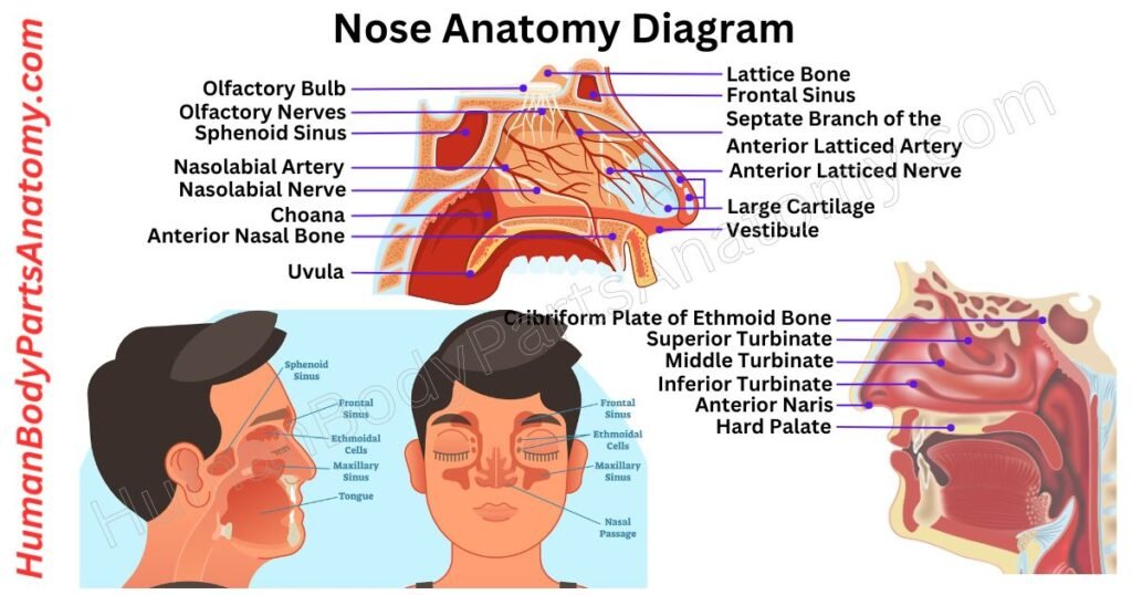
Nose Anatomy Diagram
Nose Anatomy
External Nose
A. Skeletal Framework
- Bones:
- Nasal Bones
- Frontal Process of Maxilla
- Nasal Part of Frontal Bone
- Cartilages:
- Septal Cartilage
- Lateral Nasal Cartilage
- Major Alar Cartilage
- Minor Alar Cartilage
- Vomeronasal Cartilage
B. External Openings
- Nostrils (Nares)
C. Skin and Soft Tissue
- Thin Skin
- Thick Skin
D. Muscles
- Procerus
- Nasalis
- Depressor Septi Nasi
- Levator Labii Superioris Alaeque Nasi
Internal Nose
A. Nasal Septum
- Maxillary bone (the crest)
- Perpendicular plate of ethmoid bone
- Septal nasal cartilage (ie, quandrangular cartilage)
- Vomer bone
B. Roof Bones
- Cribriform Plate of Ethmoid Bone
- Sphenoid Bone
C. Floor
- Palatine Process of Maxilla
- Horizontal Plate of the Palatine Bone
D. Lateral Wall
- Conchae (Turbinates)
- Superior Concha
- Middle Concha
- Inferior Concha
- Meatuses
- Superior Meatus
- Middle Meatus
- Inferior Meatus
E. Nasal Vestibule
F. Respiratory Region
Paranasal Sinuses
- Frontal Sinuses
- Maxillary Sinuses
- Ethmoidal Sinuses
- Sphenoidal Sinuses
Blood Supply
- Arteries:
- External Carotid Artery:
- Facial Artery
- Maxillary Artery
- Internal Carotid Artery:
- Ophthalmic Artery
- External Carotid Artery:
Venous Drainage
Nerve Supply
- Sensory:
- Ophthalmic Nerve (V1)
- Maxillary Nerve (V2)
- Olfactory Nerve (Cranial Nerve I):
- Autonomic Nerves:
- Sympathetic
- Parasympathetic
Lymphatic Drainage
- Anterior Nasal Cavity
- Posterior Nasal Cavity
External Nose Anatomy
The external nose is a prominent facial structure that leads to the nasal cavity. It helps facilitate breathing and olfaction while contributing to facial appearance.[4] Topographically, it can be divided into three units for clinical analysis: the frontal, lateral, and basal views.[4]
A – Skeletal Framework
The external nose is supported by both bones and cartilage, with bony structures forming the upper third and cartilaginous structures shaping the lower two-thirds.[4]
Bones
It is located in the upper part of the nose. The upper third of the nose contains the paired nasal bones that extend from the frontal bone caudally toward the rhinion.[4]
1. Nasal Bones
The nasal bones are paired, flat bones that form part of the facial skeleton and contribute to the bridge of the nose at the top.[4] Along with other bones (the zygomatic, maxilla, palatine, lacrimal, vomer, inferior nasal conchae, and mandible), they help shape the face.[4]
Location and Shape
The nasal bones lie on either side of the midline, between the frontal processes of the maxilla. Their lower edges connect to the nasal cartilage, shaping the upper boundary of the nasal cavity.[4]
Structure
- External Surface: It is slightly curved outward and covered by small muscles like the procerus and nasalis.[4]
- Internal Surface: It is curved inward with a groove for the anterior ethmoidal nerve.[4]
The nasal bones join with nearby bones through various connections:
- Top Border: Joins the frontal bone at the frontonasal suture.[4]
- Sides: Connect to the maxilla at the nasomaxillary suture. The nasal bones also have articulations with the lacrimal bone superolaterally and the nasomaxillary processes inferolaterally.[4]
- Bottom Border: Attaches to the lateral cartilage of the nose.[4]
- Inner Edge: Links the two nasal bones at the internasal suture and extends backward to contribute to the nasal septum.[4]
Function
The nasal bones support the structure of the nose, form the upper part of the nasal opening, and protect the nasal cavity from injury.[4]
2. Frontal Process of Maxilla
The frontal process of the maxilla is a slender, upward extension located on the front upper portion of the maxilla. It forms the anterior lacrimal crest and the ethmoidal crest, and plays a key role in shaping the lateral wall of the nasal cavity.[3][4]
Articulations of the Frontal Process:
- Anteriorly: Joins the nasal bone’s lateral edge, forming the nasomaxillary suture.[4]
- Superiorly: Meets the frontal bone’s nasal margin, creating the front maxillary suture.[4]
- Posteriorly: Connects with the lacrimal bone, contributing to the lacrimomaxillary suture.[4]
- Medially: Interfaces with the middle nasal concha of the ethmoid bone.[3]
This structure integrates with neighboring bones to support and stabilize the nasal and orbital regions, underscoring its importance in facial anatomy.
3. Nasal Part of Frontal Bone
The nasal part of the frontal bone is a small section located toward the lower mid-region of the frontal bone. It includes the nasal spine and nasal notch of the frontal bone, playing a key role in shaping the root of the nose, the bony nasal septum, and the roof of the nasal cavities.[3][4]
The nasal part forms important joints with neighboring bones:
- Inferomedially: It connects with the upper edges of the right and left nasal bones to form the frontonasal suture.[4]
- Inferolaterally: It joins the frontal processes of the right and left maxillae, creating the front maxillary sutures.[4]
- Posteroinferiorly: It articulates with the upper portion of the ethmoid bone, forming the frontoethmoidal suture.[3]
These connections contribute to the stability and framework of the nasal region.
Cartilages
Five distinct cartilages provide crucial support and shape to the external nose. These include the septal cartilage, two lateral nasal (upper lateral) cartilages, two major alar (lower lateral) cartilages, minor alar cartilages, and the vomeronasal cartilage.[4][11]
1. Septal Cartilage
The septal nasal cartilage — also known as the quadrangular cartilage — is a broad, quadrilateral structure of hyaline cartilage that forms the anterior portion of the nasal septum, dividing the nasal cavity into two chambers.[2][4][7][9]
It is positioned between the perpendicular plate of the ethmoid bone and the vomer bone, encased by a mucous membrane.[2][7] The upper edge connects to the nasal bones, and the lower edge joins the alar cartilage through fibrous tissue.[4][9]
This division into two nasal cavities helps streamline airflow. It creates turbulence in the narrow passages, which enhances the efficiency of air movement in both directions.[3][9]
Additionally, the septal nasal cartilage is crucial in shaping and aligning the nose, serving as its central structural support.[4][7][9]
2. Lateral Nasal Cartilage
The lateral nasal cartilage is a wing-shaped structure that extends outward from the septal nasal cartilage. It sits just below the nasal bones and above the major alar cartilage, with a small gap between them.[4]
These cartilages work together, with the lateral nasal cartilage and major alar cartilage curving to form a sturdy connection supported by fibrous tissue.[4]
It is made of strong yet flexible hyaline cartilage (the same type as the septal nasal cartilage). The lateral nasal cartilage connects to the septum at its upper part.[4]
This connection helps maintain the structure of the nasal cavities.[4] If the cartilage weakens or collapses, it can obstruct the inner nasal valve, reducing airflow and making it harder to breathe.[4][7]
3. Major Alar Cartilage
The major alar cartilage (lower lateral cartilage/LLC) is located on either side of the nasal tip and plays a vital role in shaping the nose and supporting airflow. These thin, hyaline cartilage structures are connected to the lateral nasal cartilage by fibrous tissue and are folded into two distinct parts: the medial crus and the lateral crus.[4]
- The medial crus forms the inner section and aligns perpendicularly with the septal nasal cartilage.[4]
- The lateral crus constitutes the outer portion, shaping the ala (the flared portion) of the nose.[4]
Together, the medial and lateral crus create an oval-shaped tip at each nostril.[4]
At the center of the nasal tip, the two sides of the major alar cartilages meet, forming a small notch called the apex of the nose. These cartilages also help form the walls of the nostrils/nares, ensuring they remain open.[4]
This structural support facilitates efficient airflow through the nasal passages, optimizing respiration by guiding air to the nasal valve.[4]
4. Minor Alar Cartilage
The minor alar cartilages are small, flexible pieces of hyaline cartilage. There are usually three to four on each side of the nose. They sit between the lateral nasal cartilage and the major alar cartilage, forming part of the nostrils’ outer edges (the ala).[4]
Also known as “accessory cartilage,” these tiny structures support and shape the nostril base. Together with the major alar cartilage, they help keep the nostrils stable and functional, maintaining their proper shape and appearance.[4]
5. Vomeronasal Cartilage
The vomeronasal cartilage is a thin piece of hyaline cartilage that joins the vomer bone with the septal nasal cartilage. It is associated with the vomeronasal organ, a component of the accessory olfactory system that detects certain chemical signals.[4][11]
This organ helps detect scents and has a lining similar to the main smelling area in the nose. The vomeronasal cartilage provides structural support, contributing to the stability and function of the nose.[4][11]
B. External Openings
The external openings or nostrils, also called anterior nasal apertures, are the two pear-shaped openings at the base of the nose.[1][4]
They serve as the entry points for air into the nasal cavity, playing a vital role in breathing and filtering particles from the air before it reaches the lungs.[1][4]
Nostrils (Nares)
Each nostril is one of the two openings in the nose that allow air and other gases to flow in and out of the nasal passages.[1]
The nostrils take turns becoming slightly congested and then decongested in a natural process called the nasal cycle, which is a physiologic alternation of resistance between the two nasal airways.[5]
A wall of tissue called the septum separates the nostrils.[2][7] Humans possess two external nostrils at the front of the nose and two internal nostrils (choanae) located at the back of the nasal cavity, which connect the nose to the throat (nasopharynx).[1][5]
Each nostril contains vibrissae — tiny hairs — that trap dust, pollen, and other particles to keep the airways clean before air reaches the lower airways.[3]
Interestingly, the brain can process different smells from each nostril, similar to how the eyes process different images, which can lead to a unique “smell rivalry” experience.
C. Skin and Soft Tissue
The skin covering the external nose varies in thickness along its length. At the upper portion (glabella to nasofrontal angle), the skin is thicker and more mobile.[4]
Over the nasal bridge, it becomes thinner and less mobile due to proximity to the underlying bone. At the nasal tip, the skin thickens again and contains numerous sebaceous (oil-producing) glands.[4]
Beneath the skin, four layers separate it from the bones and cartilage.[4] These layers include:
- A superficial fatty layer.[4]
- A fibromuscular layer connects to the facial muscles.[4]
- A deeper fatty layer.[4]
- The periosteum is a layer covering the bones.[4]
Some soft tissue areas of the nose lack cartilage support. These include regions near the sides of the septum (paraseptal area), around the lateral cartilages, the top of the nostrils, and the alae (the sides of the nostrils).[4]
D. Muscles
The muscles of the nose are part of the facial muscle group and are involved in breathing dynamics and facial expressions. These muscles include the procerus, nasalis (compressor and dilator naris), depressor septi nasi, and levator labii superioris alaeque nasi.[4] Like all facial muscles, the nasal muscles are innervated by the facial nerve (CN VII) and its branches.[4]
Superficial Muscular Aponeurotic System (SMAS)
Each muscle works independently and is interconnected through a layer of tissue called the superficial muscular aponeurotic system (SMAS). This system extends from the upper part of the nose (nasofrontal area) to the tip of the nose.[4]
At the nasal valve, the SMAS divides into two layers: a superficial layer and a deeper layer, with each layer further dividing into medial (center) and lateral (side) components. This connection allows the muscles to work together efficiently.[4]
1. Procerus
The procerus muscle, located over the bridge of the nose, plays a key role in creating wrinkles in this area. It becomes active during expressions of concentration or frowning.[4]
2. Nasalis
The nasalis muscle is composed of two distinct sections: the transverse portion (compressor naris) and the alar portion (dilator naris).[4]
- The compressor naris is responsible for narrowing and, in some cases, completely closing the nostrils.[4]
- The dilator naris (with larger posterior and smaller anterior portions) functions to flare the nostrils, enhancing airflow and supporting the nasal valves.[4]
This action enhances airflow and contributes to shaping the upper ridge of the philtrum. Additionally, the dilator naris supports the nasal valves, playing a structural role in maintaining their form and function.[4]
3. Depressor Septi Nasi
The depressor septi nasi muscle plays a key role in nasal function. Its primary job is to pull the nasal septum, columella, and nose tip downward.[4]
At the start of inhalation, this muscle contracts to stabilize the nasal septum. It works alongside the dilator naris muscle to expand the nostrils, making breathing easier.[4]
4. Levator Labii Superioris Alaeque Nasi
The levator labii superioris alaeque nasi muscle splits into two parts: a medial part and a lateral part.[4]
- The medial part connects to the cartilage of the nose (major alar cartilage) and the skin above it.[4]
- The lateral part merges with the muscles of the upper lip, specifically the levator labii superioris and the orbicularis oris.[4]
The lateral part helps lift the upper lip and makes the curve above the nasolabial fold more pronounced. The medial part pulls the side of the nostrils upward, changes the curve near the nostrils, and helps widen them.[4]
Internal Nose Anatomy
The internal nose anatomy consists of the nasal septum, turbinates (conchae), paranasal sinuses, and other supportive structures. Together, these components clean and humidify inhaled air, adjust its temperature, and produce mucus to trap dust, allergens, and pathogens, ensuring that air reaching the lungs is clean, moist, and warm.[1][2][5]
A. Nasal Septum
The nasal septum is a thin, midline wall inside the nose that separates the nasal cavity into left and right chambers, creating two nostrils. The nasal septum is composed of cartilage anteriorly and bone posteriorly.[2][7]
The anterior portion is made of quadrangular hyaline cartilage that inserts into the nasomaxillary crest of the maxilla and nasal spine. The posterior bony septum is formed by the perpendicular plate of the ethmoid bone superiorly and the vomer inferiorly, which are supported inferiorly by the maxillary and palatine bones.[1][2][7]
1. Maxillary Bone (the crest)
The maxilla (upper jawbone) is a key structure of the facial skeleton (viscerocranium). It contributes to the eye socket (orbit), nasal cavity, and palate while also supporting the upper teeth. It connects with multiple skull bones and contains the frontal, zygomatic, palatine, and alveolar processes.[3][4]
At the base of the nasal septum, the maxillary crest connects the septum to the maxilla and the palatine bones, securing the septal cartilage anteriorly and the vomer bone posteriorly, giving the nasal septum stability and structure.[1][2][3]
2. Perpendicular Plate of Ethmoid Bone
The perpendicular plate of the ethmoid bone is a thin, flat polygonal structure that extends inferiorly from the cribriform plate to help form the nasal septum. It is a vertical projection that is often slightly deviated to one side.[1][2][7]
- At the front, the perpendicular plate connects to the spine of the frontal bone and the crest of the nasal bones.[2][3][7]
- At the back, it has two parts: the upper part joins the sphenoidal crest, while the lower part connects to the vomer bone.[2][3][7]
- The bottom edge is thicker than the back edge and supports the septal nasal cartilage, a key part of the nose’s structure.[2][3][7]
Most of the plate’s surface is smooth, but near the top, there are small grooves and canals. These connect to tiny openings in the cribriform plate and carry small branches of the olfactory nerves, which are crucial for the sense of smell.[2][3][7]
3. Septal Nasal Cartilage (Quandrangular Cartilage)
The septal nasal cartilage (quadrangular cartilage) is composed of hyaline cartilage and is shaped as a broad quadrilateral, thicker at the edges than at its center. It forms the anterior portion of the septum and helps separate the nasal cavities.[1][2][7]
The anterior margin, which is thickest near the top, connects to the nasal bones and merges with the front edges of the lateral nasal cartilages. Its lower part attaches to the medial crura of the major alar cartilage through fibrous tissue.[1][4][7]
The posterior edge of this cartilage links to the perpendicular plate of the ethmoid bone. In contrast, the lower edge connects to the vomer bone and the palatine processes of the maxilla.[1][4][7]
4. Vomer Bone
The vomer is a single, unpaired facial bone situated along the midline of the skull. It forms the lower and posterior portion of the nasal septum, while the perpendicular plate of the ethmoid shapes the upper part.[1][2][3]
Its surfaces are etched with fine grooves accommodating blood vessels, including the nasopalatine groove, which transmits the nasopalatine nerve and associated vessels.[3]
The vomer connects to the sphenoid, ethmoid, left and right palatine bones, and the left and right maxillae, highlighting its structural importance in stabilizing the nasal septum and facilitating proper airflow.[1][3,1]
B. Roof Bones
The roof of the nasal cavity is formed primarily by the cribriform plate of the ethmoid bone anteriorly and the body of the sphenoid bone posteriorly.[2][3][6]
1. Cribriform Plate of Ethmoid Bone
The cribriform plate, also known as the horizontal lamina, is a delicate, spongy structure forming part of the ethmoid bone.[2][3][6]
It plays a crucial role in supporting the olfactory bulb and is perforated with numerous small openings called olfactory foramina. These foramina allow the olfactory nerves to pass through, connecting the nasal cavity to the brain for the perception of smell.[2][3][6]
The anterior edge of the cribriform plate is short, thick, and articulates with the frontal bone. Two small wing-like projections, or alae, extend from its front, fitting into depressions in the frontal bone to complete the foramen cecum.[2][3][6]
The sides of the cribriform plate are typically smooth, though they may bulge slightly due to the presence of a small air sinus within. At its medial groove, the foramina enable nerve pathways to the upper portion of the nasal septum.[2][3][6]
In contrast, the foramina, along their lateral regions, transmit nerves to the superior nasal concha. It ensures the functional connection of the olfactory system with different parts of the nasal cavity.[3]
2. Sphenoid Bone
The sphenoid bone is among the most intricate structures in the human body. Its unique shape has earned it the nickname “wasp bone.” It is positioned at the center of the skull’s base. It forms a significant part of the floor of the middle cranial fossa.[6]
This bone plays a critical role in supporting and protecting vital soft tissues, including cranial nerves and portions of the brain. It is perforated by various openings, known as foramina and canals.[6]
It acts as a pathway for blood vessels and nerves to pass between the brain and other parts of the body. The sphenoid bone’s strategic position and structural complexity make it essential for the proper functioning of the nervous and vascular systems.[6]
C. Floor
1. Palatine Process of Maxilla
The palatine process of the maxilla is a sturdy, flat bone that extends horizontally from the inner side of the maxilla. It joins with its counterpart on the opposite side at the median palatine suture, which forms a raised ridge known as the nasal crest. This crest supports the lower edge of the vomer bone.[3][6]
Along with the horizontal plate of the palatine bone and the palatine process of the incisive bone, it creates the hard palate. It is a vital structure separating the nasal cavity from the oral cavity.[3][6]
Additionally, it forms the nasal cavity floor, playing a crucial role in dividing these two spaces. As the largest component of the bony palate, it is essential for both breathing and chewing functions.[3][6]
2. Horizontal Plate of the Palatine Bone
The horizontal part of the palatine bone is a flat, rectangular structure with two main surfaces and four edges:[3][6]
- Nasal Surface (Top Side): This surface is slightly curved inward and forms the back section of the nasal cavity floor.[3][6]
- Palatine Surface (Bottom Side): It makes up the back quarter of the hard palate. It has a slightly rough and concave texture. At its back edge, there may be a small ridge where the Tensor veli palatini muscle attaches.[3][6]
- Front Edge: It is rough and notched to connect with the palatine process of the maxilla.[3][6]
- Back Edge: It is curved and free, supporting the soft palate. At its inner end, there is a small sharp projection called the posterior nasal spine, which anchors the Musculus uvulæ.[3][6]
- Side Edge: It is joined to the lower edge of the perpendicular part of the bone and has a groove for the pterygopalatine canal.[3][6]
- Inner Edge: It is thick and serrated for joining with the matching bone on the other side. When paired, their raised edges form a nasal crest, which supports the vomer bone.[3][6]
D. Lateral Wall
The lateral wall of the nasal cavity is composed of the superior, middle, and inferior turbinates (conchae). They are responsible for humidifying and warming inspired air. The middle and superior turbinates are extensions of the ethmoid bone, while the inferior turbinate is an independent bone.[1][2][3]
They communicate with the meatuses for sinus and lacrimal drainage.[3]
Conchae (Turbinates)
The nasal cavity contains three pairs of nasal conchae — curved bony structures essential for conditioning inhaled air:[1][2][3]
- Inferior Nasal Concha: The largest and most prominent concha, and the only one that is an independent bone (not part of the ethmoid). It is covered by mucosa rich in blood vessels and is the primary site for humidification and warming of incoming air.[1][2][3]
- Middle Nasal Concha: It is situated between the inferior and superior conchae. It is part of the ethmoid bone and is designed to trap airborne particles and enhance air humidification. Its variants (e.g., concha bullosa) can affect sinus drainage.[2][3][6]
- Superior Nasal Concha: The smallest concha, lying above the middle concha, just beneath the sphenoethmoidal recess. Also part of the ethmoid bone, it plays a critical role in filtering and humidifying air, and olfactory epithelium lines part of its medial surface.[2][6]
Together, these structures ensure that the air entering the respiratory system is clean, moist, and suitable for the delicate tissues of the lungs.
Meatuses
The nasal meatuses are three passageways inside the nasal cavity that direct airflow and sinus/lacrimal drainage, each located beneath a corresponding nasal concha:[2][3][6]
1. Superior Meatus
The superior meatus is the smallest and sits just below the superior nasal concha. It connects posteriorly to the sphenopalatine foramen and serves as the drainage site for the posterior ethmoidal air cells.[2][3][6]
The sphenoid sinus also communicates with the nasal cavity through the sphenoethmoidal recess, which is situated posterior and superior to the superior concha.[2][3][6]
2. Middle Meatus
The middle meatus is positioned beneath the middle nasal concha and is a major drainage pathway for the frontal sinus, anterior ethmoidal air cells, and maxillary sinuses.[2][3][6]
Its distinctive curved groove is called the hiatus semilunaris (semilunar hiatus), framed by the uncinate process below and the ethmoidal bulla above.[2][3][6]
The ethmoidal bulla is a rounded elevation containing the middle ethmoidal air cells, which drain into this region.[2][3][6]
3. Inferior Meatus
The inferior meatus is the largest and extends along the lower part of the nasal cavity, positioned beneath the inferior nasal concha. Unlike the others, it is primarily involved in tear drainage rather than sinus ventilation.[2][3][6]
The nasolacrimal duct, which carries tears from the eyes, opens into the front part of this meatus, allowing excess fluid to drain into the nasal cavity.[2][3][6]
Each meatus has a unique role in respiration, drainage, and overall nasal function, contributing to efficient airflow and sinus health.[2][3][6]
E. Nasal Vestibule
The nasal vestibule forms the frontmost part of the nasal cavity, visible externally. It is mostly surrounded by soft tissue and shaped by cartilage. The greater and lesser alar cartilages support the sides, while the flexible nasal septum forms the medial wall. A ridge called the limen nasi marks its posterior limit.[2][4]
The inner surface of the nasal vestibule is covered by a tough, protective layer of keratinized stratified squamous epithelium — differing from the respiratory epithelium lining the deeper nasal cavity.[2][4]
Inside the vestibule, vibrissae (small hairs) trap dust and particles from inhaled air before they enter the lungs.[3]
F. Respiratory Region
The respiratory region is the largest part of the nasal cavity. It plays a vital role in conditioning the air we breathe. It ensures that incoming air is warm, moist, and clean before it reaches the lungs. This region is lined with specialized tissue that traps harmful particles and produces mucus to keep the respiratory tract clear.[1][2][5]
Functions of the Respiratory Region
- Air Warming: As air passes through, blood vessels in the nasal lining transfer heat, bringing the air to body temperature.[1][2][5]
- Humidity Control: Moisture from mucus and underlying tissues saturates the air, preventing dryness in the lungs.[1][2][5]
- Air Filtration: Tiny hair-like structures (cilia) and sticky mucus capture dust, germs, and allergens, preventing them from entering the respiratory system.[1][2][5]
- Mucus Production: Goblet cells within the respiratory lining continuously produce mucus, which traps contaminants and keeps the nasal passages moist.[1][2][5]
- Mucus Clearance: Cilia in the respiratory epithelium move mucus toward the throat, where it is swallowed or expelled, keeping the airway clean.[1][2][5]
Paranasal Sinuses
The paranasal sinuses are hollow, air-filled spaces around the nose. They help make the skull lighter and warm, moisten the air we breathe, and improve the sound of our voice.[3][6]
These sinuses are found in both the braincase (neurocranium) and the facial bones (viscerocranium).[3][6] Three skull bones contain sinuses:
- Frontal bone – positioned above the eyes, forming the forehead.[3][6]
- Ethmoid bone – a complex structure between the eyes, containing multiple small air cells.[3][6]
- Sphenoid bone – located deep within the skull, behind the nasal cavity.[3][6]
The maxilla, the largest facial bone, also has sinuses, making it the only face bone with this feature.[3][6]
Each pair of sinuses is named after the bone they are in, creating four groups: frontal, ethmoid, sphenoid, and maxillary sinuses. These sinuses play an important role in breathing, voice quality, and keeping the head balanced.[3][6]
1. Frontal Sinuses
The frontal sinuses are two air-filled spaces in the frontal bone, located above the eyebrows and behind the upper nose. They are triangular in shape and drain into the middle meatus of the nose via the frontonasal duct and the infundibulum, connecting to the semilunar hiatus.[3][6]
The frontal sinuses do not develop until after age 5–6 and are typically not radiographically visible until around age 7.[6]
Sensory innervation is provided by the supraorbital nerve (a branch of the ophthalmic nerve, CN V1). Blood supply comes from the anterior ethmoidal artery, a branch of the ophthalmic artery.[3][6]
2. Maxillary Sinuses
The maxillary sinuses are the largest of the paranasal sinuses, located bilaterally in the maxillary bones. They are pyramid-shaped cavities that help lighten the skull and influence voice resonance.[3][6]
These sinuses drain mucus through maxillary ostia that open into the middle meatus via the semilunar hiatus.[6]
Sensory innervation is provided by branches of the maxillary nerve (CN V2): the anterior, middle, and posterior superior alveolar nerves. Blood supply comes from the superior alveolar branches of the maxillary artery.[3][6]
3. Ethmoidal Sinuses
The ethmoid sinuses (ethmoid air cells) consist of multiple small, air-filled compartments embedded within the ethmoid bone between the eyes and behind the nasal bridge.
Unlike other sinuses — which have one or two large cavities — the ethmoid sinuses comprise a variable number of air cells (ranging from 4 to 17 per side, with an average of approximately nine).[3][6]
These sinuses are grouped into anterior, middle, and posterior sections: the anterior group drains into the infundibulum or frontonasal duct; the middle group drains above or near the ethmoidal bulla; and the posterior group drains into the superior meatus. Their primary role is to filter, warm, and humidify inhaled air.[3][6]
4. Sphenoidal Sinuses
The sphenoidal sinuses are air-filled spaces within the sphenoid bone. It is positioned behind the nasal cavity. Their size and shape can vary, sometimes extending into the bone’s wings.[3][6]
Unlike other paired sinuses, they are asymmetrical due to an irregular bony septum that divides them unevenly. These sinuses drain into the sphenoethmoidal recess, a small space located above and behind the superior nasal concha.[3][6]
These sinuses are positioned close to several important brain structures, including:
- The optic nerves and the optic chiasm (which help with vision)[3][6]
- The pituitary gland (which controls hormones)[3][6]
- The internal carotid arteries (which supply blood to the brain)[3][6]
- The cavernous sinuses (which help drain blood from the brain).[3][6]
Their sensory innervation comes from the posterior ethmoidal nerves, which are branches of the ophthalmic division of the trigeminal nerve (CN V1). Blood supply is provided by the posterior ethmoidal arteries, stemming from the ophthalmic artery.[3][6]
Blood Supply
The nose has a rich blood supply from three main arteries: the ophthalmic artery, maxillary artery, and facial artery. These arteries originate from the carotid arteries and form a complex network beneath the nasal lining, ensuring oxygen and nutrients reach the nasal tissues.[1,2,8]
1. Ophthalmic Artery Contributions
- Anterior and posterior ethmoidal arteries: Supply the upper nasal septum, nasal roof, and nearby sinuses, including the ethmoid and frontal sinuses.[1][2]
- Dorsal nasal artery: Delivers blood to the skin over the bridge and sides of the nose.[1][2]
2. Maxillary Artery Contributions
- Sphenopalatine artery: It is the primary artery nourishing the inner nasal lining.[1][2]
- Greater palatine artery: Supports blood flow to the hard palate and nearby nasal structures.[1][2]
- Posterior lateral nasal arteries & posterior septal branches: Provide circulation to the nasal walls and septum.[1][2]
- Infraorbital artery (with superior anterior and posterior alveolar branches): Extends blood flow to adjacent facial regions.[1][2]
3. Facial Artery Contributions
- Superior labial artery: Supplies additional blood to the anteroinferior nasal septum and the nasal vestibule.[1][2][8]
- Lateral nasal and septal branches: Maintain circulation to the nostrils and surrounding skin.[8]
This intricate blood supply ensures nasal tissues remain well-nourished, promoting quick healing. However, the high vascular density also makes the nose prone to bleeding when blood vessels are damaged.[8]
Venous Drainage
The veins of the nose play a crucial role in draining blood from different regions. The angular vein is responsible for draining the sides of the nose.[10]
It receives blood from the lateral nasal veins, which extend from the nostrils (alae). This vein connects with the superior labial vein, facilitating venous return.[10]
At the upper part of the nose, small veins from the dorsum merge into the nasal arch. It is a branch of the frontal vein, which helps drain blood from the root of the nose.[10]
Woodruff’s plexus is a network of large, thin-walled veins present deeper inside the nasal cavity near the back of the inferior meatus.[10]
These veins are large, thin-walled, and have little surrounding tissue, like muscle or fibers. The mucous membrane covering them is also thin and has very few structures.[10]
Lymphatic Drainage
The lymphatic system of the nose follows a clear drainage pattern. The surface lymphatic vessels run alongside veins, while the deeper ones follow arteries.[10]
- The front part of the nasal cavity, including its walls, drains lymph and external nasal skin into the submandibular lymph nodes.[10]
- The deeper parts of the nasal cavity and the paranasal sinuses drain into the upper deep cervical lymph nodes, either directly or via the retropharyngeal lymph nodes.[10]
Additionally, the back portion of the nasal floor is likely to drain into the parotid lymph nodes.[10]
FAQ’s
The nose has two main parts: the external nose (nostrils, nasal tip, nasal bridge) and the internal nose (nasal cavity, septum, turbinates, and sinuses). Together, they help with breathing, smelling, filtering air, and voice resonance.[1][4]
The nasal septum is the midline partition of cartilage and bone that divides the nose into left and right nasal passages. It maintains balanced airflow and provides structural support. A deviated septum — which may be congenital or post-traumatic — can cause nasal blockage and significant breathing difficulty.[2][7]
Nasal turbinates (conchae) are curved bony structures covered with vascular mucosa. They warm, humidify, and filter inhaled air, protecting the lower respiratory tract from dust, allergens, and dry air.[1][2][3]
The nose filters dust and pathogens using nasal hairs and mucus, warms cold air via its vascular mucosa, and adds moisture before air reaches the lungs. Mucociliary clearance sweeps trapped particles toward the throat. This makes nasal breathing physiologically superior to mouth breathing.[1][5]
The nose detects smells using olfactory receptor neurons located in the olfactory region at the roof of the nasal cavity. These neurons are stimulated by odor molecules in inhaled air and send signals along the olfactory nerves (CN I) through the cribriform plate to the olfactory bulb and central nervous system.[1][5]
The nasal cavity and paranasal sinuses act as resonating chambers for sound, helping shape voice quality and tone. Nasal blockage or sinus congestion alters this resonance and changes how a person’s voice sounds.[3][6]
Nasal congestion occurs when blood vessels in the nasal lining swell due to inflammation, infection, allergies, or structural factors such as enlarged turbinates or a deviated nasal septum, reducing the cross-sectional area of the nasal passages and hindering airflow.[5][3]
Yes. Structural abnormalities in the nose can lead to chronic sinusitis, sleep-disordered breathing (including sleep apnea), respiratory difficulty, headaches, and reduced sense of smell (hyposmia or anosmia). Adequate nasal airway function is closely linked to overall respiratory and sleep health.[1][3][5]
References-
- Kucybała I, Janik KA, Ciuk S, et al. (2023). Anatomy, Head and Neck, Nasal Cavity. In: StatPearls [Internet]. Treasure Island (FL): StatPearls Publishing; 2023.
Available from: https://www.ncbi.nlm.nih.gov/books/NBK544232/ — PMID: NBK544232 - Svider PF, Husain Q, et al. (2023). Anatomy, Head and Neck, Nose Bones. In: StatPearls [Internet]. Treasure Island (FL): StatPearls Publishing; 2023.
Available from: https://www.ncbi.nlm.nih.gov/books/NBK541117/ — PMID: NBK541117 - Drake RL, Vogl W, Mitchell AWM. (2023). Anatomy, Head and Neck, Nose Paranasal Sinuses. In: StatPearls [Internet]. Treasure Island (FL): StatPearls Publishing; 2023.
Available from: https://www.ncbi.nlm.nih.gov/books/NBK499826/ — PMID: NBK499826 - Hohman MH, Hadlock TA. (2023). Anatomy, Head and Neck, Nose. In: StatPearls [Internet]. Treasure Island (FL): StatPearls Publishing; 2023.
Available from: https://www.ncbi.nlm.nih.gov/books/NBK532870/ — PMID: NBK532870 - Eccles R. (2023). Physiology, Nasal. In: StatPearls [Internet]. Treasure Island (FL): StatPearls Publishing; 2023.
Available from: https://www.ncbi.nlm.nih.gov/books/NBK526086/ — PMID: NBK526086 - Henson B, Edens MA. (2023). Anatomy, Head and Neck, Sinus Function and Development. In: StatPearls [Internet]. Treasure Island (FL): StatPearls Publishing; 2023.
Available from: https://www.ncbi.nlm.nih.gov/books/NBK532926/ — PMID: NBK532926 - Watters K, Brar S, Yapa S. (2022). Septoplasty. In: StatPearls [Internet]. Treasure Island (FL): StatPearls Publishing; 2022.
Available from: https://www.ncbi.nlm.nih.gov/books/NBK567718/ — PMID: NBK567718 - Klinginsmith M, Hohman MH, Katrib J. (2025). Nasal Septal Fracture. In: StatPearls [Internet]. Treasure Island (FL): StatPearls Publishing; 2025.
Available from: https://www.ncbi.nlm.nih.gov/books/NBK555912/ — PMID: NBK555912 - Downs BW, Sauder CL. (2023). Septal Perforation. In: StatPearls [Internet]. Treasure Island (FL): StatPearls Publishing; 2023.
Available from: https://www.ncbi.nlm.nih.gov/books/NBK537208/ — PMID: NBK537208 - Fakoya AO, Hohman MH, et al. (2024). Anatomy, Head and Neck, Nasal Concha. In: StatPearls [Internet]. Treasure Island (FL): StatPearls Publishing; 2024.
Available from: https://www.ncbi.nlm.nih.gov/books/NBK546636/ — PMID: 31536243 - Bruintjes TD, et al. (2023). Clinical Significance of Human Vomeronasal Organ.
https://pmc.ncbi.nlm.nih.gov/articles/PMC10039832/
PMCID: PMC10039832
https://pmc.ncbi.nlm.nih.gov/articles/PMC6050168/
Read More-
Human Body-
- Human Anatomy: Guide to Bones, Muscles, Organs, Systems, Functions & Diagram
- Human Skeleton Anatomy: All 206 Bones Explained with Functions & Diagrams
- Human Muscle Anatomy: Validated Guide to Every Major Muscles & Functions
Head, Face & Senses-
- Skull Anatomy: Parts of the Skull, Structure, Cranial, Facial Bones & Functions
- Mouth Anatomy: Guide on Parts of Mouth, Lips, Palate, Gums & Oral Cavity
- Eye Anatomy: Parts of the Eye, Cornea, Lens, Retina, Optic Nerve & Diagram
- Ear Anatomy: Parts of the Ear, Outer, Middle & Inner Ear & Structures
Brain & Nervous System-
- Brain Anatomy: Parts of the Brain, Structure, Functions & Regions Explained
- The 4 Lobes of the Brain: Complete Guide with Locations & Functions
Spine & Back-
- Cervical Spine Anatomy: C1–C7 Vertebrae, Muscles & Nerves Explained
- Spine Anatomy: Parts of the Spine, Vertebrae, Curves, Spinal Cord & Diagram
- Neck Muscle Anatomy: Guide with Key Muscles, Groups, Functions & Diagrams
- Rib Cage Anatomy: Ribs, Sternum, Thoracic, Vertebrae & Functions Explained
Organs-
- Pancreas Anatomy: Parts of Pancreas, Structure, Location, Functions & Role
- Stomach Anatomy: Parts of Stomach, Regions, Layers & Digestive Function
- Heart Anatomy: Guide on Parts of Heart, Chambers, Valves & Blood Flow
- Liver Anatomy: Key Parts of Liver, Functions, Lobes, Segments & Diagram
- Kidney Anatomy: Guide on Parts of Kidney, Structure, Functions & Diagram
Upper Limb-
- Shoulder Anatomy: Parts of the Shoulder, Bones, Joint Structure & Diagram
- Wrist Anatomy: Parts of the Wrist, 8 Carpal Bones, Tendons & Diagram
- Hand Anatomy: Parts of the Hand, Bones, Muscles with Functions & Diagram
- Arm Anatomy: Parts of Arm, Bones, Muscles & Joints with Functions & Diagram
Lower Limb-
- Hip Muscle Anatomy: Guide on Key Muscle Groups, Names, Functions & Diagram
- Hip Bone Anatomy: Parts of Hip Bone, Ilium, Pubis, Functions & Diagram
- Leg Anatomy: Parts of the Leg, Bones, Muscles & Lower Leg with Functions
- Knee Anatomy: Parts of Knee, Bones, Ligaments, Cartilage & Joint Structure
- Thigh Muscle Anatomy: Key Muscle Groups, Names, Functions & Diagram
Medical Disclaimer
All content on HumanBodyPartsAnatomy.com is educational and based on verified, peer-reviewed medical sources. Articles are authored or reviewed by qualified medical or biomedical professionals to ensure accuracy.
This website does not provide medical advice, diagnosis, or treatment. Always consult a licensed healthcare professional for personal medical guidance.
No commercial or promotional interests influence the medical content published on this site.