
📅 Published on January 13, 2025 | 🕒 Last updated on January 1, 2026
Overview of Human Spine Anatomy
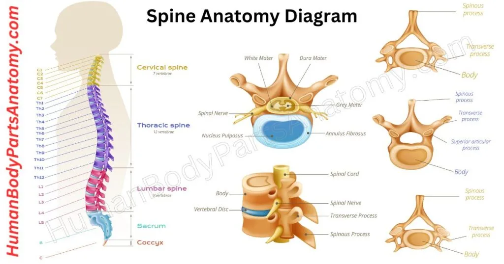
The human spine plays one of the most vital roles in the human body. It supports posture, enables movement, and protects the spinal cord.[1] It also forms the main communication pathway between the brain and the rest of the body, helping the brain control movement, sensation, reflexes, and vital body functions.[2] The spine anatomy consists of 33 vertebrae—including 24 movable (presacral) vertebrae (7 cervical, 12 thoracic, and 5 lumbar) along with the fused sacrum and coccyx—stacked to create a strong yet flexible column.[1] Intervertebral discs, located between the vertebrae, contain a gel-filled center that cushions spinal motion.[3] These discs act like shock absorbers, reducing bone friction and allowing smooth, pain-free actions such as walking, bending, and twisting.[4]
Ligaments connect the vertebrae and maintain spinal stability, while tendons attach muscles to the spine to generate movement and strength.[1] Additionally, facet joints link adjacent vertebrae to guide and control spinal flexibility.[5]
In this article, we’ll explore the anatomy of the spine, its major parts, functions, and importance for everyday movement. A simple labeled diagram will also be provided to help you easily visualize and understand how the spine works.
Spine Anatomy Diagram
Anatomy of the Spine
Regions of the Spine
- Cervical Spine
- Thoracic Spine
- Lumbar Spine
- Sacrum
- Coccyx
Curvatures of the Spine
- Cervical Curve
- Thoracic Curve
- Lumbar Curve
- Sacral Curve
Vertebrae Anatomy
- Body (Centrum)
- Vertebral Arch
- Pedicles
- Laminae
- Vertebral Foramen
- Spinous Process
- Transverse Processes
- Articular Processes
Facet Joints
Intervertebral Discs
- Annulus Fibrosus
- Nucleus Pulposus
Spinal Cord
Nerve Roots
Ligaments of the Spine
- Anterior Longitudinal Ligament (ALL)
- Posterior Longitudinal Ligament (PLL)
- Ligamentum Flavum
- Interspinous Ligaments
- Supraspinous Ligament
- Nuchal Ligament
- Intertransverse Ligaments
Regions of the Spine Anatomy
The human spine is made up of 33 small bones called vertebrae, which fit together to support the upper body and protect the spinal cord.[1] The spine helps us stand tall, hold up our heads, and move freely. These 33 bones are grouped into five regions — cervical, thoracic, lumbar, sacral, and coccygeal — which together keep our body strong and flexible.[1]
What is the Cervical Spine?
The cervical spine consists of seven vertebrae (C1 to C7) arranged in a column from the base of the skull to the upper back.[6] Each vertebra plays a key role in supporting the neck and enabling movement. The first two vertebrae are highly specialized:
- Atlas (C1): A ring-shaped bone that directly supports the skull and allows you to hold your head upright.[6]
- Axis (C2): It is positioned just below the atlas. It provides a pivot point that makes side-to-side “no” head rotation possible.[6]
The vertebrae are connected at the back by facet joints, which guide neck movements like bending, extending, and twisting.[5]
Between each vertebra lies an intervertebral disc, which acts as a cushion to absorb shock, protect the bones, and maintain flexibility.[3]
The cervical spine is surrounded by muscles, ligaments, and tendons that provide stability and strength. Also, the nerves transmit signals between the brain and the rest of the body.[6]
The spinal cord runs through the central canal of these vertebrae. It makes the cervical spine a critical part of the nervous system.[2]
Key Functions of the Cervical Spine
- Protecting the Spinal Cord: The cervical vertebrae form a bony canal that safeguards the spinal cord, preventing damage as signals travel from the brain to the body.[2]
- Supporting and Moving the Head: Despite weighing about 10–13 pounds, the head is fully supported by the cervical spine. It allows a wide range of motions, including forward (flexion), backward (extension), sideways bending (lateral flexion), and rotation.[6]
- Protecting Blood Vessels: Most cervical vertebrae (C1–C6) have transverse foramina that transmit the vertebral arteries (and accompanying veins/sympathetic plexus), which contribute significant blood flow to the brain.[7]

Read More – Cervical Spine Anatomy: Vertebrae, Muscles, Nerves, Functions & Diagrams
Read More –
- How to perform a Cervical spine CT scan?
- How to perform a Cervical MRI scan?
- Mobility of the Cervical Spine in Patients With Unspecific Neck Pain.
- Cervical Spine Motion in Older People.
What is the Thoracic Spine?
The thoracic spine is the middle section of the backbone and is composed of 12 vertebrae (T1–T12) extending from the base of the neck and extending down to the bottom of the rib cage.[1][4] This region is the longest part of the spine.
Together with the rest of the spinal column, these vertebrae form a strong, protective tunnel for the spinal cord, which carries vital signals between the brain and the body.[4]
Structure of the Thoracic Spine
- Vertebrae: The thoracic vertebrae are sturdy, interconnected bones that provide structure and allow controlled movements, such as twisting and limited bending.[4]
- Intervertebral Discs: These discs are located between each vertebra. They are soft, cushion-like discs that absorb shock, reduce friction, and enhance spinal flexibility.[3]
- Supporting Tissues: Muscles, tendons, ligaments, and nerves surround the thoracic spine. They help maintain posture, protect the spinal cord, and enable smooth movement.[1]
- Rib Attachments: Unlike the cervical or lumbar regions, most thoracic vertebrae connect to the ribs, forming the ribcage that safeguards the heart, lungs, and other vital organs.[4]
Key Functions of the Thoracic Spine
- Protection: It shields the spinal cord and branching nerves within the vertebral canal.[2]
- Ribcage Support: It connects to the ribs and creates a stable cage that protects the chest organs.[4]
- Stability & Balance: It works with the ribcage to keep the body upright and stable.[1]
- Movement: It allows safe twisting and side bending. Although forward and backward bending are limited in this region, the thoracic spine provides the greatest rotation compared to other spinal sections.[4]
- Breathing: Its flexibility helps the ribcage expand and contract, playing a vital role in effective inhalation and exhalation.[4]
Read More –
- How to perform a Thoracic spine CT scan?
- How to perform a Thoracic spine X-ray?
- Thoracic Spine Manipulation for Individuals With Low Back Pain.
- Effect of Thoracic Spine Mobilization on Sympathetic Nervous Systems.
What is the Lumbar Spine?
The lumbar spine, also called the lower back, is one of the most important parts of the spine. It is made up of five strong vertebrae (L1 to L5), which are the largest and thickest bones in the spinal column.[1][5] These vertebrae are located below the thoracic spine (mid-back, 12 vertebrae) and above the sacrum, a triangular bone formed by five fused vertebrae.
Structure of the Lumbar Spine
- Strong vertebrae: The lumbar bones are larger, heavier, and stronger than the bones in the neck and mid-back.[5]
- Weight-bearing design: Their size and strength allow them to support most of the body’s weight.[5]
- Muscle and ligament connections: They provide attachment points for major muscles and ligaments, which help with posture, stability, and movement.[1][5]
Key Functions of the Lumbar Spine
The lumbar spine is important for both movement and protection. Its main functions are:
- Supports Body Weight – Distributes weight from the upper body to the pelvis and legs, especially during lifting or physical activities.[5]
- Enables Movement – Provides flexibility for bending forward, backward, twisting, and side-to-side motion.[5]
- Protects Nerves – Safeguards the nerve roots and cauda equina, preventing damage to the nervous system.[2]
- Controls Leg Functions – Lumbar nerves help manage leg movement, balance, reflexes, and sensory functions.[2]
Why the Lumbar Spine Matters
The lumbar spine is strong yet flexible, designed to balance support, movement, and nerve protection. Without it, everyday activities like sitting, standing, bending, and lifting would not be possible.
Keeping your lumbar spine healthy is essential for mobility, stability, and overall quality of life.[5]
Read More –
- How to perform a Lumbar spine CT scan?
- How to perform a Lumbar MRI scan?
- How to do a disk replacement – lumbar spine?
- Classification of lumbar spine disorders using large language models and MRI segmentation.
What is the Sacrum?
The sacrum is a large, triangular bone located at the base of the spine.[6] It forms through the fusion of five sacral vertebrae (S1–S5) between the ages of 18 and 30.[6][12] It is positioned at the back of the pelvic cavity and connects to four other bones.[12]
The side projections of the sacrum, called alae (or wings), link with the ilium through the sacroiliac joints, forming an L-shaped connection.[12]
Its upper portion joins the last lumbar vertebra (L5). At the same time, the lower end articulates with the coccyx, also known as the tailbone. This connection is supported by small bony extensions called the sacral and coccygeal cornua.[13]
The sacrum’s structure accommodates nearby pelvic organs with three distinct surfaces. The bone is generally concave, curving inward toward the pelvis.
The sacral alae (wings) form the lateral surfaces that articulate with the ilia to transmit weight to the pelvis.[12]
Its broad uppermost region, called the sacral base, tilts forward, creating a prominent inward ridge known as the sacral promontory.
The midsection of the sacrum curves outward toward the back, increasing the space within the pelvic cavity for functional and anatomical needs.[13]
Read More –
What is the Coccyx?
The coccyx, the tailbone, is the small, curved bone at the bottom of your spine. You might not think about it much—until you accidentally fall on it! Despite its size, the coccyx is important in supporting your body and connecting muscles and ligaments.[1][6]
The name “tailbone” comes from its history. Long ago, humans had tails, and the coccyx is a leftover from that time. It’s considered a vestigial part of the body, meaning it’s not essential for survival but is a reminder of our evolutionary past.
Your bones act like your body’s framework, providing support and stability. The coccyx works with the pelvis to help balance your weight when you sit down.[6] Together, they form a tripod that keeps you steady.
The tailbone is also an anchor point for several important muscles, including:
- Gluteus Maximus: The largest muscle in your butt.
- Levator ani: Muscles that help support the pelvic floor.[6]
- Muscles around the anus.
In addition to muscles, the coccyx supports tendons and ligaments that connect to nearby structures. Nerves around the coccyx provide sensation to the area, making it a small but significant part of your body.
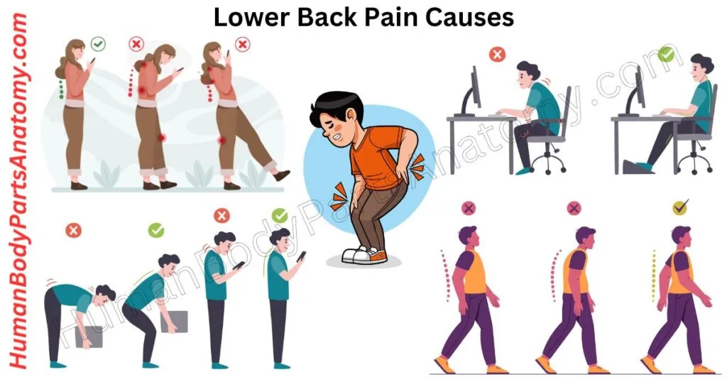
Read More – Lower Back Pain Causes & Symptoms: Common Reasons & Risk Factors
Read More –
- What are the Tailbone Disorders?
- What is tailbone trauma?
- Tailbone pain: How can I relieve it?
- What is tailbone trauma? How to make aftercare?
Curvatures of the Spine
The spine has four natural curves: cervical, thoracic, lumbar, and sacral. These curves, along with the intervertebral discs, act like shock absorbers. They help spread out the stress on your back from daily movements like walking or more vigorous activities like running and jumping.[1][3]
What is a Cervical Curve?
The cervical curve is the natural inward curve of the neck. It forms part of the spine’s overall S-shape when viewed from the side. This curve plays a crucial role in maintaining the body’s biomechanical efficiency and supporting various functions.[1][6]
- Cushioning impacts: It helps absorb the head’s weight and reduces stress on the spine.[6]
- Providing support: The curve ensures the neck stays stable and maintains proper structure.[6]
- Allowing movement: It enables smooth and normal neck movements, like bending and turning.[6]
- Easing tension: The curve helps balance tension in the spinal cord and brainstem for better function.[6]
Read More –
- The association between cervical spine curvature and neck pain.
- Know everything about the Cervical Curve.
What is a Thoracic Curve?
The thoracic curve, also called kyphosis, is the natural outward bend of the upper spine located behind the chest. This curve plays a key role in keeping the body balanced and upright.
In a healthy spine, the curve is usually between 20 and 40 degrees.[4] If it becomes steeper than 45 degrees, it may signal a problem that needs medical care.
Read More –
- Right thoracic curvature in the normal spine.
- Left thoracic curve patterns and their association with disease.
What is the Lumbar Curve?
The lumbar curve, also called lordosis, is the inward arch of the lower spine that naturally forms to support the body’s structure.[5] This gentle curve plays a vital role in absorbing the impact of activities like walking and jumping, protecting the spine, and maintaining balance.[5]
However, if the curve becomes too deep, it can affect your posture and may cause pain or discomfort.
Read More –
- Lumbar curve, trunk muscles, and line of gravity with different heel heights.
- Lumbar lordosis in acute and chronic low back pain patients.
What is a Sacral Curve?
The sacral curve refers to the natural backward bend of the sacrum, a triangular bone at the base of the spine. This curvature forms during fetal development as a primary structure, shaping the human pelvis.[12]
It plays a crucial role in supporting abdominal organs. It contributes to the distinct structure of the pelvic region, which is essential for upright posture and locomotion.[12]

Read More – Lower Back Pain Treatments: Effective Relief Options, Tests & Expert Tips
Read More –
- Pelvic Incidence Is Associated With Sacral Curvature, Sacroiliac Joint Angulation, and Sacral Ala Width.
- Anatomy and clinical significance of sacral variations
Vertebrae Anatomy
Body (Centrum)
The vertebral body is the large front part of a vertebra, designed to support the body’s weight. It is strong yet lightweight, offering maximum support with minimal bone mass.
Vertebral bodies are linked by soft, cushion-like intervertebral discs, forming a flexible column that supports the trunk and head.[4][16] This structure also absorbs forces from muscle contractions during movement.
Vertebral bodies are shaped like cylinders and vary slightly across different parts of the spine. Their width generally increases from the neck (C2) down to the lower back (L3) because each lower vertebra supports more weight.[4][16]
In the lumbar region, the last two vertebrae may vary in width. However, from the first sacral vertebra down to the tailbone’s tip (coccyx), the width decreases steadily.
Read More –
Vertebral Arch
The vertebral arch is a bony structure positioned behind the body of each vertebra. It surrounds an opening called the vertebral foramen. The vertebral arch is composed of two primary components on each side: the pedicles and the laminae.[9][16]
- Pedicles: The pedicles are robust, short projections that extend backward from the upper part of the vertebral body on each side. It is positioned at the junction where the posterior and lateral surfaces of the vertebral body meet. They serve as structural bridges connecting the vertebral body to the vertebral arch.[9][16]
- Laminae: The laminae are broad, flat plates that extend from the pedicles, angling backward and inward. These plates join at the midline, forming the roof of the vertebral arch. Their upper edges and the lower portions of their front surfaces are textured to provide attachment points for the ligamenta flava, which help maintain the stability and flexibility of the spinal column.[9][16]
Read More –
- MRIs Are Less Accurate Tools for the Most Critically Worrisome Pedicles Compared to CT Scans
- Pedicle Morphometry of Lumbar Vertebrae: Male, Taller, and Heavier Specimens Have Bigger Pedicles
Vertebral Foramen
The vertebral foramen is an opening located within each vertebra of the spine. It serves as a passageway for the spinal cord and associated structures. Its front boundary is formed by the back surface of the vertebral body, while the vertebral arch defines its sides and back.[9][10]
Although this feature is present in all vertebrae, its size and shape differ across regions of the spinal column.
- In the cervical spine, the foramen is large and triangular, accommodating the thick spinal cord in this area.
- In the thoracic spine, the foramen is smaller and circular, reflecting the narrower cord region.
- The foramen is triangular and larger than in the thoracic region but smaller than in the cervical.
It provides space for the expanding nerve roots in the lower back.
Read More –
- The vertebral foramen: a report concerning its contents
- Tortuous vertebral artery triggering vertebral foramen expansion and radiculopathy in a 19-year-old patient: a case report
Spinous Process
The spinous process is a bony bump that sticks out from the back of each vertebra in the spine. It plays an important role in supporting the spine and helping it move.[16] Muscles and ligaments attach to these bony projections, making it easier for the spine to bend, twist, and rotate.
Read More –
- Anatomy and Pathologies of the Spinous Process.
- Lumbar Spinous Process Impaction Injuries Caused by Extension Stress in Adolescent Athletes: A Report of Two Cases.
- Spinous Process Fractures in Osteoporotic Vertebral Fractures: A Cross-Sectional Study.
Transverse Processes
The transverse process is a small bony projection on each side of a vertebra in the spine. It is located where two parts of the vertebra, the pedicle and lamina, meet.[16]
These projections provide attachment points for muscles and ligaments, helping to stabilize the spine. They also assist in movements like bending sideways and rotating the body.
By connecting to these supportive structures, the transverse processes contribute to the spine’s strength, flexibility, and proper posture.[16]
Read More –
- Variations of spinous and transverse process length in the human lumbar spine.
- Accessory articulation of cervical vertebral transverse process: a rare case.
Articular Processes
Articular processes are bony projections on each vertebra that contribute to the spine’s movement and flexibility. It is positioned where the lamina meets the pedicle. Every vertebra features two superior and two inferior articular processes. These structures form joints with adjacent vertebrae, enabling controlled motion and stability.[5]
- Superior Articular Processes: These extend upward and are angled slightly backward and outward, allowing them to connect with the inferior processes of the vertebra above.
- Inferior Articular Processes: These project downward and are oriented forward, with a slight inward tilt, aligning with the superior processes of the vertebra below.
Together, these processes form the facet joints, which play a crucial role in maintaining spinal alignment while allowing a range of movements, such as bending and twisting.
Read More –
Facet Joints in Spine Anatomy
These joints are small connections between the bones (vertebrae) in your spine and are located on both sides of each vertebra. It links the bones together and helps your spine move and bend.[5]
Without them, your movements would feel stiff and restricted. These joints belong to a group called synovial joints, like your knees or elbows.
Synovial joints allow smooth movement because the ends of the bones are covered with a slippery layer called articular cartilage.[5] This cartilage cushions the bones and reduces rubbing when you move.
Each facet joint is surrounded by a soft tissue covering called the joint capsule. This capsule contains strong ligaments that hold the joint in place and a special liquid called synovial fluid.
The fluid acts like oil in a machine, keeping the joints lubricated so they can move easily and without friction.
Read More –
- Lumbar Facet Joint Disease: What, Why, and When?
- Facet Joints of the Spine: Structure-Function Relationships, Problems and Treatments, and the Potential for Regeneration.
- Facet Joint Syndrome: Pathophysiology, Diagnosis, and Treatment.
Intervertebral Discs in Spine Anatomy
These Intervertebral discs are cushion-like pads located between the bones of the spine (vertebrae). They are flat, round, and about half an inch thick. These discs have two main parts:
- Nucleus Pulposus: This is the jelly-like center of the disc. It contains much water, which gives the disc flexibility and helps it absorb shocks.[3]
- Annulus Fibrosus: This is the tough outer layer of the disc, made up of strong, stretchy fibers arranged in layers, like elastic bands.[3]
Discs work like tiny shock absorbers. The outer fibers of the annulus keep the bones of the spine together while the jelly-like nucleus acts as a cushion, allowing smooth movement. When you move, the nucleus helps the vertebrae roll over each other, like a ball bearing.
The nucleus is mostly fluid, which changes throughout the day. At night, when you lie down, the discs absorb fluid and “plump up.” During the day, as you stand and move, the fluid is gradually pushed out.
As we age, our discs lose their ability to hold fluid. It makes them thinner and less flexible, which is why people often get shorter as they grow older.[7]
Read More –
- What is the Intervertebral disc disease?
- Self-healing injectable multifunctional hydrogels for intervertebral disc disease.
Spinal Cord in Spine Anatomy
The spinal cord is a soft, tube-shaped structure made of nerve tissue. These nerves run inside the backbone and link the brain with the rest of the body. It begins at the base of the brain, just below the back of the head, and extends down to the lower back.
Although it passes through the spine, the spinal cord itself is shorter and ends around the upper part of the lower back.[2][8] After this point, the nerves continue as a bundle called the cauda equina (Latin for “horse’s tail”).
In adults, the spinal cord measures about 45 cm (18 inches) in men and 43 cm (17 inches) in women. Its thickness is not uniform—it is widest in the neck (cervical region) and lower back (lumbar region), and thinnest in the middle (thoracic region).[14]
The surrounding bones of the spine provide strong protection for this vital nerve pathway.
Function
- The spinal cord’s main job is to carry messages between your brain and your body. It sends signals from your brain to your muscles to control movement and brings information from your body’s senses back to your brain.[2][14]
- It also manages simple, automatic actions called reflexes, like quickly pulling your hand away from something hot.[2]
- Some parts of the spinal cord can handle basic movement patterns, like the rhythm needed for walking, without direct help from your brain.[2]
Read More –
- What are the Spinal Cord Diseases?
- What are the Spinal Cord Injuries?
- Overview of Spinal Cord Disorders.
- What is Spinal cord trauma?
- Different Spinal Cord Injury Rehabilitation Basics.
Nerves in Spine Anatomy
The spinal cord serves as a communication highway, linking the brain to the rest of the body through a network of nerve fibers.
These fibers branch into pairs of nerve roots that exit through small openings (foramina) between the vertebrae. Each segment of the spinal cord connects to specific regions of the body, which is why injuries to different parts of the spinal cord cause distinct effects.[2]
- Nerves from the cervical spine control the upper chest, arms, and shoulders.
- The thoracic spinal nerves manage the chest and abdominal areas. Lumbar spine nerves extend to the legs and regulate bowel and bladder functions.
These nerves play a crucial role in coordinating muscle movements and regulating the activity of organs.
In addition to controlling movement, spinal nerves carry sensory signals to the brain. They enable you to feel sensations such as pressure, temperature, and pain.
When tissues in your body are injured, the nerves transmit pain signals to the brain as a warning system. If the nerves themselves are damaged, it can result in symptoms like pain, numbness, or tingling along the nerve’s pathway.[2]
This intricate system keeps the body in sync, ensuring that both movement and sensation are well-regulated.
Read More –
Ligaments of the Spine Anatomy
Anterior Longitudinal Ligament (ALL)
The anterior longitudinal ligament is a strong, wide band of fibrous tissue that spans the front surfaces of the vertebral bodies and intervertebral discs.
It begins at the occipital bone near the foramen magnum and attaches to the anterior tubercle of the atlas (C1). From there, it continues downward, anchoring to the front (pelvic) surface of the upper sacrum.[16][12]
This ligament consists of multiple layers and plays a key role in stabilizing the spine. It supports the connections between vertebral bodies and intervertebral discs while uniquely serving as the sole ligament that prevents excessive backward bending, or hyperextension, of the spine.[16][12]
Read More –
Posterior Longitudinal Ligament (PLL)
The posterior longitudinal ligament runs along the back surfaces of the vertebral bodies within the vertebral canal. It connects to both the vertebral bodies and intervertebral discs, stretching from the axis (C2) down to the sacrum.[10]
At its upper end, it continues as the tectorial membrane, extending into the base of the skull. Unlike the broader and stronger anterior longitudinal ligament, the posterior longitudinal ligament is narrower and less robust.
Its main functions are to limit excessive forward bending (hyperflexion) of the spine and to reduce the risk of the nucleus pulposus protruding backward through an intervertebral disc.[10]
Read More –
Ligamentum Flavum
The ligamenta flava are strong, flexible bands that connect the arches of neighboring vertebrae in the spine. Made mostly of yellow elastic tissue, they are located at the back of the vertebral canal.[11][16][17]
Each ligament stretches from the upper edge of one vertebra to the lower edge of the vertebra above it, meeting in the middle to form a continuous band.
Their main function is to support the spine during movement. They prevent the vertebrae from separating too much when you bend forward and help the spine return smoothly to its natural upright position.[11][16][17]
Interspinous Ligaments
The interspinous ligaments are thin bands of tissue that link the spinous processes of neighboring vertebrae. They stretch from the base to the tip of each spinous process, connecting with the ligamenta flava at the front and the supraspinous ligament at the back.[16]
Supraspinous Ligament
The supraspinous ligament is a strong, cord-like structure that links the tips of the spinous processes from the C7 vertebra to the sacrum. It transitions into the nuchal ligament in the cervical region.[16]
This ligament plays a vital role in stabilizing the spine by limiting excessive forward bending (flexion) and preventing the spinous processes from moving apart during such movements.[16]
Nuchal Ligament
The nuchal ligament is a strong ligament located at the back of the neck. It stretches from the base of the skull down to the spinous process of the seventh cervical vertebra (C7).
It attaches to key points, including the external occipital protuberance, the back edge of the foramen magnum, the posterior tubercle of the first cervical vertebra (C1), and the tips of the spinous processes of the other cervical vertebrae.[16] At the lower end, it connects with the tip of C7 and continues into the supraspinous ligament.
This ligament plays an important role in supporting the head and neck. It helps prevent the head from bending too far forward (excessive flexion) and assists in bringing it back to its normal upright position.[16]
In addition, the nuchal ligament serves as an attachment site for several neck and shoulder muscles, which adds strength, stability, and movement to the upper body.[16]
Intertransverse Ligaments
The intertransverse ligaments are thin bands of connective tissue linking the transverse processes of neighboring vertebrae.[16] They span from the top edge of one vertebra’s transverse process to the bottom edge of the one above it.
Unlike many other spinal ligaments, they lack clear medial and lateral boundaries and often merge with nearby muscles. Their primary role is to restrict excessive side-bending movements of the spine, helping maintain stability during lateral flexion.[16]
Read More –
- A Pilot Study on the Age-Dependent, Biomechanical Properties of Longitudinal Ligaments in the Human Cervical Spine
- Anatomic morphology and clinical significance of intraforaminal ligaments of the cervical spine
FAQ’s
The human spine is divided into five main regions: cervical (neck), thoracic (mid-back), lumbar (lower back), sacral, and coccygeal (tailbone). It consists of 33 vertebrae, which protect the spinal cord and support body movement.[1][5][6]
There are 33 vertebrae in total—7 cervical, 12 thoracic, 5 lumbar, 5 sacral (fused), and 4 coccygeal (fused). In adults, these lower segments fuse to form a total of 26 movable bones.[1][5]
The spine supports the body’s weight, maintains posture, and protects the spinal cord. It also enables flexibility and movement, like bending, twisting, and rotating the torso.[1][5]
The spine is the bony structure made of vertebrae, while the spinal cord is the soft bundle of nerves that runs inside it, transmitting signals between the brain and the rest of the body.[2][8]
The lumbar spine (lower back) is the most commonly affected area due to its role in supporting most of the body’s weight and its involvement in frequent movement and lifting.[5][12]
Spinal discs are cushion-like pads located between vertebrae. They act as shock absorbers, prevent bones from rubbing together, and allow smooth flexibility and motion in the spine.[3][7]
Maintain good posture, exercise regularly, lift with your legs (not your back), maintain a healthy weight, and ensure ergonomic seating. Stretching and strengthening your core muscles also protects spinal health.[5][15]
A misaligned spine can cause nerve compression, muscle pain, posture problems, and restricted movement. Chiropractic adjustments, physical therapy, and posture correction can help restore alignment and relieve symptoms.[5][15]
References-
- DeSai C, et al. (2023). Anatomy, Back, Vertebral Column. StatPearls. NCBI Bookshelf. https://www.ncbi.nlm.nih.gov/books/NBK525969/
- Waxenbaum JA, et al. (2023). Physiology, Spinal Cord. StatPearls NCBI. Bookshelf. https://www.ncbi.nlm.nih.gov/books/NBK544267/
- MedlinePlus / U.S. National Library of Medicine (NLM) — Intervertebral disk: MedlinePlus Medical Encyclopedia. Updated Aug 27, 2024. https://medlineplus.gov/ency/imagepages/19469.htm
- Waxenbaum JA, et al. (2023). Anatomy, Back, Thoracic Vertebrae. StatPearls. NCBI Bookshelf. https://www.ncbi.nlm.nih.gov/books/NBK459153/
- Institute for Quality and Efficiency in Health Care (IQWiG). (2025). In brief: How does the spine work? InformedHealth.org. NCBI Bookshelf. https://www.ncbi.nlm.nih.gov/books/NBK279468/
- Rahman S, Das J. (2023). Anatomy, Head and Neck: Cervical Spine. StatPearls. NCBI Bookshelf. https://www.ncbi.nlm.nih.gov/books/NBK557516/
- Vo N, et al. (2016). Molecular Mechanisms of Biological Aging in Intervertebral Discs. J Bone Miner Res, 31(6):1139-50. PMCID: PMC4988945. https://pmc.ncbi.nlm.nih.gov/articles/PMC4988945/
- Ganapathy MK, et al. (2024). Neuroanatomy, Spinal Cord Morphology. StatPearls. NCBI Bookshelf. https://www.ncbi.nlm.nih.gov/books/NBK545206/
- Peabody T, et al. (2023). Anatomy, Back, Vertebral Canal. StatPearls. NCBI Bookshelf. https://www.ncbi.nlm.nih.gov/books/NBK557587/
- Aryal V, et al. (2023). Anatomy, Back, Posterior Longitudinal Ligament. StatPearls. NCBI Bookshelf. https://www.ncbi.nlm.nih.gov/books/NBK560691/
- Sayit E, et al. (2013). Dynamic Changes of the Ligamentum Flavum in the Cervical Spine Assessed with Kinetic Magnetic Resonance Imaging. Global Spine Journal. PMC / NIH. https://pmc.ncbi.nlm.nih.gov/articles/PMC3854599/
- Sattar MH, et al. (2023). Anatomy, Back, Sacral Vertebrae. StatPearls. NCBI Bookshelf. https://www.ncbi.nlm.nih.gov/books/NBK551653/
- Kim DW, et al. (2014). Morphologic Diversities of the Sacral Canal in Children. PMCID: PMC4099238. https://www.ncbi.nlm.nih.gov/pmc/articles/PMC4099238/ — PMID: 24779232
- Mortell T, Mortezaei A, Samrid R, Keshavarzi S, Inoue S, Kikuchi K, Iwanaga J, Dumont A, Tubbs R. (2025). Comprehensive Review of the Cervical Ligamenta Flava. Neurospine Journal. PMC / NIH. https://pmc.ncbi.nlm.nih.gov/articles/PMC11961527/
- U.S. National Library of Medicine. (2024). Taking Care of Your Back at Home. MedlinePlus. https://medlineplus.gov/ency/article/002119.htm
- Johns Hopkins Medicine Staff. (2025). Facts About the Spine, Shoulder, and Pelvis. Johns Hopkins Medicine. https://www.hopkinsmedicine.org/health/treatment-tests-and-therapies/facts-about-the-spine-shoulder-and-pelvis
- Tortonese D, et al. (2023). Ligamentum flavum analysis in patients with lumbar discus hernia and lumbar spinal stenosis. Diagnostics (Basel), https://pmc.ncbi.nlm.nih.gov/articles/PMC9992359/
Read More-
Lower Limb
- Hip Bone Anatomy – Complete Guide with Parts, Names, Functions & Diagram
- Complete Guide on Leg Anatomy with Parts, Functions & Diagram
- Complete Guide to Thigh Muscle Anatomy: Learn Parts, Names & Diagram
- Knee Anatomy: Complete Guide to Parts, Names, Functions & Diagram
- Femur Anatomy: Complete Guide with Parts, Names, Functions & Diagram
- Hip Muscle Anatomy – Complete Guide with Parts, Names, Functions & Diagram
Upper Limb
- Complete Guide to Finger Anatomy with Parts, Names, Functions & Diagram
- Complete Guide to Forearm Anatomy: Parts, Names, Functions & Diagram
- Comprehensive Guide to Arm Anatomy: Parts, Names & Diagram
- Comprehensive Guide to Hand Anatomy: Parts, Functions & Diagram
- Ultimate Guide to Bicep Anatomy: Parts, Names, Functions & Diagram
- Shoulder Anatomy: Ultimate Guide to Parts, Names, Functions & Diagram
- Wrist Anatomy: Ultimate Guide to Parts, Names, Functions & Diagram
- Complete Guide to Nail Anatomy with all Parts, Names & Diagrams
Human Head
- Skull Anatomy: Complete Guide with Parts, Names, Functions & Diagram
- Ultimate Guide to Eye Anatomy: Parts, Structure, Functions & Diagram
- Tongue Anatomy: Complete Guide with Parts, Names, Functions & Diagram
- Mouth Anatomy: Complete Guide with Parts, Names, Functions & Diagram
- Complete Guide to Tooth Anatomy: Learn Parts, Names & Diagram
- Ultimate Guide to Ear Anatomy: Parts, Structure, Functions & Diagram
Organs
- Kidney Anatomy: Complete Guide with Parts, Names, Functions & Diagram
- Liver Anatomy: Complete Guide with Parts, Names, Functions & Diagram
- Heart Anatomy: Complete Guide with Parts, Names, Functions & Diagram
Medical Disclaimer
All content on HumanBodyPartsAnatomy.com is educational and based on verified, peer-reviewed medical sources. Articles are authored or reviewed by qualified medical or biomedical professionals to ensure accuracy.
This website does not provide medical advice, diagnosis, or treatment. Always consult a licensed healthcare professional for personal medical guidance.
No commercial or promotional interests influence the medical content published on this site.